Received: 5 July, 2007. Accepted: 12 October, 2007. Invited Review
Biological and Molecular Characterization of Olive latent virus 1
Maria do Rosário Félix
1,3*•
Joana M. S. Cardoso
2,3•
Solange Oliveira
2,3•
Maria Ivone E. Clara
1,31 Departamento de Sanidade Animal e Vegetal, Universidade de Évora, Apartado 94, 7002-554 Évora, Portugal
2 Departamento de Biologia, Universidade de Évora, Apartado 94, 7002-554 Évora, Portugal
3 ICAM, Instituto das Ciências Agrárias Mediterrâneas, Universidade de Évora, Apartado 94, 7002-554 Évora, Portugal
Corresponding author: * [email protected]
ABSTRACT
Olive latent virus 1 (OLV-1) belongs to the Necrovirus genus, Tombusviridae family and is pathogenic to olive, citrus and tulip plants. It is
easily mechanically transmissible to indicator plants causing necrotic lesions and can be transmitted through the soil into the plant roots in the absence of biological vectors. Infected cells contain virus aggregates, inclusions made up of excess of viral coded peptides and extensive vesiculation in the cytoplasm. The virions are isometric with ca. 30 nm, possess a monopartite single-stranded positive-sense RNA genome sized 3700 nt with 5 open reading frames (ORFs) and small inter cistronic regions. ORF 1 encodes a polypeptide with a molecular weight of 23 kDa and the read through of its amber stop codon results in ORF 1 RT that encodes the virus RNA dependent RNA polymerase with 82 kDa. ORF2 and ORF3 encode two small peptides, with 8 kDa and 6 kDa, respectively, which appear to be involved in the virus cell-to-cell movement. ORF 4 is located in the 3′-terminal and encodes a protein with 30 kDa identified as the viral coat protein. The complete genomic sequences of two well characterized OLV-1 isolates (obtained from citrus and olive) are similar, revealing an overall nucleotide sequence identity of 95%. The electrophoretic profile of the dsRNAs recovered from infected tissues exhibits three major species with ca. 3.7, 1.5, and 1.3 kbp. Application of molecular techniques based on PCR and on dot blot hybridiza-tion has been successfully used for routine diagnosis of OLV-1 infechybridiza-tions.
_____________________________________________________________________________________________________________
Keywords: OLV-1, olive, molecular characterization, necrovirus, virus diagnosis
Abbreviations: bp, base pair; CP, coat protein; dsRNA, double stranded RNA; ELISA, Enzyme linked immunosorbent assay; kb, kilo
base; nt, nucleotide; OMMV, Olive mild mosaic virus; ORF, open reading frame; PCR, polymerase chain reaction; RdRp, RNA dependent RNA polymerase; RT-PCR, reverse transcription polymerase chain reaction; ssRNA, single stranded RNA; TNV-A, Tobacco
necrosis virus A; TNV-D, Tobacco necrosis virus D
CONTENTS
INTRODUCTION... 170
VIRUS PROPERTIES...171
Biological and physico-chemical properties ...171
Cytopathology ...171
VIRAL DISSEMINATION ...172
GENOME ORGANIZATION AND EXPRESSION...172
PHYLOGENETIC ANALYSIS ...173
DIAGNOSIS ...173
Dot blot hybridization...175
Reverse Transcription–Polymerase Chain Reaction ...176
CONCLUDING REMARKS ...176
ACKNOWLEDGEMENTS ...176
REFERENCES...176
_____________________________________________________________________________________________________________
INTRODUCTION
Olive latent virus 1 has an apparently restricted natural host
range. It was first detected in olive (Olea europaea L.) trees growing in Southern Italy that showed either no symptoms or occasional fasciation and bifurcation of stems (Gallitelli and Savino 1985). The virus was recovered by means of mechanical inoculation of olive flower extracts onto herba-ceous indicator plants where it caused, mainly, local lesions (Table 1). Following virus purification and characterization it was revealed that it had physico-chemical properties simi-lar to those of necroviruses but differed serologically from them. Thus, it was proposed as a new species of the
Necro-virus genus (Gallitelli and Savino 1985; Martelli et al.
1996). In 1995, similar isolates were found in olive in
Jor-dan, in Portugal (Félix and Clara 2002) and successively in other countries of the Middle East, being likely that the virus is widely distributed wherever the crop is grown (Table 1). However, its economic importance is yet to be ascertained.
In addition to olive, in Turkey OLV-1 was detected in several citrus species where it appeared symptom less, as well as, in many plants affected by the ‘chlorotic dwarf dis-ease’, that is characterized by leaf malformation, chlorotic flecking in the interveinal areas and oak leaf patterns (Çinar
et al. 1993; Martelli et al. 1996). In Portugal several isolates
were obtained from Galega vulgar, Cordovil de Serpa and Verdeal Alentejana cultivars showing low vigor, leaf chlo-rosis (Fig. 1A) and, occasionally, no symptoms. Recently, a virus recovered from tulips, in Japan, exhibiting mottling
and yellow streak leaf symptoms (Kanematsu et al. 2001) was identified as an OLV-1 isolate (Pare P) based on a notably high homology of an amplified viral coat protein (CP) genomic sequence with that of the citrus isolate, and on the lack of serological relationship with other necro-viruses.
Since year 2000, OLV-1 is recognized by the Interna-tional Committee on Taxonomy of Viruses (van Regen-mortel et al. 2000), as a distinct and definitive species with-in the Necrovirus genus of the Tombusviridae family.
Here, an up-to-date overview is provided focusing on OLV-1 properties, cytology, molecular genome organiza-tion and expression strategies in comparison with that of other members of Tombusviridae, using the isolates from citrus (Grieco et al. 1996) and from the olive cultivar ‘Ga-lega vulgar’, in Portugal, (GM6) (Félix et al. 2005b) as mo-dels, since they are the only ones studied in sufficient detail.
VIRUS PROPERTIES
Biological and physico-chemical properties
OLV-1 can infect a range of herbaceous hosts (Table 1) where it causes local lesions ranging from chlorotic to nec-rotic or reddish, as in Gomphrena globosa, except in N.
benthamiana, where it is reported to also induce systemic
mosaic (Gallitelli and Savino 1985; Merciega et al. 1996). In our work with the Portuguese olive isolate, GM6, syste-mic leaf mosaic seldom occurs but preliminary evidence shows that mixed inoculation with Olive mild mosaic virus (OMMV) results in systemic invasion of N. benthamiana by both necroviruses (Félix et al. 2006b). OLV-1 inoculation of young tobaccos at 4 to 6 leaf stage, employing
medium-high inocula, leads to a complete necrosis with the plant dying prematurely within a few days.
The virus is easily purified by means of butanol and chloroform clarification of infected plant extracts, followed by polyethylene glycol precipitation and recovery through differential centrifugation cycles. Partial purified virus sus-pensions, when ultracentrifuged in sucrose density gradients, sediment as a single component, with ca. 111 S, which is infectious (Gallitelli and Savino 1985). Transmission elec-tron microscopy of uranyl acetate treated purified prepara-tions shows a homogeneous population of isometric parti-cleswith a 30 nm diameter and an outline that appears roun-ded to slightly angular depending on whether the particles are free or closely packed (Fig. 1B). Similar size and mor-phology are also shared by the necrovirus TNV-A, TNV-D (Merciega et al. 1996) and OMMV (Cardoso et al. 2004).
Gel electrophoresis analysis of the viral capsid dissoci-ated in the presence of SDS and mercaptoethanol shows it to be made of a major polypeptide with an apparent mole-cular mass of 32 kDa. The virus is moderately immuno-genic. Serological analysis by Ouchterlony’s gel double dif-fusion, using polyclonal antibodies raised against the OLV-1 strain (olive, Italian) showed it to be unrelated to several tombusvirus tested (Gallitelli and Savino 1985) but indistin-guishable from the citrus Turkish isolate and from a few others from Italian, Jordanian and Portuguese olives (Mer-ciega et al. 1996; Félix and Clara 2002). These observations indicate that the virus capsid is a conserved feature among isolates present in geographically distant regions. On the other hand, ELISA tests using OLV-1 GM6 antigen did not react with OMMV specific antibodies nor with a commer-cial mixture of specific immunoglobulins for eleven dif-ferent TNV serotypes (Loewe Phytodiagnostic, Germany), demonstrating a total lack of serological relationship with those necroviruses.
Analysis of the nucleic acid fraction extracted from OLV-1 preparations by a standard SDS-phenol treatment re-vealed it to consist of a single stranded RNA molecule ca. 1.4 × 10⁶ Da (ca. 3.7 kb) infectious to N. benthamiana. On the other hand, dsRNA fractions, extracted from infected tissues by means of phenol, cellulose chromatography of the aqueous phase and serial elutions with appropriate buf-fers, were found to contain three major classes of dsRNA. Their sizes, 2.6 × 10⁶, 1.05 × 10⁶ and 0.94 × 10⁶ Da,
indi-cate that they are the replicative forms of the full length viral RNA and of two smaller subgenomic RNAs, respec-tively (Merciega et al. 1996; Pantaleo et al. 1999). Compa-rative analysis of the OLV-1 RNAs related with those of TNV-A, TNV-D (Merciega et al. 1996) and OMMV (Car-doso et al. 2004) shows them to be similar in number and size.
Cytopathology
Significant ultrastructural changes occur in cells of OLV-1
Table 1 Host range, symptoms and world distribution of OLV-1.
Natural hosts Experimental hosts
Plant Associated symptoms Plant species Induced symptoms
Geographical distribution (first report)
Olive (several cultivars)
Tree low vigour, stem fasciation and bifurcation, leaf chlorosis; no symptoms
Nicotiana benthamiana Leaf local necrosis and
systemic mosaic or necrotic local lesions only Citrus
(lemon, lime, tangelo, grapefruit and orange)
Leaf deformation and chlorotic flecking; no symptoms
Tulip Leaf mottling and yellow streaking
Celosia cristata, Chenopodium amaranticolor, Ch. quinoa, Cucumis sativus, Cucurbita pepo, Datura stramonium, Gomphrena globosa, Momordica balsamina, Nicotiana cavicola, N. clevelandii, N. glutinosa, N. megalosiphon, N. occidentalis, N. rotundifolia, N. rustica, N. tabacum, Ocymum basilicum, Petunia hybrida, Phaseolus aureus, Ph. vulgaris and Vigna unguiculata
Local lesions
Italy (Gallitelli and Savino 1985) Jordan (Martelli et al. 1995) Turkey (Martelli et al. 1996) Portugal (Félix and Clara 2002) Japan (Kanematsu et al. 2001) Lebanon (Fadel et al. 2005) Syria (Al Abdullah et al. 2005)
A B
Fig. 1 Symptoms exhibited by leaves of an OLV-1 infected olive tree
showing mild to intense chlorosis (A), electron micrograph of a purified preparation of OLV-1 (GM6 olive isolate) negatively stained in 2% aqueous uranyl acetate, Bar = 100 nm (B).
infected hosts as compared with that of non-inoculated con-trol plants, when observed under electron microscope. Major alterations are: i) presence of electron dense rounded particles, sized about 22 nm in diameter, interpreted as vir-ions. They occur in the cytoplasm of mesophyll paren-chyma cells and in conducting tissues, either scattered or in ordered paracrystaline arrays, and often in association with filaments of proteinaceous nature; ii) extensive vesiculation of the cell membrane system, from which numerous vesi-cles appear to derive, with variable sizes up to 100 nm in diameter. These can be observed within the nuclear enve-lope likely originated from the inner and/or the outer mem-brane of the nucleus, freely scattered in the cytoplasm, aggregated near the dyctiosomes, and inside the vacuole appearing to protrude from the tonoplast. The most part of such abundant vesicular structures contain a fine network of fibrils, usually interpreted as nucleic acid folded strands; iii) cytoplasmic inclusions made up of parallel filaments with a criss-cross pattern or helical structure, often mingled with virus-like particles. Occasionally, these filaments were also seen inside the cell nucleous; iv) small electron dense amor-phous inclusions scattered throughout the cytoplasm; v) anomalous thickening of the cell wall due to deposition of electron lucent callose-like material between that structure and the cell plasma membrane (Castellano et al. 1987).
Generally, the described cell alterations are seen in both locally and systemically infected N. benthamiana as well as in C. quinoa. For the most part, the role of the newly in-duced structures in the infection process or in cell defence, if any, is not known. However, recent immunocytochemical studies using gold labelled antibodies against an
Escheri-chia coli expressed movement protein of the virus,
design-nated p8, revealed their association (identification) with the fibrous cytoplasmic inclusions made up of the thin fila-ments above mentioned. Thus, it is suggested that these in-clusions may represent a form of accumulation of the OLV-1 encoded p8 (Castellano et al. 2005). Further application of the gold immunolabeling technology permitted the iden-tification of the electron dense amorphous inclusions as ex-cess of viral CP accumulating in the cell cytoplasm (Panta-leo et al. 2006).
VIRAL DISSEMINATION
It is generally accepted that olive viruses are disseminated through infected vegetative material, that is, rooted stem cuttings used for plant propagation. Recent studies have shown that OLV-1 is detectable by sensitive RT-PCR ana-lysis in whole olive flowers (Lobão et al. 2002), pollen (Saponari et al. 2002), fruit pulp (Félix, unpublished) as well as in a high percentage (over 80%) of seedlings origin-ated from seeds of an infected olive tree (Saponari et al. 2002), demonstrating the virus seed transmission. Epide-miologically these data are relevant as they indicate that OLV-1 may be spreading among field olive plants by means of ovule fertilization with infected pollen and by means of grafting olive cultivars that are ‘recalcitrant’ to rooting onto seedlings originated from infected seeds, a practice in use in some olive growing regions.
Laboratory experiments using N. benthamiana as plant model showed that leaf inoculation with OLV-1 (the Portu-guese GM6 isolate) results in local lesions, as expected, and in downwards symptom less invasion of plant roots fol-lowed by virus release into surrounding soil. The virus is then capable of invading healthy roots of new N.
bentha-miana plants grown in that soil or in a liquid substrate
containing the virus (Félix et al. 2006b), in the absence of any biological vector. However, in such cases, OLV-1 infec-tion is restricted to the root system, rarely extending to the tobacco lower leaf, as clearly shown by ELISA and RT-PCR (Félix et al. 2004). It is likely that this mode of dissemina-tion also occurs in olive fields and especially in nurseries, where thousands of stem cuttings are closely packed in a rooting substrate, under a high moisture environment, for ca. two months before transplantation to individual containers.
Plant root proximity and moisture flow are conditions highly conducive to virus spread.
So far there are no indications that biological vectors, other than plant parts, are involved in OLV-1 transmission. Thus, effective avoidance of virus spreading relies mainly upon the use of virus free vegetative material and of non contaminated plant growth substrates in the nurseries.
GENOME ORGANIZATION AND EXPRESSION
The OLV-1 genome consists of a single molecule of ssRNA of messenger sense with ca. 3.7 kb. The genome of two iso-lates, citrus and GM6 olive, have been fully sequenced and characterized (Grieco et al. 1996; Félix et al. 2005b). It is not capped at the 5′ terminus nor polyadenylated at the 3′ terminus.
The viral RNA contains five open reading frames (ORF) and the strategies employed for genome expression includes stop codon suppression, overlapping of reading frames and subgenomic messenger RNAs. In the olive GM6 isolate ORF 1, which follows a non coding leader sequence of 60 nt, begins at the first AUG codon in position 61 and extends to the UAG stop codon in position 666, encoding a 23 kDa (p23) peptide. The read through of that leaky amber termination codon generates an 82 kDa (p82) product encoded by ORF 1 RT completely overlapping that of ORF 1. Located in the central region of the virus genome ORF 2 and ORF 3 encode two small peptides with 8 kDa (p8) and 6 kDa (p6), respectively, whereas ORF 4 occupies the 3′ end of the genome and codes for a 30 kDa protein. The schematic representation of the viral RNA organization is shown in Fig. 2 and the relative position of the five ORFs and other genomic features of the two OLV-1 isolates are compared in Table 2.
The read through portion of the 82 kDa peptide contains the GDD (glycine-aspartic acid-aspartic acid) motif and other typical polymerase sequences suggesting that p82 is the virus RdRp (Poch et al. 1989). Both p23 and p82 are ex-pressed directly from the virus genome and are indispensa-ble for its replication, as inferred from site directed mutage-nesis (Pantaleo et al. 1999). The p8 and p6 products, ex-pressed from a bicistronic subgenomic RNA (sgRNA1) 1519 nt long, are involved in facilitating the virus cell-to-cell movement. Additionally, p6 contains a predicted trans-membrane motif that may have a trans-membrane docking func-tion (Castellano et al. 2005). The p30 is the viral CP which is translated from a 1237 nt sgRNA2. Besides its role in the capsid formation p30 appears to assist the citrus isolate in its systemic spread in infected N. benthamiana plants.
In the CP ‘shell’ domain of three OLV-1 isolates (citrus, GM6 olive and tulip) the ‘S’ signature consensus pattern, consisting of 26 amino acids and conserved among small icosahedral viruses is easily recognized (Falquet et al. 2002). It was found that in all those isolates the amino acid leucine occupies the 17th position in the signature sequence,
contrary to that exhibited by other necrovirus species. Thus the sequence is now proposed to be enunciated as follows:
[FYW]-x-[PSTA]-x(7)-G-x-[LIVM]-x-[LIVM]-x-[FYWIL]-x(2)-D-x(5)-P (Félix et al. 2005a).
Single strand conformation polymorphism (SSCP) ana-lysis of a ca. 750 nt sequence comprising about 90% of the CP gene, which was generated through the application of specific RT-PCR (Fig. 3A) (see Diagnosis) to a set of 14 olive OLV-1 isolates, showed 5 distinct electrophoretic pro-files consisting of 1, 2 or 3 bands corresponding to different conformations of the amplicon molecules, four of which are shown in Fig. 3B (Félix et al. 2006a). SSCP analysis is con-sidered adequate to reveal genetic diversity as it detects a single nucleotide change in DNA fragments sized up to ca. 700 nt (Rubio et al. 1996). The variability observed within that amplified sequence suggests the occurrence of mixed infections, in some cases. That finding is not surprising given the century long life span of the olive host that allows plenty of opportunity for mutation and genetic exchange to take place within the virus natural population andaccumu-
lation of new variants within the tree. Concerning the above 14 isolates no correlation was found between the SSCP electrophoretic pattern observed and neither the host loca-tion nor the type of olive cultivar from which the viral iso-lates were derived (Félix et al. 2006a). Interestingly, the SSCP electrophoretic profile corresponding to the GM6 isolate showed only one band indicating, on one hand, that its viral population is homogeneous in the part of the ge-nome analysed and, on the other hand, that different (com-plementary) sequences can exhibit stable conformations with identical mobility in the gel.
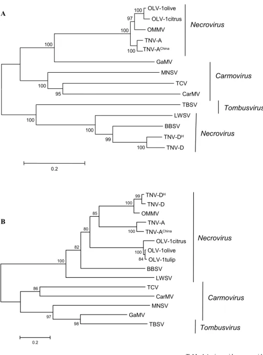
PHYLOGENETIC ANALYSIS
Multiple alignment and phylogenetic analysis of the RdRp sequences from OLV-1 isolates revealed significantly grea-ter sequence similarity with that of OMMV and TNV-A iso-lates than with that of TNV-D isoiso-lates and other
Tombusvi-ridae analysed, indicating that the RdRps of necrovirus
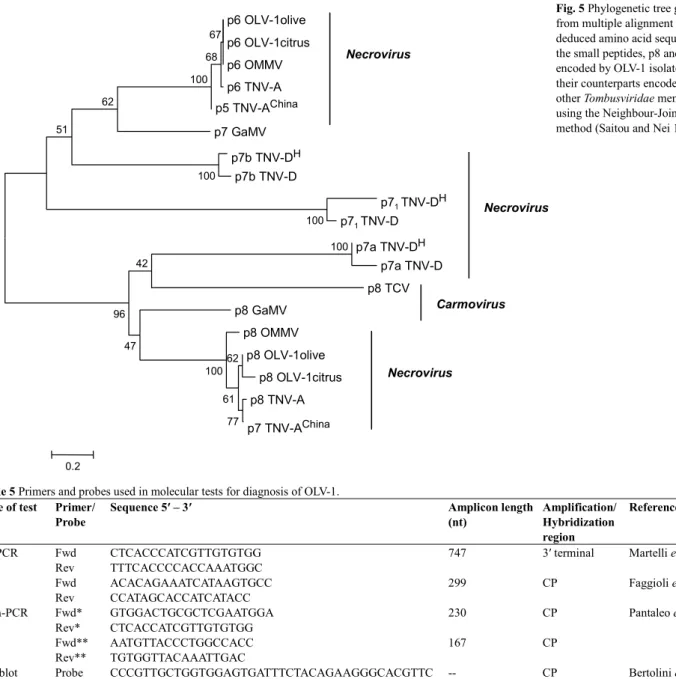
form two distinct clusters (Fig. 4A and Table 3). On the other hand, similar analysis of CP sequences from the same viruses showed a very high sequence similarity among the three OLV-1 isolates, in contrast with that of other necro-virus, namely OMMV, although the necrovirus CPs appear as a single cluster (Fig. 4B and Table 3). Concerning the OLV-1 GM6 isolate small proteins, p6 and p8, it is remar-kable the extensive sequence similarity with those of other
OLV-1 isolates, of OMMV and of TNV-A isolates, as op-posed to that of other necro- and carmovirus analysed (Fig.
5 and Table 4).
In this respect it is interesting to note the case of OMMV that originated from RNA recombination events, probably via a mechanism of replicase-driven template switching mode, where TNV-D RNA function as ‘donor’ and OLV-1 RNA as ‘acceptor’ template. OMMV shares high homology in about two thirds (RdRp, p6 and p8) and one third (CP), with OLV-1 and TNV-D, respectively (Car-doso et al. 2005). OMMV was originally recovered from an olive tree of the cultivar ‘Galega vulgar’, in Portugal, and initially identified as a TNV-D isolate (designated GP) based on ELISA reactions and on the nucleotide sequence of the CP gene (Félix and Clara 2002; Cardoso et al. 2004; Louro 2004), but was later recognized as a distinct necro-virus species based on the complete genome organization, sequencing and comparison with those of other small plant RNA viruses (Cardoso et al. 2004). More recently OMMV was found in Holland appearing to be involved in the Au-gusta complex disease of tulips (Pham et al., unpublished).
Besides OMMV, also GaMV appears to have derived from recombination occurred between a necro- and a tom-busvirus (Ciuffreda et al. 1998), which may explain its rela-tive position in the phylogenetic trees here presented (Figs.
4, 5).
These and other data give support to the idea that the necrovirus is not a homogeneous genus and that some of its species need more research for a better understanding of its taxonomic standing.
DIAGNOSIS
The economic importance of OLV-1 is not yet clearly known. That virus infection is associated with leaf mild mosaic and low vigour of olive trees (Fig. 1A), chlorosis and dwarfing of citrus (Martelli et al. 1996), leaf mottle and yellow streaks in tulips (Kanematsu et al. 2001) but OLV-1 also occurs asymptomatic in these hosts (Table 1). The in-creasing international demand for olive plants and legisla-tion enacted by the European Union, as the Directive 93/48 dated 23-06-93 Conformitas Agraria Communitatis, require that all propagative material of olive produced in nurseries must be free of all viruses. This led to the development of sensitive diagnosis techniques to assist in selection, im-provement and sanitary certification of olive planting mate-rial, particularly in Italy, Portugal and Spain. Reliable virus detection is also needed in epidemiological studies and in establishing strategies for virus disease control.
Fig. 2 Schematic representation of the genomic organization of OLV-1. The thick
line represent the (+) strand RNA genome. The boxes represent the ORFs encoding proteins p23, p82, p8, p6 and p30 kDa. Rt – read through, RdRp – RNA dependent RNA polymerase, CP – coat protein.
Table 2 Comparative genome organization features of OLV-1 isolates.
Properties GM6 (olive) isolate Citrus isolate
Genome full length 3702 3699
5′ NCR1 base composition (60 nt) 36.7% A, 23.3% C, 18.3% G, 21.7% U 35% A, 20% C, 21.6% G, 23.4% U ORF 1 61 to 666 nt 61 to 666 nt ORF 1 RT 666 to 2232 nt 666 to 2232 nt ORF 2 2219 to 2437 nt 2216 to 2443 nt ORF 3 2443 to 2610 nt 2443 to 2613 nt ORF 4 2635 to 3444 nt 2635 to3447 nt
ICR1 (ORF 2 – ORF 3) 5 nt 2 nt
ICR (ORF 3 – ORF 4) 24 nt 21 nt
Size overlap ORF 1 RT – ORF 2 14 nt 17 nt
3′ NCR 258 nt 253 nt
1 ICR – Inter cistronic region; NCR – Non coding region
M S 1 2 3 4
A B
Fig. 3 Agarose gel electrophoretic analysis of the 750 nt amplicon obtained by RT-PCR applied to olive stem tissues using OLV-1 speci-fic primers. M - 1kb Plus DNA Ladder (Invitrogen), S – OLV-1 infected
sample (A). Electrophoretic SSCP profiles of the 750 nt amplicon ob-tained by RT-PCR applied to several naturally OLV-1 infected Portuguese olive cultivars. Numbers above the lanes indicate four different SSCP pat-terns (B).
OLV-1olive OLV-1citrus OMMV TNV-A TNV-AChina GaMV MNSV TCV CarMV TBSV LWSV BBSV TNV-DH TNV-D 100 99 100 100 95 100 100 100 100 97 100 0.2 Necrovirus Necrovirus Carmovirus Tombusvirus TNV-DH TNV-D OMMV TNV-A TNV-AChina OLV-1citrus OLV-1olive OLV-1tulip BBSV LWSV TCV CarMV MNSV GaMV TBSV 98 97 86 84 100 100 99 100 100 82 85 80 0.2 Necrovirus Carmovirus Tombusvirus A B
Fig. 4 Phylogenetic tree generated
from multiple alignment of deduced amino acid sequence of RdRp (A), and of CP (B), encoded by OLV-1 isolates, with their respective counterparts encoded by other
Tombusviridae members , using the
Neighbour-Joining method (Saitou and Nei 1987).
Table 3 Amino acid sequence identity of RdRp and CP encoded by
OLV-1 GM6 genome with their counterparts from other Tombusviridae.
Sequence identity of proteins (%) Virus / isolate RdRp CP OLV-1citrus 97.3 87.7 OLV-1tulip na 98.5 OMMV 92.1 43.3 TNV-A 90.7 41.5 TNV-AChina 90.4 41.1 TNV-D 34.1 42.5 TNV-DH 35.3 42.2 LWSV 33.3 28.2 BBSV 34.8 36.3 GaMV 51.1 13.9 MNSV 37.8 14.6 TCV 36.3 14.6 CarMV 32.0 13.4 TBSV 31.5 14.6
na- not available
NCBI of proteins accession number: OLV-1 (GM6 olive) p8 (AAZ43262) and p6 (AAZ43263); OLV-1citrus p8 (NP_043909) and p6 (NP_043910); OMMV p8 (YP_224018) and p6 (YP_224019); TNV-A p8 (NP_056826) and p6 (NP_056827); TNV-ACh – China isolate p7 (AAT69239) and p5 (AAT69240); TNV-DH – Hungary
isolate p71 (NP_608313), p7a (NP_608314) and p7b (NP_608315); TNV-D p71
(BAA00787), p7a(BAA00788) and p7b (BAA00789); BBSV p5 (NP_758812), p7a (NP_758813) and p7b (NP_758814); LWSV p11 (NP_044742) and p6 (NP_044743); MNSV p7a (NP_041229) and p7b (NP_041230); TCV p8 (NP_620722); GaMV p8 (NP_044734) and p7 (NP_044735).
Table 4 Amino acid sequence identity among the small peptides p8 and p6
encoded by the OLV-1 GM6 genome with their counterparts from other
Tombusviridae.
Virus/isolate Peptide Identity (%)
Virus/isolate Peptide Identity
(%) OLV-1olive p8 100 OLV-1olive p6 100 OLV-1citrus p8 93.2 OLV-1citrino p6 100 OMMV p8 87.6 OMMV p6 100 TNV-A p8 89.0 TNV-A p6 98.2 TNV-ACh p7 91.7 TNV-ACh p5 98.2 TNV-DH p7 1 10.5 TNV-DH p7a 7.6 TNV-DH p7a 20.5 TNV-DH p7b 16.4 TNV-D p71 6.7 TNV-D p7a 6.1 TNV-D p7a 20.5 TNV-D p7b 17.9 BBSV p5 12.3 BBSV p7a 7.9 BBSV p7a 23.2 BBSV p7b 20.0 LWSV p11 13.0 LWSV p6 21.0 MNSV p7a 28.7 MNSV p7a 9.2 TCV p8 15.7 MNSV p7b 27.4 GaMV p8 43.2 GaMV p7 36.2
NCBI of proteins accession number: OLV-1 (GM6 olive) p8 (AAZ43262) and p6 (AAZ43263); OLV-1citrus p8 (NP_043909) and p6 (NP_043910); OMMV p8 (YP_224018) and p6 (YP_224019); TNV-A p8 (NP_056826) and p6 (NP_056827); TNV-ACh – China isolate p7 (AAT69239) and p5 (AAT69240); TNV-DH – Hungary
isolate p71 (NP_608313), p7a (NP_608314) and p7b (NP_608315); TNV-D p71
(BAA00787), p7a(BAA00788) and p7b (BAA00789); BBSV p5 (NP_758812), p7a (NP_758813) and p7b (NP_758814); LWSV p11 (NP_044742) and p6 (NP_044743); MNSV p7a (NP_041229) and p7b (NP_041230); TCV p8 (NP_620722); GaMV p8 (NP_044734) and p7 (NP_044735).
Main difficulties faced when deciding on a suitable de-tection technique reside on the irregular distribution of the virus within the canopy, specially in woody crops as olive and citrus, the low antigen titre and the presence of com-pounds i.e. phenols, polysaccharides, oils and pigments in certain tissues. In addition, all these factors vary throughout the year season interfering with the outcome of some detec-tion tests. Thus time of sampling and type of tissue to be selected are very important in diagnosis. Concerning the olive plant it was found that scrapings of cortical tissues taken from two year stems, in the spring, are very good OLV-1 sources, although fruits and shoots collected in the autumn are also adequate (Faggioli et al. 2005; Varanda 2005).
Recently, dot blot hybridization and various formats of RT-PCR tests were successfully optimized for the direct detection of OLV-1, in single or in mixed infections of olive tissues.
Dot blot hybridization
In these tests the viral target is spotted onto nylon mem-branes and, under appropriate conditions, hybridized to OLV-1 specific riboprobes, designed complementary to a region of the virus genome (Table 5) and labelled with a
non-radioactive marker, as digoxigenin (dig), which is later detected by chemiluminescence or by ELISA (Grieco et al. 2000). Of the several substrates tested, crude olive leaf extracts, total nucleic acids recovered from olive cortical tissues with the RNeasy Plant Extraction Kit (Qiagen), and denatured dsRNA preparations, the latter proved to be the only one allowing successful detection of OLV-1. The sensitivity of the dig-riboprobes used in these reactions was shown to be very high, detecting up to a 1 pg of the RNA target, revealing them very specific and reliable, with poten-tial to be routinely used in plant sanitary selection prog-rammes.
A similar hybridization technique is applicable to the final reaction product of RT-PCR assay (described below) specific for OLV-1. In this case, the final amplicon is de-natured, spotted onto a nylon membrane and hybridized with 3′ dig-labelled probes internal to that PCR product. The label is then colourimetrically evidenced by anti-dig-alkaline phosphatase conjugated antibody, using nitroblue tetrazolium and bromochloroindolyl phosphate as the en-zyme substrate (Bertollini et al. 2001). This procedure has a sensitivity 10 times higher than that of monospecific RT-PCR, and it works best when the viral target is the total RNA fraction recovered from infected tissues through the use of RNeasy Plant mini kit. Although more laborious this
Table 5 Primers and probes used in molecular tests for diagnosis of OLV-1.
Type of test Primer/
Probe
Sequence 5′ – 3′ Amplicon length
(nt) Amplification/ Hybridization region Reference Fwd CTCACCCATCGTTGTGTGG Rev TTTCACCCCACCAAATGGC
747 3′ terminal Martelli et al. 1996 Fwd ACACAGAAATCATAAGTGCC RT-PCR Rev CCATAGCACCATCATACC 299 CP Faggioli et al. 2005 Fwd* GTGGACTGCGCTCGAATGGA Rev* CTCACCATCGTTGTGTGG 230 CP Fwd** AATGTTACCCTGGCCACC RT-n-PCR Rev** TGTGGTTACAAATTGAC 167 CP Pantaleo et al. 2001 Dot blot hybridization
Probe CCCGTTGCTGGTGGAGTGATTTCTACAGAAGGGCACGTTC -- CP Bertolini et al. 2001 -- not applicable; CP, coat protein; Fwd, forward; Rev, reverse; *, **, primers used in the first and second PCR amplification reactions, respectively.
Fig. 5 Phylogenetic tree generated
from multiple alignment of deduced amino acid sequences of the small peptides, p8 and p6, encoded by OLV-1 isolates, with their counterparts encoded by other Tombusviridae members, using the Neighbour-Joining method (Saitou and Nei 1987). p6 OLV-1olive p6 OLV-1citrus p6 OMMV p6 TNV-A p5 TNV-AChina p7 GaMV p7b TNV-DH p7b TNV-D p71 TNV-DH p71 TNV-D p7a TNV-DH p7a TNV-D p8 TCV p8 GaMV p8 OMMV p8 OLV-1olive p8 OLV-1citrus p8 TNV-A p7 TNV-AChina 100 100 100 67 68 100 62 51 42 96 47 100 77 62 61 0.2 Necrovirus Necrovirus Necrovirus Carmovirus
procedure has the advantage of avoiding use of the toxic ethidium bromide employed in gel electrophoresis to reveal the presence of the viral related amplicon.
Reverse Transcription–Polymerase Chain Reaction
RT-PCR in its various formats, conventional two-step or single step, monospecific or multiplex or nested PCR (RT-n-PCR) have been successfully applied to diagnose OLV-1 in single and mixed infections aiming at the certification of propagative material. The type of substrates suitable to detect OLV-1 in olive tissues include crude plant sap, total RNA preparations obtained from cortical stem tissues or from fruits by using commercial extraction kits, and dsRNA fractions obtained from cortical tissues or fruits (Grieco et
al. 2000; Bertollini et al. 2001; Varanda et al. 2006). Crude
plant sap is not always adequate as a substrate to use in RT-n-PCR assays whereas total RNA extracted with the com-mercial kit is the best and easiest method to prepare the tar-get to obtain reliable results.
Typically, a RT-PCR uses 100 mg to 1 g of tissue pow-dered in liquid nitrogen of which a 0.1 g sample is taken and further extracted with the RNeasy Plant mini kit to obtain total RNA preparation (Faggioli et al. 2005). Several primers specific to OLV-1 (Table 5) have been designed to amplify particular sequences of the genome and used rou-tinely in RT-PCR tests to diagnose the virus directly in tis-sues from olive trees (Fig. 5A). In addition to high speci-ficity, RT-PCR also exhibits high sensitivity detecting as little as 10 fg of OLV-1 RNA (Grieco et al. 2000).
This technique has been successfully applied in virus surveys to detect single and multiple targets. For instance, OLV-1 was the 2nd and the 5th most prevalent olive virus in
Lebanon and in Syria, respectively, and at one site in each country, its presence reached 20 % of the sampled trees (Al Abdullah et al. 2005; Fadel et al. 2005). In Portugal, RT-PCR analysis of a collection of mechanically transmitted virus from naturally infected olive tissues revealed that 60% were OLV-1, which is most prevalent in the south (Félix, unpublished).
The development of a multiplex RT-PCR to detect the 3 necrovirus infectious to olive, OLV-1, TNV-D and OMMV, using either total RNA or ds RNA fractions extracted from stems or fruits, allowed the viral survey of a field collection of olive clones in the northeast of Portugal. Data obtained showed that only one tree had OLV-1 among the 35 that were necrovirus infected (Félix et al. 2003; Varanda et al. 2006). In this limited survey it was not possible to distin-guish between TNV-D and OMMV infections as the pri-mers used in the PCR hybridize within the CP domain and both viruses share extensive identity (ca. 77%) in that gene (Cardoso et al. 2005).
The RT-n-PCR technique is much more sensitive than RT-PCR but the experimental procedure presents high risks of contamination, as it involves two separate rounds of PCR reactions, rendering it difficult to be routinely used (Panta-leo et al. 2001). To overcome these Bertollini et al. (2003) adapted a device developed by Olmos et al. (1999) that is a microtube containing a specially adjusted plastic tip inside that allows the complete RT-n-PCR reaction to be carried out in a single closed tube. This avoids or greatly reduces the risk of outside contaminations. The application of such procedure was successful in simultaneously diagnosing four different olive viruses and its sensitivity proved to be at least 100 times higher than that of multiplex RT-PCR (Ber-tollini et al. 2003).
CONCLUDING REMARKS
OLV-1 is a typical necrovirus as shown by its biological and biochemical properties, genome organization and gene ex-pression strategy. It infects two major socio economic crops, olive and citrus, and is present in many groves of Medit-erranean and Middle East countries. The virus is often
asso-ciated to olive decline and citrus dwarfism but its impact in the yield and/or quality of plant products is not known and should be investigated as it is very widespread.
OLV-1 is a plant virus exhibiting several features that contribute for its success in nature, namely its capacity to multiply in different plant genera and to remain viable in the soil, subsequently infecting healthy hosts via their root system. Lacking known fungus, nematode or arthropode vectors, OLV-1 relies for survival on the natural spreading or human trading of infected seeds, rootstocks, scions or bulbs. This emphasizes the importance of developing and use of sensitive OLV-1 diagnostic techniques to assist in plant breeding and sanitary selection programmes to pre-vent or greatly restrict viral dissemination, as required by European Union legislation.
An interesting feature of the OLV-1 genome is its poten-tial ability to act as a ‘parental’ molecule, together with that of TNV-D, to give rise to a new recombinant virus as is the case of OMMV. The recent finding of a virus infecting tulips showing extensive genomic identity with that of OMMV reveals that this virus species is more widespread than anticipated and that additional hosts may emerge. Whether this is a result of selection pressure favouring ge-netic recombination between the two necrovirus species is speculative at this point but very interesting to investigate further.
ACKNOWLEDGEMENTS
The authors thank to the graduate students working in our Plant Virus Laboratory (ICAM/University of Évora) whose efforts con-tributed to our research in this field. The second author is recipient of a PhD fellowship from Fundação para a Ciência e a Tecnologia - FCT (SFRH/BD/16673/2004). This work was partially supported by research grants from the Portuguese Ministries of Agriculture and of Science, Technology and Higher Education.
REFERENCES
Al Abdullah A, El Beaino T, Saponari M, Hallak H, Digiaro M (2005)
Preli-minary evaluation of the status of olive-infecting viruses in Syria. Bulletin
OEPP/EPPO 35, 249-252
Bertolini E, Olmos A, Martínez MC, Gorris MT, Cambra M (2001)
Single-step multiplex RT-PCR for simultaneous and clourimetric detection of six RNA viruses in olive trees. Journal of Virological Methods 96, 33-41
Bertolini E, Olmos A, López MM, Cambra M (2003) Multiplex nested
reverse transcription-polymerase chain reaction in a single tube for sensitive and simultaneous detection of four RNA viruses and Pseudomonas
savasta-noi pv. savastasavasta-noi in olive trees. Phytopathology 93, 286-292
Cardoso JMS, Félix MR, Clara MIE, Oliveira S (2005) The complete
ge-nome sequence of a new necrovirus isolated from Olea europaea L. Archives
of Virology 150, 815-823
Cardoso JMS, Félix MR, Oliveira S, Clara MIE (2004) A Tobacco necrosis
virus D isolate from Olea europaea L.: Viral characterization and coat protein
sequence analysis. Archives of Virology 149, 1129-1138
Castellano MA, Di Franco A, Martelli GP (1987) Electron microscopy of two
olive viruses in host tissues. Journal of Submicroscopic Cytology 19, 495-508
Castellano MA, Loconsole G, Grieco F, Di Sansebastiano GP, Martelli GP
(2005) Subcellular localization and immunodetection of movement proteins of Olive latent virus 1. Archives of Virology 150, 1369-1381
Çinar A, Kersting U, Onelge N, Kormaz S, Sas G (1993) Citrus virus and
virus-like diseases in Eastern Mediterranean region of Turkyie. Proceedings
12th Conference IOCV, Riverside 1993, USA, pp 397-400
Ciuffreda P, Rubino L, Russo M (1998) Molecular cloning and complete
nuc-leotide sequence of Galinsoga mosaic genomic RNA. Archives of Virology
143, 173-180
Fadel C, Digiaro M, Choueiri E, El Beaino T, Saponari M, Savino V, Mar-telli GP (2005) On the presence and distribution of olive viruses in Lebanon.
Bulletin OEPP/EPPO 35, 33-36
Faggioli F, Ferretti L, Albanese G, Sciarroni R, Pasquini G, Lumia V, Barba M (2005) Distribution of olive tree viruses in Italy as revealed by one-step
RT-PCR. Journal of Plant Pathology 87, 49-55
Falquet L, Pagni M, Bucher P, Hulo N, Sigrist CJ, Hofman K, Bairoch A
(2002) The PROSITE database, its status in 2002. Nucleic Acids Research 30, 235-238
Félix MR, Cardoso JMS, Clara MIE (2006b) Single Strand Conformation
Polymorphism analysis of Olive latent virus 1 isolates from Olea europaea L..
Proceedings of the Olivebioteq 2006 - Second International Seminar “Bio-technology and Quality of Olive Tree Products around the Mediterranean
Basin”, Sicily, Italy, Vol II, pp 243-246
Félix MR, Cardoso JMS, Oliveira S, Clara MIE (2003) Detection of Tobacco
necrosis virus and Olive latent virus 1 in Olea europaea L. by multiplex
RT-PCR. Phytopathologia Mediterranea 42, 298
Félix MR, Cardoso JMS, Oliveira S, Clara MIE (2005a) Viral properties,
pri-mary structure and phylogenetic analysis of the coat protein of Olive latent
virus 1 isolate from Olea europaea L. Virus Research 108, 195-198
Félix MR, Cardoso JMS, Varanda CMR, Oliveira S, Clara MIE (2005b)
Complete nucleotide sequence of an Olive latent virus 1 isolate from olive trees. Archives of Virology 150, 2403-2406
Félix MR, Clara MIE (2002) Two Necrovirus isolates with properties of Olive
latent virus 1 and of Tobacco necrosis virus from olive in Portugal. Acta Hor-ticulturae 586, 725-728
Félix MR, Varanda C, Cardoso JMS, Clara MIE (2004) Soil transmission of
an olive isolate of Olive latent virus 1. Proceedings of the 15th International
Plant Protection Congress, Beijing, China, 447
Félix MR, Varanda C, Cardoso JMS, Clara MIE (2006a) Plant root uptake
of Olive latent virus 1 and Olive mild mosaic virus in single and mixed infec-tions. Proceedings of the 12th Congress of the Mediterranean
Phytopatho-logical Union, Rhodes, Greece, pp 516-517
Gallitelli D, Savino V (1985) Olive latent virus 1, an isometric virus with a
sin-gle RNA species isolated from olive in Apulia, Southern Italy. Annals of
Ap-plied Biology 106, 295-303
Grieco F, Alkowni R, Saponari M, Savino V, Martelli GP (2000) Molecular
detection of olive viruses. Bulletin OEPP/EPPO 30, 469-473
Grieco F, Savino V, Martelli GP (1996) Nucleotide sequence of the genome of
a citrus isolate of Olive latent virus 1. Archives of Virology 141, 825-838
Kanematsu S, Taga Y, Morikawa T (2001) Isolation of Olive latent virus 1
from tulip in Toyama Prefecture. Journal of General Plant Pathology 67, 333-334
Lobão DL, Félix MR, Clara MIE, Oliveira S, Leitão FA, Serrano JF (2002)
Detection of Olive latent virus 1 in Olea europaea L. tissues by reverse trans-cription-polymerase chain reaction. XIII Congresso Nacional de Bioquimíca, Lisboa, Portugal, p 102
Louro TJFM (2004) Caracterização de um isolado viral de Olea europaea L.,
produção de anticorpos policlonais específicos, e aplicação de métodos de diagnóstico. MSc Thesis, Universidade de Évora, Évora, Portugal, 91 pp
Martelli GP, Sabanadzovic S, Savino V, Abu-Zurayk AR, Masannat M
(1995) Virus-like disease and viruses of olive in Jordan. Phytopathologia
Me-diterranea 34, 133-136
Martelli GP, Yilmaz MA, Savino V, Baloglu S, Grieco F, Güldür ME, Greco
N, Lafortezza R (1996) Properties of a citrus isolate of Olive latent virus 1, a
new necrovirus. European Journal of Plant Pathology 102, 527-536
Merciega V, Boscia D, Savino V (1996) Comparison of five isolates of Olive
latent virus 1. Phytopathologia Mediterranea 35, 1-8
Olmos A, Cambra M, Esteban O, Gorris MT, Terrada E (1999) New device
and method for capture, reverse transcription and nested PCR in a single closed-tube. Nucleic Acids Research 27, 1564-1565
Pantaleo V, Grieco F, Castellano MA, Martelli GP (1999) Synthesis of
infec-tious transcripts of Olive latent virus 1: genes required for RNA replication and virus movement. Archives of Virology 144, 1071-1079
Pantaleo V, Saponari M, Gallitelli D (2001) Development of a nested PCR
protocol for detection of olive-infecting virus in crude extracts. Journal of
Plant Pathology 83, 143-146
Pantaleo V, Grieco F, Di Franco A, Martelli GP (2006) The role of the
C-ter-minal region of Olive latent virus 1 coat protein in the host systemic infection.
Archives of Virology 151, 1973-1983
Poch O, Sauvaget I, Delarue M, Tordo N (1989) Identification of four
con-served motifs among the RNA-dependent polymerase encoding elements.
The EMBO Journal 8, 3867-3874
Rubio L, Ayllón MA, Gerri J, Pappu H, Niblett C, Moreno P (1996)
Differ-entiation of Citrus tristeza Closterovirus (CTV) isolates by single-strand con-formation polymorphism analysis of the coat protein gene. Annals of Applied
Biology 129, 479-489
Saitou N, Nei N (1987) The neighbour-joining method: A new method for
re-constructing phylogenetic trees. Molecular Biology and Evolution 4, 406-442
Saponari M, Savino V, Martelli GP (2002b) Trasmissioni per seme dei virus
dell’olivo. Frutticoltura 4, 103-105
van Regenmortel MHV, Fauquet CM, Bishop DHL, Carstens EB, Estes MK, Lemon SM, Maniloff J, Mayo MA, McGeoch DJ, Pringle CR, Wickner RB (2000) Virus Taxonomy, Academic Press, San Diego, California,
USA, 1162 pp
Varanda C (2005) Avaliação das técnicas de diagnóstico viral baseadas em
iso-lamento de dsRNA e RT-PCR, para certificação de uma colecção de clones da cv. ‘Negrinha de Freixo’ (D.O.) de Olea europaea L. MSc Thesis, Univer-sidade de Évora, Évora, Portugal, 65 pp
Varanda C, Félix MR, Leitão F, Sismeiro R, Clara MIE (2006) Application
of reverse transcription – polymerase chain reaction to screen a collection of clones of Olea europaea L. for the presence of necrovirus (Tombusviridae).
8th Conference of the European Foundation for Plant Pathology & British
Society for Plant Pathology Presidental Meeting 2006. Sustainable Disease Management: The European perspective, Copenhagen, Denmark, p 21