, a New Family of Transcriptionally
Active Retrotransposons from the Olive Fruit
Fly, with Y Chromosome Preferential
Distribution
Konstantina T. Tsoumani1, Elena Drosopoulou2, Kostas Bourtzis3,4,5, Aggeliki Gariou-Papalexiou6, Penelope Mavragani-Tsipidou2, Antigone Zacharopoulou6, Kostas D. Mathiopoulos1*
1Department of Biochemistry and Biotechnology, University of Thessaly, Larissa, Greece,2Department of Genetics, Development and Molecular Biology, Aristotle University of Thessaloniki (AUTH), Thessaloniki, Greece,3Insect Molecular Genetics Group, IMBB, Vassilika Vouton, 71110 Heraklion, Crete, PO Box 1527, Greece,4Department of Environmental and Natural Resources Management, University of Patras, Agrinio, Greece,5Insect Pest Control Laboratory, Joint FAO/IAEA Division of Nuclear Techniques in Food and Agriculture, Vienna, Austria,6Department of Biology, Division of Genetics, Cell and Developmental Biology, University of Patras, Patras, Greece
*kmathiop@bio.uth.gr
Abstract
Sex chromosomes have many unusual features relative to autosomes. The in depth explo-ration of their structure will improve our understanding of their origin and divergence (degen-eration) as well as the evolution of genetic sex determination pathways which, most often are attributed to them. In Tephritids, the structure of Y chromosome, where the male-determining factor M is localized, is largely unexplored and limited data concerning its sequence content and evolution are available. In order to get insight into the structure and organization of the Y chromosome of the major olive insect pest, the olive flyBactrocera oleae, we characterized sequences from a Pulse Field Gel Electrophoresis (PFGE)-isolated Y chromosome. Here, we report the discovery of the first olive fly LTR retrotransposon with increased presence on the Y chromosome. The element belongs to theBEL-Pao superfam-ily, however, its sequence comparison with the other members of the superfamily suggests that it constitutes a new family that we termedAchilles. Its ~7.5 kb sequence consists of the 5’LTR, the 5’non-coding sequence and the open reading frame (ORF), which encodes the polyprotein Gag-Pol.In situhybridization to theB.oleaepolytene chromosomes showed thatAchillesis distributed in discrete bands dispersed on all five autosomes, in all centro-meric regions and in the granular heterochromatic network corresponding to the mitotic sex chromosomes. The between sexes comparison revealed a variation inAchillescopy num-ber, with male flies possessing 5–10 copies more than female (CI range: 18–38 and 12–33 copies respectively per genome). The examination of its transcriptional activity demon-strated the presence of at least one intact active copy in the genome, showing a differential level of expression between sexes as well as during embryonic development. The higher expression was detected in male germline tissues (testes). Moreover, the presence of
a11111
OPEN ACCESS
Citation:Tsoumani KT, Drosopoulou E, Bourtzis K, Gariou-Papalexiou A, Mavragani-Tsipidou P, Zacharopoulou A, et al. (2015)Achilles, a New Family of Transcriptionally Active Retrotransposons from the Olive Fruit Fly, with Y Chromosome Preferential Distribution. PLoS ONE 10(9): e0137050. doi:10.1371/journal.pone.0137050
Editor:Ruslan Kalendar, University of Helsinki, FINLAND
Received:May 18, 2015
Accepted:August 13, 2015
Published:September 23, 2015
Copyright:© 2015 Tsoumani et al. This is an open access article distributed under the terms of the Creative Commons Attribution License, which permits unrestricted use, distribution, and reproduction in any medium, provided the original author and source are credited.
Data Availability Statement:Data are available from Genbank under accession number KT280063.
Funding:This work was partially supported by a grant from the Hellenic General Secretariat of Research and Technology (99 ED529), the Pythagoras II grant from the Greek Ministry of National Education and Religious Affairs and the two Graduate Programs of the Biochemistry and Biotechnology Department of the University of Thessaly (‘Biotechnology - Nutrition and Environment’
Achilles-like elements in different species of the Tephritidae family suggests an ancient ori-gin of this element.
Introduction
Sex chromosomes have many unusual features compared to autosomes and since long have been the subject of genetic, cytological and recently molecular research. Sex chromosomes are implicated in multiple biological processes involving sex determination, epigenetic chromo-some-wide regulation of gene expression, distribution of genes on chromosomes, genomic con-flict, local adaptation, speciation and, not least, genome evolution (for review see [1]).
Current evolutionary models suggest that sex chromosomes originated from an ordinary pair of autosomes after one of them acquired a male-determining factor on a proto-Y. Massive gene loss on the proto-Y would follow, generating a typical Y chromosome in which the few remaining genes were mostly shared with the X [2–4]. The suppressed recombination between nascent X and Y chromosome, matched with male-limited transmission, led to the degenera-tion of the Y chromosome. This process occurred as a result of the inserdegenera-tion of transposable elements and other non-coding sequences on the Y, chromosomal rearrangements, accumula-tion of sexually antagonistic mutaaccumula-tions and silencing of all or most of the genes present on the proto-Y [2,5].
Although this theory is very well-supported in mammals and other groups (e.g., [6]), differ-ent theories have been proposed for the evolution of dipteran Y chromosomes, suggesting that they are either degenerated Xs or neo-Ys or of a non-canonical origin [7]. Comparative studies in 37 dipteran species uncovered a tremendous hidden variation in dipteran sex chromosomes suggesting a dynamic process in which the transition between autosomes and sex chromo-somes is highly labile [8,9]. Besides such fundamental differences, Y chromosomes do share several common features. Firstly, the presence of a male-determining factor in several dipterans that may have a common origin and may show a high level of sequence conservation. Secondly, they appear strongly heterochromatic, gene-poor and plentiful of highly repetitive DNA. This last set of characteristics, common to most animal Ys, make them intractable to current sequencing technologies [10,11].
Y chromosomes are also considered a sink of repetitive sequences, such as transposable ele-ments (TEs) which are known to make up a significant proportion of the genomes of all organ-isms and play an important role in their evolution [12–14]. About 77% of theD.melanogaster annotated heterochromatin is occupied by fragmented and nested TEs and other repetitive DNAs [15] and in spite of their low copy number they are responsible for more than 50% of naturally-occurring mutations with major morphological effects [16]. The vast majority of TE’s correspond to retrotransposons, which include mobile elements that transpose through an RNA intermediate. LTR-retrotransposons generally contain one or two segments of coding region that are homologous to thegagandpolregions of retroviruses. Within thepolregion are short stretches of highly conserved amino acids [17] associated with the Gag/Pol protease reverse transcriptase, RNase H or integrase domains.True LTR retrotransposons (excluding infectious and non-infectious retroviruses) can be further divided into three superfamilies based on their sequence similarity and various structural features:Copia,GypsyandBEL/Pao [18]CopiaandGypsy-like elements are present in almost all eukaryotic genomes. In contrast, theBEL/Paosuperfamily contains fewer elements that have been identified only recently [19]. While elements from theCopiaandGypsysuperfamilies are widespread in eukaryotic The funders had no role in study design, data
collection and analysis, decision to publish, or preparation of the manuscript.
genomes,BEL/Paoelements are only present in metazoan genomes, suggesting a later appear-ance in eukaryote evolution or a recent loss in several major eukaryotic lineages [20].
Transposable elements can directly affect gene expression by inserting either into protein-coding genes or their regulatory elements. Alternatively, transposable elements can induce large-scale chromatin structural changes (i.e., heterochromatinization of chromosomal regions) which can result in the simultaneous silencing of a large number of genes [21–23]. Thus large-scale structural changes during evolution of Y chromosomes could be attributed to some extent to the accumulation of repetitive sequences and transposable elements. Indeed, a lot of reports from a broad range of taxa suggest the TE-accumulation on sex chromosomes. A high percentage ofD.melanogasterY chromosome comprises of retrotransposons [10,24]. Similarly, a massive accumulation of retrotransposons was revealed in the neo-Y chromosomal regions ofD.miranda[25–27]. Noticeably,mtanga, an LTR retrotransposon that is distributed in clusters and is actively expressed on the Y chromosome, has been also characterized in the malaria mosquitoAnopheles gambiae[28]. Further molecular analysis of Y chromosome-derived sequences of this species provided evidence supporting the enrichment of TEs along this chromosomal region [29].
Except from the well-studiedD.melanogaster, where most of the information is generated and most of the theories are built, Y chromosome structure and evolution in Tephritids is largely unexplored. Tephritidae is a very large family of fruit flies of major economic impor-tance in agriculture [30]. Cytogenetic analyses of several Tephritid species have revealed the existence of six pairs of chromosomes in their metaphase karyotype, including a pair of largely heterochromatic sex chromosomes, with the male being the heterogametic sex (detailed review in [31]). Sex chromosomes remain under-replicated (albeit transcriptionally active [32] and tend to form a granular heterochromatic network in polytene spreads [33,34]. Relative compar-isons of chromosome length in various Tephritids have demonstrated a considerable size vari-ability of the Y chromosome which, in many cases, is a very small, dot-like chromosome (e.g., Bactrocera oleae,B.dorsalis,B.tryoni,Anastrepha ludensandA.obliqua). The small size of these Y chromosomes offers an opportunity to quantitatively separate them in a Pulsed Field Gel Electrophoresis (PFGE) and subsequently analyse their sequence.
In terms of sequence content of the Y chromosome of Tephritids very little is known. In Cer-atitis capitata, the best studied member of the Tephritidae family, only some specific and Y-enriched repetitive sequences have been isolated [35–37], supporting the repetitive and degener-ate structure of its Y chromosome. Analysis of a series of Y chromosome deletions in male trans-location lines of this species defined also the male-determining region on the long arm of the Y chromosome [38]. Clearly, these fully viable and fertile mutants maintain their maleness factor in their truncated Y chromosome, being depleted from‘unnecessary’sequences. A few more repetitive sequences have also been isolated and analyzed inB.oleae[34,39].
In order to get insight into the structure and organization of the Y chromosome of the major olive insect pest, the olive flyB.oleae, we isolated sequences from a PFGE-isolate Y chro-mosome. Here, we report the characterization of the first olive fly LTR retrotransposon that defines a new BEL/Pao family with increased presence on the Y chromosome. We termed this newB.oleaeretrotransposon familyAchilles, after the greatest ancient Greek warrior of the Trojan War.
Materials and Methods
Ethics statement
Flies and DNA preparation
The source ofB.oleaewas the‘Demokritos’strain, that is maintained in our laboratory.B. oleaegenomic DNA was extracted from adult flies using the Wizard Genomic DNA extraction kit (Promega, Madison, WI, USA) and quantified spectrophotometrically.
Isolation of Y-enriched sequences
Pulse field gel electrophoresis. About 2000B.oleaeeggs were collected 24–48 hours after oviposition. Eggs were dechorionated in 50% bleach solution (50% Clorox; final concentration: 2.5% hypochlorite) with gentle agitation. After about one minute, the bleach solution was drawn off and eggs were washed three times in distilled water, once in Hank’s balanced salts solution (HBSS) and resuspended in 1 ml HBSS. Eggs were crushed in a 2-ml Kontes Dounce tissue grinder and an equal volume (1 ml) of 1% pulsed field certified agarose (Bio Rad) melted in HBSS was added, quickly mixed and dispensed in PFGE plug mold. Agarose plugs were solidified at 4°C for 20 minutes and then lysed in 20 ml ESP (0.1M EDTA, 1% SDS and 50μg/ ml proteinase K) at 50°C for 48 hours with a lysis solution change after ~24 hours. Plugs were finally washed and maintained in 0.5M EDTA.
Egg-containing agarose plugs were washed in 1xTBE solution prior to PFGE electrophore-sis. One quarter to a full plug was used for electrophoreelectrophore-sis. Genomic DNA was separated in a CHEF DR II apparatus (BioRad) under the same conditions used for the separation of Dro-sophila’s chromosome 4 [40], i.e., 0.7% agarose in 1xTBE at 50V for 180 hours at 15°C with a linearly ramped switch time from 2500 to 4500 sec.S.pombechromosomes were used as size markers. In addition to a high molecular zone near the electrophoresis wells, an extra smeary zone was observed in the area of 3–5 Mb. This area was excised from the gel and the agarose was removed with the use of agarase. DNA was concentrated by ethanol precipitation. Initially, this material was used inin situhybridization in order to verify its origin. Subsequently, it was used for preparation of Y-enriched genomic libraries.
Y-enriched library preparation and screening. About 100 ng of Y-DNA was fragmented into small pieces by digestion with the frequent cutter restriction enzymeMboI, digestion prod-ucts were cloned into theBamHI site of the pBluescript vector and directly plated on LB agar plates. Five hundred randomly selected white colonies were isolated, restricted withPvuII, elec-trophoresed on agarose gels and sandwich transferred onto replicate nylon membranes. Mem-branes were pre-hybridized in the presence of high excess female genomic DNA in order to suppress female-specific signals and signals from highly repetitive elements: 70μg of female gDNA was added, corresponding to an approximate 1000-fold excess with regard to the radio-active probe. Each set of replicate membranes was subsequently hybridized with a radioradio-actively labeled probe from whole genomic male and whole genomic female DNA, respectively, also in the presence of 1000-fold excess female gDNA. Seventeen clones whose hybridization signal with the male probe was considerably more intense than that with the female probe were con-sidered for further analysis. Of them, clone pFF5 is analyzed in this publication.
Library screening, subcloning and sequencing
Preselected library fractions of an adult olive fly library inλDASH II [41,42] was screened with
analysis. Approximately 2.5μg of phage DNA was digested by the enzymesEcoRI andHindIII which generate 0.9, 1.9, 1.35, 4,8 and 6.0-kb-long fragments, separated on 1% agarose gels and transferred onto Hybond-N+ nylon membranes (Amersham Biosciences) using neutral trans-fer. Southern hybridization was performed according to standard protocols described by [43] at 60°C using 20 ng/ml of the biotin-labeled probe Achill400. DNA restriction fragments of the lambda cloneF443were subcloned into pBluescript SKII+ and then sequenced by Macrogen Inc (Korea). DNA sequences were assembled using Omiga (International Biotech Inc., CT) and searched for homologies using BLAST [44] programs. To ensure that no small fragments were missing from the junctions of the subclones p443-0.9E and p443-1.9H and subclo-nesp443-1.9H and p443-1.35EH, the amplified in-between regions based on the flanking sequences (S1 Table), were further cloned and sequenced.
An approximately 6.0 kb region upstream of the subcloned fragments was amplified by long PCR and its sequence was determined by primer walking (S1 Fig). After an initial round of sequencing from a known sequence at one end of the template, each subsequent round was ini-tiated from a new primer, which was based on the end of the sequence obtained from the previ-ous reaction (S2 Table).
The 3’coding sequence as well as its downstream region that was missing from the analyzed phage clone were determined using a modified protocol of Pearce et al.[45] for the rapid isola-tion of LTR sequences (S2 Fig). Genomic DNA was partially digested withTaqI and the appro-priate adapters (Taq1 adapter5’-GACGATGGATCCTGAG, Taq2 adapter5’-CGCTCAGGAT CCAT) were ligated according to standard procedures [46]. Subsequently two semi-nested
PCRs were performed, with nested primers specific toAchillessequences and to the adapters (S3 Table), prior to subcloning and sequence determination.
Chromosome preparations and
in situ
hybridization
Spread preparations of mitotic and polytene chromosomes were made from the brain (cerebral ganglia) and the salivary glands, respectively, of third instar larvae and young pupae (1–2 days old) [34]. Probes were labeled with digoxigenated dUTP (Dig-11dUTP) using the random priming method. Hybridization was performed at 62°C and signals were detected with specific antibodies (ROCHE Diagnostics, Mannheim, Germany) according to [47]. Detailed descrip-tion of the pretreatment of chromosome preparadescrip-tions, hybridizadescrip-tion, detecdescrip-tion and image analysis are presented in [31,48].
RNA isolation and qRT-PCR expression analysis
For the RNA extractions were used: 1) two virgin female and two virgin male (i.e., two biologi-cal replicates per sex) adultB.oleaeflies from the“Demokritos”laboratory strain, 2) two indi-vidual eggs (two biological replicates) from the various time points during the embryonic developmental stages (as described in [49]), 3) two pools of five pairs of testes and two pools of five ovaries (two biological replicates per tissue) before mating. The total RNA of each sample was isolated using TRIsure-reagent (Bioline, London, UK) according to the manufacturer's instructions and subsequently treated with TURBO DNA-free DNase (Ambion1
, USA) to remove any residual DNA contamination. 1μg of total RNA was used as a template for random priming reverse transcriptions (RT) with the MMLV Reverse Transcriptase (GeneON, Ger-many). Reverse transcription was conducted at 42°C for 50 min and 70°C for 15 min. Standard amplifications of a 0.4 kb intron crossing fragment of BoEST_175 were carried out to test for DNA contamination using the primers epic175F and epic175R (S1 Table,[50]).
transcriptase region (RT) ofAchilles(S1 Table). qPCR reactions were carried out in a total vol-ume of 20μl consisting of 1μl of template DNA, 1x of the iTaq Universal SYBR Green Super-mix (Biorad, Gaithesburg, MD) and 400nM of each primer (S1 Table). The thermal cycling conditions were: 95°C 10 min, 95°C 10 s, 53°C 10 s, 72°C 10 s for 40 cycles. qPCR was carried out on CFX Connect Real-Time PCR Detection System and data analyzed using the CFX Man-ager™software. No template control (NTC) was also included in each experimental run as neg-ative control to verify that no reagent contamination had occurred by the target DNA. Triplicate reactions were conducted in each assay and expression values were calculated rela-tively to the housekeepingrpl19gene.
TE copy number estimation
To evaluate the copy number of the element in the olive fly genome, an approach based on absolute Real-time qPCR using SYBR Green I dye was performed as described in detail previ-ously [51,52]. The absolute quantity of the element in the genomic DNA is obtained by inter-polating the Ct value of the target sequence (Achilles-RT) against the standard curve generated by the dilution series of a standard plasmid. Each PCR reaction was performed using as tem-plate either the cloned RT (standards) or genomic DNA (unknowns). Initially, a series of the 1.350 kbEcoRI/HindIII fragment containing theAchilles-RT dilutions were prepared (1.071 fg, 21.437 fg, 8.575 pg, 171.5 pg) and used as an external standard for PCR. The fragment was gel isolated by digesting the recombinant plasmid p443-1.35EH with the respective enzymes. The starting copy number of the standard in each dilution was obtained based on mass, concentra-tion and size parameters according to the equaconcentra-tion[53]:
DNAðcopyÞ ¼6:2210
23
ðcopies=molÞ DNA massðgrÞ
DNA sizeðbpÞ 660ðgr=molÞ
Subsequently, the Ct values measured by the qPCR for each standard dilution automatically generated the standard curve (measured Ct values againstAchilles-RT copies). The genomic samples were determined by three replicates in each experiment. Finally, theAchillescopies in the unknown male or female genomic DNA sample (0.75 ng per reaction) were determined by interpolating its Ct value against the logarithm of their initial template copy numbers of the standard curve. Both the standard plasmid and the unknown DNA were PCR amplified with the same primer set under identical reaction conditions.
Sequence alignments
To explore the phylogenetic relationship ofAchilleswith other elements of theBEL/Paofamily, the reverse transcriptase (RT) amino acid sequences were used in multiple sequence alignments with CLUSTALX [54]. Phylogenetic trees were constructed on the basis of the neighbor-joining method using MEGA v.2.1 program [55] using the neighbor-joining method. Bootstrap values were estimated from 1000 pseudoreplicates.
Results
Screening of a Y-enriched library and isolation of a retrotransposon
related sequence
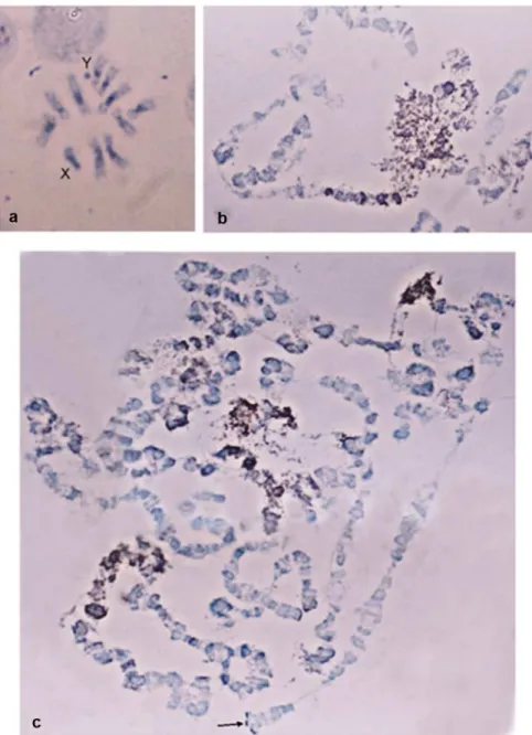
Hybridization signal was detected on the entire Y chromosome, as well as ona large part of the X chromosomes and the centromeric regions of all autosomes (Fig 1a). On the polytene spreads hybridization was observed on the heterochromatic network corresponding to the sex chromosomes, as well as on the centromeric regions and dispersed bands of the polytene ele-ment (Fig 1b and 1c).These results demonstrate that the PFGE isolated material included sequences preferentially located on the Y chromosome and on heterochromatic regions throughout the chromosomes. Most likely, these sequences are repetitive elements known to accumulate in heterochromatic and peri-centromeric regions.
Fig 1. Cytogenetic distribution of partially Y-enriched libraries with preference towards the male DNA.
In situhybridization toB.oleaea) metaphase & b, c) polytene chromosomes revealed that the element is dispersed throughout the genome and also located at heterochromatic regions.
Subsequently, this material was used to prepare Y-enriched libraries. Screening of these Y libraries with male and female genomic DNA as probes yielded several clones with differential male preference. One of these clones, pFF5, showed a strong hybridization signal with male DNA compared to female genomic DNA (S3 Fig). Based on this pattern of preferential hybrid-ization in male DNA, together with the fact that this clone was identified using a Y-enriched probe, we speculated that this might represent a repeats equence on the Y chromosome. Sequence analysis indicated agag-encoding region with identity to Pao-like retrotransposons [57].
In order to isolate full-length copies of the putative retrotransposon, a PCR product of clone pFF5 was used as a probe to screen aλDASHIIB.oleaegenomic library. Four putative
retro-transposon-containing lambda phage clones were identified and mapped using restriction enzymes and analyzed by Southern blotting. PhageF443 that contained the largest retrotran-sposon sequence was further analyzed extensively. Sequencing ofF443 subclones revealed the coding region of the putative retrotransposon, while a contiguous DNA sequence correspond-ing to the 5’region of the element was obtained by primer-walking.
Structural organization of
Achilles
and its position within the phylogeny
of LTR retrotransposons
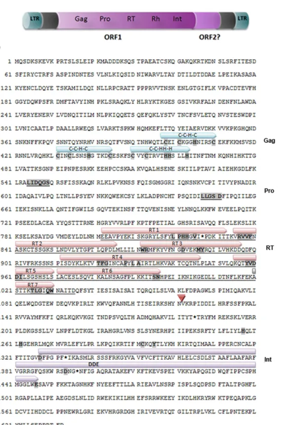
The partial genomic sequence of the novelB.oleaeretrotransposon characterized here was comprised of 7,487 bp and was isolated from a phage clone that did not contain the intact ele-ment. In particular, the 3’end of the ORF as well as the 3’LTR sequence were absent. The 5’LTR sequence was determined to be 672 bp long, flanked by the dinucleotides TG and CA found at its ends. Downstream of this putative 5’LTR region, a primer binding site (PBS-like) sequence was identified (S4 Fig) which shared significant homology to theD.melanogaster Tyr-tRNA gene, showing complementarity to 18 nucleotides of the 3’end of the respective tRNA that could be used to prime the element’s minus strand replication. The typical LTR poly-A transcription termination signal AATAAA was not detected. However, an ATTAAA motif was detected between 285 and 290 bp that could be used alternatively as a polyadenyla-tion signal [58]. Direct and indirect repeats forming a complex structure were also identified within the 5’UTR spanning the region between the LTR and the ORF (S5 Fig). Similar struc-tures are common within theBEL/Paoas well as theGypsyfamily [57,59]. One large open read-ing frame (ORF) was detected between positions 2588 bp and 7486 bp of the total sequence. Thisin silico-identified ORF, encodes a Gag-Pol precursor of 1632 amino acids, which is then cleaved to the respective proteins without an intervening termination codon. No envelope pro-tein ORF was detected within the analyzed sequence. The 5’to 3’order of the domains in the polymerase ORF that encode for the different proteins indicated that the novel retrotransposon could be classified as an LTR retrotransposon following the arrangement of theBEL/Pao super-family [59]. Nonetheless, the maximum similarity with the other members of theBEL/Pao superfamily is at a maximum of 49% (amino acid level, reverse transcriptase [RT] putative pro-tein). According to Wicker et al [18], a similarity level below 80% would justify a separate fam-ily of elements within a defined superfamfam-ily. Therefore, the novelB.oleaeelement should constitute a new family ofBEL/PaoLTR retroelements, that we denominate BEL-Achilles_Bo. The accession number given to this new element is KT280063. Therefore, based on standard nomenclature rules, the element’s name should be RLB_Achilles_KT280063 (denoting Retroe-lement Class I, LTR order,BEL-Paosuperfamily,Achillesfamily, and its accession number). For the sake of simplicity, henceforth the element will be referred to asAchilles.
many LTR retrotransposons were identified in the GAG protein. Additionally, a third Cys motif C-X2-4-C-X3-4-HH-X3-4-H (CCHHH), which is proposed to be a specific Pao-like feature
[61], was also detected. Thepolgene domains are localized downstream of thegaggene, includ-ing the enzymatic domains of protease, reverse transcriptase and integrase (Fig 2).
The PRO domain encoding a potential aspartic acid protease was recognized by the typical conserved amino acid residues“LIDQGS”and“LLGSD”[62]. Following in order, the seven characteristic motifs of the reverse transcriptase (RT) were detected, as indicated inFig 2b. Immediately downstream of the RT domain, a possible deletion was identified and the RNAse H (RH) motifs could not be found. Finally, the integrase activity domain (INT) was successfully defined. At its amino end, INT contains the conserved residues of the zinc-finger binding motif H-X4-H-X29-C-X2-C which is implicated in the recognition of the LTR sequences. In
general, its central catalytic domain is composed of invariant aspartate (D) and glutamate (E) residues separated by 35 amino acids [17].Achilles’INT contains 52 residues between D and E, which is within the reported range of the internal residues inPao-like elements [61].
Several stop codon mutations were found within the coding region, which disrupt the reverse transcriptase domain. In addition, there were missing parts from the RNaseH domain that could not be identified somewhere else within the sequence as a consequence of an internal rearrangement, suggesting its deletion. In order to test if this internal deletion is attributed only to the deduced sequence fromF443, flanking primers to the deleted RH region were used for PCR amplification on genomic and phage DNA, respectively. The differences in length between the derived PCR products were indicative of the presence of RH in an intact element somewhere else in theB.oleaegenome (S6 Fig).
The comparison of the amino acid sequences coding for the RT domain has been widely used to generate phylogenetic relationships and to infer the classification of retrotransposons [63]. Therefore, a multiple sequence alignment of the RT domain was conducted in order to examine the relationship ofAchillesto five representatives from theBEL/Paofamily (MAX, GATE,BEL,Paoandninja). The constructed phylogenetic tree placedAchilleswithin theBEL/ Paofamily of LTR retrotransposons (Fig 3) and revealed that its closest relative was theBEL element.Achillesalso grouped closely withPaoandninja, whereas it is clearly distinct from the copia-like retrotransposon, which served as an outgroup.
Genomic distribution of
Achilles
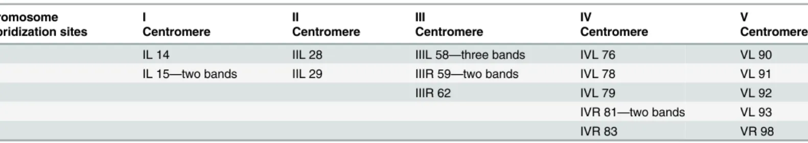
In order to study the chromosomal localization of the element, a digoxygenin-labelled probe of thegaggene was used as probe. Thein situhybridization of theAchilles-fragment on the poly-tene chromosome preparations showed a dispersed hybridization pattern. Multiple hybridiza-tion signals were identified in the centromeric regions of all five autosomes and in the granular heterochromatic network representing the under-replicated sex chromosomes. Moreover, about twenty two discrete bands dispersed on all polytene elements were identified as well (Table 1andFig 4).
Fig 2. Organization and structure of the putative intactAchilleselement.a) ORF1 encodes a Gag-Pol polyprotein consisting of 1632 aa. b) Conceptual translation of theAchillescoding region. The highly conserved amino acids are denoted by bold letters and highlighted in grey. The underlined amino acids and/or the above of them arrows represent the identifiable motifs shared with other retrotransposons. Their designations are shown on the right. The position marked with the red triangle (▽) indicates the deletion of the RH region. Asterisks (*) refer to the stop mutations within the coding sequences.
number, with male flies possessing 5–10 copies more than female (CI range: 18–38 and 12–33 copies, respectively).
Species distribution of
Achilles
Homology tBLASTn searches withAchillesas a query yielded homologous sequences of Cerati-tis capitataandBactrocera dorsalis. The above result indicated that this element may be present in other Tephritidae species as well. Several representatives of the Tephritidae family were used to explore the host range ofAchilles, includingAnastrepha serpentinaandAn.striata,C. capi-tata,Bactrocera oleae,B.correctaandB.dorsalis. TheD.melanogasterwas also included in the analysis due toAchilleshomology withMAX. Genomic amplification of the RT domain in the 7 species tested confirmed its presence within the examined genomes (Fig 5). These data sug-gest that this element might have been present in their common ancestor. However, the lack of amplification in evolutionarily more distant Dipteran species, such asD.melanogaster, does not exclude the presence of the element in these genomes too; rather, this may be a result of insufficient hybridization of the primers used during the PCR amplification due to sequence divergence. Nevertheless, data ofAchillesdistribution within related genomes are preliminary and there is need to investigate the intra-family relations more thoroughly to assume its evolu-tionary fate.
Transcriptional activity of
Achilles
In order to assess potential transcriptional activity ofAchilles, a relative quantitative RT-PCR was performed with specific primers. qRT-PCR products were detected in both male and Fig 3. Phylogenetic analysis ofAchilles.The RT domains among representative members of theBEL/Pao
family (MAX, GATE, BEL, Pao, ninja) were aligned with the corresponding region ofAchillesusing the ClustalW. The N-J tree was generated using as outgroup the copia element.Achillesis more related to the
BELelements. The numbers at the nodes are the bootstrap values based on 1000 pseudoreplications.
doi:10.1371/journal.pone.0137050.g003
Table 1. Cytogenetic sites ofAchillesas revealed byin situhybridization onB.oleaepolytene chromosomes.
Chromosome I II III IV V
Hybridization sites Centromere Centromere Centromere Centromere Centromere
IL 14 IIL 28 IIIL 58—three bands IVL 76 VL 90
IL 15—two bands IIL 29 IIIR 59—two bands IVL 78 VL 91
IIIR 62 IVL 79 VL 92
IVR 81—two bands VL 93
IVR 83 VR 98
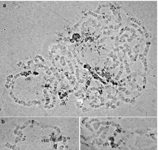
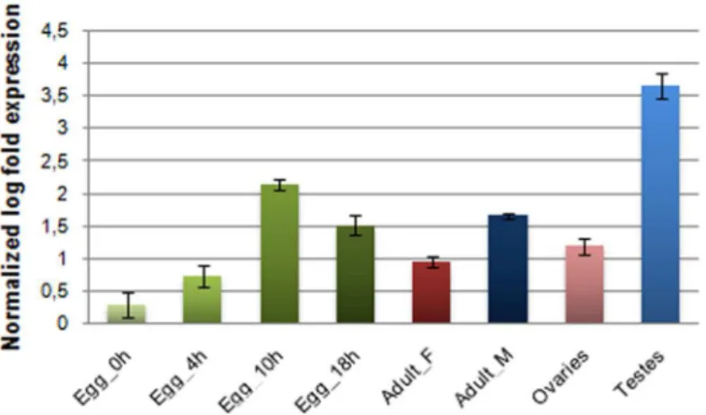
female samples, demonstrating a differential level of expression between sexes (Fig 6). The detected transcriptional activity should result from an intact copy and not the impaired copy under study. Interestingly, the BLAST hits generated by tBLASTn homology searches of Achil-lescorresponded to sequences generated by RNAseq or published EST datasets, thus to active Fig 4.In situhybridization of a digoxigenin-labelledAchilles-probe onB.oleaepolytene
chromosomes.a) Polytene nucleus, b) part of polytene chromosome III, c) part of polytene chromosome IV. Spread preparations of polytene chromosomes were made from the salivary glands of third instar larvae as described by Drosopoulou et al. (1995). Centromeres are indicated by“c”and the corresponding
chromosomes are demonstrated by their number (I-V). Multiple heterochromatic signals are indicated by multiple arrows (in Panel a). Discrete euchromatic signals are indicated by the number of the respective chromosomal division (Panels a, b, c).
doi:10.1371/journal.pone.0137050.g004
Table 2. Analysis data to estimateAchillescopy number by qPCR using a reference standard curve.
Sample concentration (ng)a Calculated copy numberb Copy number per haploid genomec CI for mean
Male 0.75 5.93E+04 27.82±3.72d 12–33
Female 0.75 4.75E+04 23.02±4.33d 18
–37
aInitial template concentration of theB.oleaegenomic DNA (ng) used at the qPCR reactions.
bMean copy number ofAchilleswhich was estimated for the initial template copies of the genomic DNA (a) based on the standard curve. cB.oleaehaploid genome size: 0.352 pg
dStandard Error (SE) for the triplicate measurements (n = 3)
elements. For example, similarity was obtained to a maleC.capitataEST dataset (JK832499.1, e-value: 9e-43), which is in accordance to our results.
Besides the entire insect body, germline tissues (male testes and female ovaries) as well as embryos at various developmental stages were examined for transcriptional activity. Although transcription was confirmed among the samples tested,Achillesexpression levels showed con-siderable variability (Fig 6). The highest expression was observed in testes. In all cases, however, preferential male expression was affirmed. Similar observations were reported by [65] regard-ing a potentially active heterochromatic copy of theGATEelement whose transcription was suggested to be regulated via an RNAi mechanism in testes.
Discussion
Molecular and genetic studies, combined with new information from whole transcriptome analyses, have given new perspectives towards the understanding of genome structure of the olive fruit flyB.oleae([49,66] and references therein).
Achillesis the first characterized retrotransposon in theB.oleaegenome. Partial sequence of this novel element was first identified in a library enriched with material derived from the Y chromosome of the olive fly (Fig 1). Initially a differential hybridization approach was imple-mented, using as probes labeled total DNA from both sexes, in order to screen for clones Fig 5. Species distribution ofAchilles.Genomic PCR amplfication ofAchilles-RT in in the 7 diptera species tested using AchillF2/R2 primer set. [Dm:Drosophila melanogaster, As:Anastrepha serpentina, Ast:
Anastrepha striata, Cc:Ceratitis capitata, Bo:Bactrocera oleae, Bc:Bactrocera correctaand Bd:Bactrocera dorsalis.]
doi:10.1371/journal.pone.0137050.g005
Fig 6. Expression profile ofAchillesin specific tissues of both sexes as well as in embryos at various developmental stages.Expression levels were detected by relative qRT-PCR in a) individual eggs collected at different time points during embryonic development and b) in two different tissues per gender of 7thday adult virgin flies: total bodies of females (Adult_F) and males (Adults_M) and the germline tissues testes and ovaries. Standard error of the mean of two biological replicates per time point is depicted in bars.
preferentially hybridizing to the male probe. Based on the kinetics and dynamics of DNA hybridization of a total genome-derived probe, only highly or moderately repetitive DNA sequences would hybridize. Thus, this approach targeted male-specific repetitive DNA or sequences that are highly amplified on the Y chromosome but may be present elsewhere in the genome as well. The overall premise was that the analysis of the neighborhood around such sequences could shed light on the structural organization of the olive fly Y chromosome.
Indeed, during the analysis of the lambda phage clones that contained the putative repetitive element, we ran across sequences such as aB.oleaespecific centromeric satellite repeat, BoR300 [52]. Wong and Choo [67] argued against the hypothesis that retrotransposons may be a common source for centromeric DNA by providing the evolutionary templates on which tandem repeats have evolved. In fact, apart from the presence of anAchilles-fragment between the repeat arrays along the heterochromatic centromeres, we have no further data to claim any association ofAchilleswith the BoR300 repeats.
The novel retrotransposon shares sequence homology and structural features characteristic of the LTR-retrotransposons of theBEL/Paosuperfamily. In contrast to the indiscernible simi-larities at the nucleotide level betweenAchillesand otherBEL/Paoelements, the conservation at the amino acid level enabled further molecular characterization. The different conservation between the two levels can be a consequence of diverse codon usage pattern of each species [50,68] that is probably influencing the evolutionary rate of such elements.
Achillesdisplayed an RNA binding motif characteristic ofgag BEL/Paoelements followed by domains of enzymatic action protease, reverse transcriptase (RT), RNAseH, and integrase, in that order (Fig 2). It was also found to be related to other discrete families but tends to cluster withBELretrotransposons. Notwithstanding such similarities,Achilles’differentiation from its closest relatives at the nucleotide and protein level would define a separate family of retrotranspo-sons within the BEL/Pao superfamily.The particular element that we analyzed in detail seemed to be defective. However, future genome sequencing will allow an assembled consensus sequence of the likely active element, as well as the discovery of other members of theAchillesfamily.
TheAchillescopy isolated from phage cloneF443 is clearly inactive due to accumulated stop mutations. However, a qRT-PCR approach demonstrated the presence ofAchilles tran-scripts. While this finding indicates potential mobility ofAchillesat this point in evolutionary time, further studies need to be conducted in order to explore this scenario. In order to explore the transcriptional activity in diverse developmental stages ofB.oleae,Achillestranscripts were sought in embryonic through adult tissues. Indeed, different levels of expression were observed in the various tissues tested, with the germline tissues of males possessing the most abundant transcripts (Fig 6). The functional role of the detected transcriptional activity during embryo-genesis is still an open issue and may be indicative of a probable involvement ofAchillesin cell differentiation and organogenesis. The exact role of retrotransposon transcripts in gonadal somatic cells is still obscure. Clearly, however, the expression and retrotransposition in germ line would be a more effective strategy for retrotransposons to ensure their inheritance to the next generation. In that regard, retrotransposons may use this path to obtain additional access to gametes, as somatic expression may detour the host defense against retrotransposons in the germ line. The reported transcript abundance of LTR retrotransposons and RNAi-related genes in the gonadal somatic cells ofD.melanogasterembryos, led to the hypothesis that the transcripts are inactivated by a post-transcriptional regulatory mechanism [69]. The same authors proposed that LTR-retrotransposon transcripts would act as a“trigger”for their subse-quent processing by RNAi pathway to produce siRNA, which in turn silences the retrotranspo-sons in the following developmental stages.
correspond to Y-specific integrations. We consider that the calculated copy number range is in reasonable agreement with thein situhybridization results, which allowed the physical map-ping of the retrotransposon onB.oleaepolytene chromosomes. Thein situhybridization data reveal an interspersion pattern of the element throughout the insect’s polytene chromosomes (Fig 4andTable 1).Achillesis concentrated at non-polytenized heterochromatic regions but some copies were found to be located on discrete bands of polytenized elements as well. Similar distribution was also observed inD.melanogaster MAXelement mapping on polytene as well as on mitotic chromosomes [57].
Hybridization signals ofAchilleson these regions strongly suggest that these chromosomes may either act as a refuge or a trap forAchillescopies. Since in heterochromatic and pericentro-meric regions recombination is suppressed and there is no selection against insertional muta-tions. Therefore, the accumulation of a considerable amount of copies in such genetically inert areas could be indicative of their reduced impact on genes or genome. Thus, it represents another example demonstrating that these genomic regions comprise favorable refuges for ret-roelements [45].
In addition, there is evidence that the presence of transposons themselves can contribute to the formation of heterochromatin. For example, there is compelling evidence that the accumu-lation of TEs, especially retrotransposons, triggered Y chromosome degeneration inD. miranda[27]. Several molecular studies in different taxa demonstrated that during early evolu-tion of the chromosomes, a sequence reshaping within new sex chromosome regions is occur-ring through modifications of the chromatin structure and the insertion of repetitive DNA sequences [23]. These morphologically and genetically induced alterations together with the absence of recombination favor the differentiation of sex chromosomes and lead to genetic ero-sion of the Y chromosome. Sex determination mechanisms, on the other hand, appear to be a labile feature and demonstrate a wide variety within Diptera [70]. UnlikeD.melanogaster, in which male development is determined by the X chromosome dosage, in Tephritids sexual dif-ferentiation relies on the male-determining factor, M, which is linked to the Y chromosome [38,71,72].Interestingly, in the midge Chironomus the male sex determiner has been associated with a single heterochromatic band consisted of highly repetitive DNA and transposable ele-ments [73,74]. Therefore, the in depth exploration of sex chromosomes structure will improve our understanding of their origin and divergence (degeneration) as well as the evolution of genetic sex determination pathways which are attributed to them. Furthermore, due to the fact that sexual differentiation and male fertility comprise a crucial part of insect control strategies, such as the sterile insect technique (SIT), the manipulation of sequences involved in these pro-cesses is of particular interest. In this context, the analysis of the Tephritidae sex chromosomes can contribute towards the establishment of new genetic tools for sex separation strategies, since only males are the active component for SIT and effective population control [75].
its functional and structural impact on Y-linked genomic regions (such as, for example, the maleness factor) still evade. While such questions are being considered, Achilles’heel still remains well protected.
Supporting Information
S1 Fig. Primer walking approach based on restriction map ofF443.a) The upstream region of 6.0 kb was initially amplified with long PCR. b) Subsequently, the sequence of the amplifica-tion product was determined by primer walking. The designed primers as well as the order of their use regarding the sequencing of the 3’end are schematically represented on the Fig. The primers used for the sequencing of the 5’end are reported inS2 Table.
(TIF)
S2 Fig. Schematic representation of the modified protocol used for the isolation of the 3’ coding region.a) Partial digestion of genomic DNA with the restriction endonucleaseTaqI. b) Isolation of the fragments with the desirable length and ligation of the adapters Taq-AD. c) PCR amplification using the primers 443–0.9 F1 and TaqI AP. d) Semi-nested PCR on the pre-viously amplified region using the primers 443–0.9 F1 and TaqI AP. e) Cloning and sequencing of the amplicons.
(TIF)
S3 Fig. Screening of partially Y-enriched libraries.Clone pFF5 with the highest differential hybridization to male DNA disclosed a fragment of a putative repetitive element.
(TIF)
S4 Fig. Sequence and structural characteristics of the LTR and the PBS.Flanking inverted repeats of the 674 bp long 5’LTR are shown boxed. Immediately following the 50LTR the puta-tive PBS (677–690 bp) complementary to the 30end of tRNA-Tyr is located. A clearly identifi-able putative TATA box was not found, thus not indicated.
(TIF)
S5 Fig. Dot plot comparison ofAchilles5’region versus itself.The analyzed region spanned
the 5’LTR (including 5’UTR) until the beginning of theAchillesORF and was aligned using the program Omiga 2.0. The central black line corresponds to identical residues. Additional similarities of shorter regions (blue or red dots) are due to repeats found in LTRs.
(TIF)
S6 Fig. PCR amplification of the RH region.The genomic amplification product is larger (2500 bp) than that of the phage (1900 bp), suggesting the presence of the RH region in the geno-mic DNA, unlike theF443 DNA were RH was deleted. The other bands in Lane 1 most likely correspond to non-intactAchilleselements in the genome. Lane 1, genomicB.oleaeDNA; Lane 2,F443 phage DNA; Lane 3, negative control. L: molecular weight marker SM0331 (GeneON). (TIF)
S7 Fig. Estimation of copy number using the standard curve method of an external control plasmid sample.Standard curve generated from the control plasmid DNA containing RT domain ofAchilles. Fluorescent threshold values (CT) were plotted against the logarithm of the starting copy number to produce a linear function. The slope and intercept of the curve were calculated from the linear equation describing the standard curve.For the construction of the curves, serial 10-fold dilutions of plasmid DNA were used (1.071 fg, 21.437 fg, 8.575 pg, 171.5 pg) converted to copy numbers.
S1 Table. Primer sequences and parameters of the standard PCR (1), RT-PCR (2), absolute qPCR (3) and relative qPCR assay (4).
(DOCX)
S2 Table. Primer sequences used in PCR reactions to amplify the upstream 6.0 kb region (Long PCR), as well in the primer-walking approach.
(DOCX)
S3 Table. Primer sequences and parameter used in the initial standard PCR and the subse-quent semi-nested PCR reactions to detect the downstream 3’region ofAchilles.
(DOCX)
Author Contributions
Conceived and designed the experiments: KDM AZ KB. Performed the experiments: KTT ED KB AGP PMT KDM. Analyzed the data: KTT KB KDM AZ PMT. Contributed reagents/mate-rials/analysis tools: KDM AZ KB PMT. Wrote the paper: KTT ED KB PMT AZ KDM.
References
1. Kaiser VBV, Bachtrog D. Evolution of sex chromosomes in insects. Annu Rev Genet. 2010; 44: 91–112. doi:10.1146/annurev-genet-102209-163600PMID:21047257
2. Rice W. Evolution of the Y sex chromosome in animals. Bioscience. 1996; 46: 331–343. Available:
http://www.jstor.org/stable/1312947.
3. Charlesworth B. The evolution of chromosomal sex determination and dosage compensation. Curr Biol. 1996; 6: 149–62. Available:http://www.ncbi.nlm.nih.gov/pubmed/8673462. PMID:8673462
4. Charlesworth B, Charlesworth D. The degeneration of Y chromosomes. Philos Trans R Soc London B Biol Sci. 2000; 355: 1563–1572. Available:http://rstb.royalsocietypublishing.org/content/355/1403/ 1563.abstract. PMID:11127901
5. Bachtrog D. A dynamic view of sex chromosome evolution. Curr Opin Genet Dev. 2006; 16: 578–585. doi:10.1016/j.gde.2006.10.007PMID:17055249
6. Skaletsky H, Kuroda-Kawaguchi T, Minx PJ, Cordum HS, Hillier L, Brown LG, et al. The male-specific region of the human Y chromosome is a mosaic of discrete sequence classes. Nature. 2003; 423: 825–37. doi:10.1038/nature01722PMID:12815422
7. Bernardo Carvalho A, Koerich LBL, Clark AAG, Carvalho A, Koerich LBL, Clark AAG. Origin and evolu-tion of Y chromosomes: Drosophila tales. Trends Genet. 2009; 25: 270–7. doi:10.1016/j.tig.2009.04. 002PMID:19443075
8. Vicoso B, Bachtrog D. Reversal of an ancient sex chromosome to an autosome in Drosophila. Nature. 2013; 499: 332–5. doi:10.1038/nature12235PMID:23792562
9. Vicoso B, Bachtrog D. Numerous transitions of sex chromosomes in Diptera. PLoS Biol. 2015; 13: e1002078. doi:10.1371/journal.pbio.1002078PMID:25879221
10. Carvalho AB, Dobo BA, Vibranovski MD, Clark AG. Identification of five new genes on the Y chromo-some of Drosophila melanogaster. Proc Natl Acad Sci. 2001; 98: 13225–30. doi:10.1073/pnas. 231484998PMID:11687639
11. Hoskins R, Carlson J, Kennedy C. Sequence finishing and mapping of Drosophila melanogaster het-erochromatin. Science (80-). 2007; Available:http://www.sciencemag.org/content/316/5831/1625. short.
12. Kidwell MG, Lisch D. Transposable elements as sources of variation in animals and plants. Proc Natl Acad Sci U S A. 1997; 94: 7704–11. Available:http://www.pubmedcentral.nih.gov/articlerender.fcgi? artid=33680&tool = pmcentrez&rendertype = abstract. PMID:9223252
13. Kidwell MMG. Transposable elements and the evolution of genome size in eukaryotes. Genetica. 2002; 115: 49–63. Available:http://link.springer.com/article/10.1023/A:1016072014259. PMID:
12188048
genome_organization_and_evolution_the_case_of_Drosophila/links/02bfe513f546f79551000000.pdf. PMID:16093655
15. Smith C, Shu S, Mungall C, Karpen G. The Release 5.1 annotation of Drosophila melanogaster hetero-chromatin. Science (80-). 2007; Available:http://www.sciencemag.org/content/316/5831/1586.short. 16. Eickbush T, Furano A. Fruit flies and humans respond differently to retrotransposons. Curr Opin Genet
Dev. 2002; 12: 669–674. Available:http://www.sciencedirect.com/science/article/pii/ S0959437X02003593. PMID:12433580
17. Kulkosky J, Jones KS, Katz RA, Mack JP, Skalka AM. Residues critical for retroviral integrative recom-bination in a region that is highly conserved among retroviral/retrotransposon integrases and bacterial insertion sequence transposases. Mol Cell Biol. 1992; 12: 2331–8. Available:http://www.
pubmedcentral.nih.gov/articlerender.fcgi?artid=364405&tool = pmcentrez&rendertype = abstract. PMID:1314954
18. Wicker T, Sabot F, Hua-Van A, Bennetzen JL, Capy P, Chalhoub B, et al. A unified classification sys-tem for eukaryotic transposable elements. Nat Rev Genet. Nature Publishing Group; 2007; 8: 973–82. doi:10.1038/nrg2165PMID:17984973
19. Xiong Y, Burke WD, Eickbush TH. Pao, a highly divergent retrotransposable element from Bombyx mori containing long terminal repeats with tandem copies of the putative R region. Nucleic Acids Res. 1993; 21: 2117–23. Available:http://www.pubmedcentral.nih.gov/articlerender.fcgi?artid=
309473&tool = pmcentrez&rendertype = abstract. PMID:8389039
20. De la Chaux N, Wagner A, Chaux de la N, Wagner A. BEL/Pao retrotransposons in metazoan genomes. BMC Evol Biol. 2011; 11: 154. doi:10.1186/1471-2148-11-154PMID:21639932
21. Gatti M, Pimpinelli S. Functional elements in Drosophila melanogaster heterochromatin. Annu Rev Genet. 1992; 26: 239–75. doi:10.1146/annurev.ge.26.120192.001323PMID:1482113
22. Dorer D, Henikoff S. Expansions of transgene repeats cause heterochromatin formation and gene silencing in Drosophila. Cell. 1994; 77: 993–1002. Available:http://www.sciencedirect.com/science/ article/pii/0092867494904391. PMID:8020105
23. Steinemann S, Steinemann M. Y chromosomes: born to be destroyed. Bioessays. 2005; Available:
http://onlinelibrary.wiley.com/doi/10.1002/bies.20288/full.
24. Pimpinelli S, Berloco M, Fanti L, Dimitri P, Bonaccorsi S, Marchetti E, et al. Transposable elements are stable structural components of Drosophila melanogaster heterochromatin. Proc Natl Acad Sci U S A. 1995; 92: 3804–8. Available:http://www.pubmedcentral.nih.gov/articlerender.fcgi?artid=42050&tool = pmcentrez&rendertype = abstract. PMID:7731987
25. Steinemann M, Steinemann S. Preferential Y chromosomal location of TRIM, a novel transposable ele-ment of Drosophila miranda, obscura group. Chromosoma. 1991; 101: 169–79. Available:http://www. ncbi.nlm.nih.gov/pubmed/1665124. PMID:1665124
26. Steinemann M, Steinemann S. Degenerating Y chromosome of Drosophila miranda: a trap for retro-transposons. Proc Natl Acad Sci U S A. 1992; 89: 7591–5. Available:http://www.pubmedcentral.nih. gov/articlerender.fcgi?artid=49756&tool = pmcentrez&rendertype = abstract. PMID:1323846
27. Steinemann M, Steinemann S. The enigma of Y chromosome degeneration: TRAM, a novel retrotran-sposon is preferentially located on the Neo-Y chromosome of Drosophila miranda. Genetics. 1997; 145: 261–6. Available:http://www.pubmedcentral.nih.gov/articlerender.fcgi?artid=1207793&tool = pmcentrez&rendertype = abstract. PMID:9071582
28. Rohr CJBC, Ranson H, Wang X, Besansky NNJ. Structure and evolution of mtanga, a retrotransposon actively expressed on the Y chromosome of the African malaria vector Anopheles gambiae. Mol Biol Evol. 2002; 19: 149–62. Available:http://mbe.oxfordjournals.org/content/19/2/149.short. PMID:
11801743
29. Krzywinski J, Sangaré D, Besansky NJ. Satellite DNA from the Y chromosome of the malaria vector Anopheles gambiae. Genetics. 2005; 169: 185–96. doi:10.1534/genetics.104.034264PMID:
15466420
30. White I, Elson-Harris M. Fruit flies of economic significance: their identification and bionomics. Bull Entomol Res. 1992; 82: 433–433. Available:http://www.cabdirect.org/abstracts/19921161954.html. 31. Mavragani-Tsipidou P, Zacharopoulou A, Drosopoulou E, Augustinos AA, Bourtzis K, Frantisek M.
Tephritid Fruit Flies (Diptera). In: Sharakhov Igor V., editor. Protocols for Cytogenetic Mapping of Arthropod Genomes. Boca Raton, Florida: CRC Press, Taylor & Francis Group; 2014. Available:
https://www.google.com/books?hl = el&lr=&id = goxqBAAAQBAJ&oi = fnd&pg=PA1&dq=Tephritid +Fruit+Flies+(Diptera).+Protocols+for+Cytogenetic+Mapping+of+Arthropod+Genomes&ots = of0M7n2UDi&sig=7Y36INA2HHZXh9GF5bEaDNdu-D8.
33. Bedo DG. Polytene chromosome mapping in Ceratitis capitata (Diptera: Tephritidae). Genome. NRC Research Press Ottawa, Canada; 1987; 29: 598–611.
34. Drosopoulou E, Nakou I, Síchová J, Kubíčková S, Marec F, Mavragani-Tsipidou P, et al. Sex chromo-somes and associated rDNA form a heterochromatic network in the polytene nuclei of Bactrocera oleae (Diptera: Tephritidae). Genetica. 2012; 140: 169–80. doi:10.1007/s10709-012-9668-3PMID:
22825842
35. Anleitner JE, Haymer DS. Y enriched and Y specific DNA sequences from the genome of the mediterra-nean fruit fly, Ceratitis capitata. Chromosoma. 1992; 101: 271–278. doi:10.1007/BF00346005PMID:
1533581
36. Zhou Q, Untalan PM, Haymer DS. Repetitive A-T rich DNA sequences from the Y chromosome of the Mediterranean fruit fly, Ceratitis capitata. Genome. 2000; 43: 434–8. Available:http://www.ncbi.nlm. nih.gov/pubmed/10902705. PMID:10902705
37. Torti C, Gomulski LM, Moralli D, Raimondi E, Robertson HM, Capy P, et al. Evolution of different sub-families of mariner elements within the medfly genome inferred from abundance and chromosomal dis-tribution. Chromosoma. 2000; 108: 523–32. Available:http://www.ncbi.nlm.nih.gov/pubmed/10794574. PMID:10794574
38. Willhoeft U, Franz G. Identification of the sex-determining region of the Ceratitis capitata Y chromo-some by deletion mapping. Genetics. 1996; 144: 737–45. Available:http://www.pubmedcentral.nih. gov/articlerender.fcgi?artid=1207564&tool = pmcentrez&rendertype = abstract. PMID:8889534
39. Gabrieli P, Gomulski LM, Bonomi A, Siciliano P, Scolari F, Franz G, et al. Interchromosomal duplica-tions on the Bactrocera oleae Y chromosome imply a distinct evolutionary origin of the sex chromo-somes compared to Drosophila. PLoS One. 2011; 6: e17747. doi:10.1371/journal.pone.0017747
PMID:21408187
40. Locke J, McDermid HE. Analysis ofDrosophila chromosome4 using pulsed field gel electrophoresis. Chromosoma. 1993; 102: 718–723. doi:10.1007/BF00650898PMID:8149812
41. Lagos D, Ruiz MF, Sánchez L, Komitopoulou K. Isolation and characterization of the Bactrocera oleae genes orthologous to the sex determining Sex-lethal and doublesex genes of Drosophila melanogaster. Gene. 2005; 348: 111–121. doi:10.1016/j.gene.2004.12.053PMID:15777677
42. Kakani EG, Trakala M, Drosopoulou E, Mavragani-Tsipidou P, Mathiopoulos KD. Genomic structure, organization and localization of the acetylcholinesterase locus of the olive fruit fly, Bactrocera oleae. Bull Entomol Res. 2013; 103: 36–47. doi:10.1017/S0007485312000478PMID:22967668
43. Sambrook J, Fritsch E, Maniatis T. Molecular cloning. 1989; Available:http://nhjy.hzau.edu.cn/kech/ jycz/jczs/ml-introduction/Content.pdf.
44. Altschul SF, Gish W, Miller W, Myers EW, Lipman DJ. Basic local alignment search tool. J Mol Biol. 1990; 215: 403–10. doi:10.1016/S0022-2836(05)80360-2PMID:2231712
45. Pearce S, Stuart‐Rogers C. Rapid isolation of plant Ty1‐copia group retrotransposon LTR sequences for molecular marker studies. Plant. 1999.
46. Sambrook J, Fritsch EF, Maniatis T. Molecular Cloning: A Laboratory Manual. Cold Spring Harbor labo-ratory press. New York. Cold Spring Harbor; 1989.
47. Drosopoulou E, Scouras Z. Theβ-tubulin gene family evolution in theDrosophila montium subgroup of themelanogaster species group. J Mol Evol. 1995; Available:http://link.springer.com/article/10.1007/ BF01215176.
48. Zambetaki A, Zacharopoulou A, Scouras ZG, Mavragani-Tsipidou P. The genome of the olive fruit fly Bactrocera oleae: localization of molecular markers by in situ hybridization to the salivary gland poly-tene chromosomes. Genome. 1999; 42: 744–751.
49. Sagri E, Reczko M, Tsoumani KT, Gregoriou M-E, Harokopos V, Mavridou A-M, et al. The molecular biology of the olive fly comes of age. BMC Genet. 2014; 15 Suppl 2: S8. doi: 10.1186/1471-2156-15-S2-S8PMID:25472866
50. Tsoumani KT, Augustinos AA, Kakani EG, Drosopoulou E, Mavragani-Tsipidou P, Mathiopoulos KD. Isolation, annotation and applications of expressed sequence tags from the olive fly, Bactrocera oleae. Mol Genet Genomics. 2011; 285: 33–45. doi:10.1007/s00438-010-0583-yPMID:20978910
51. Tsoumani KT, Mathiopoulos KD. Genome size estimation with quantitative real-time PCR in two Tephri-tidae species: Ceratitis capitata and Bactrocera oleae. J Appl Entomol. 2012; 136: 626–631.
52. Tsoumani KT, Drosopoulou E, Mavragani-Tsipidou P, Mathiopoulos KD. Molecular characterization and chromosomal distribution of a species-specific transcribed centromeric satellite repeat from the olive fruit fly, Bactrocera oleae. PLoS One. 2013; 8: e79393. doi:10.1371/journal.pone.0079393PMID:
53. Soleimani V, Baum B, Johnson D. Quantification of the retrotransposon BARE-1 reveals the dynamic nature of the barley genome. Genome. 2006; Available:http://www.nrcresearchpress.com/doi/abs/10. 1139/g05-119.
54. Thompson JD, Gibson TJ, Higgins DG. Multiple sequence alignment using ClustalW and ClustalX. Curr Protoc Bioinformatics. 2002;Chapter 2: Unit 2.3. doi:10.1002/0471250953.bi0203s00
55. Kumar S, Tamura K, Jakobsen IB, Nei M. MEGA2: molecular evolutionary genetics analysis software. Bioinformatics. 2001; 17: 1244–5. Available:http://www.ncbi.nlm.nih.gov/pubmed/11751241. PMID:
11751241
56. Mavragani-Tsipidou P, Karamanlidou G, Zacharopoulou A, Koliais S, Kastritisis C. Mitotic and polytene chromosome analysis in Dacus oleae (Diptera: Tephritidae). Genome. 1992; 35: 373–378. Available:
http://www.ncbi.nlm.nih.gov/pubmed/1624130. PMID:1624130
57. Marsano R, Marconi S. MAX, a novel retrotransposon of the BEL-Pao family, is nested within the Bari 1 cluster at the heterochromatic h39 region of chromosome 2 in Drosophila. Mol Genet. 2004.
58. Benachenhou F, Jern P, Oja M, Sperber G, Blikstad V, Somervuo P, et al. Evolutionary Conservation of Orthoretroviral Long Terminal Repeats (LTRs) and ab initio Detection of Single LTRs in Genomic Data. Ahmed N, editor. PLoS One. 2009; 4: e5179. doi:10.1371/journal.pone.0005179PMID:19365549
59. Frame I, Cutfield J, Poulter R. New BEL-like LTR-retrotransposons in Fugu rubripes, Caenorhabditis elegans, and Drosophila melanogaster. Gene. 2001; 263: 219–230. Available:http://www.
sciencedirect.com/science/article/pii/S0378111900005679. PMID:11223261
60. Covey SN. Amino acid sequence homology in gag region of reverse transcribing elements and the coat protein gene of cauliflower mosaic virus. Nucleic Acids Res. 1986; 14: 623–33. Available:http://www. pubmedcentral.nih.gov/articlerender.fcgi?artid=339453&tool = pmcentrez&rendertype = abstract. PMID:2418414
61. Abe H, Ohbayashi F, Sugasaki T, Kanehara M, Terada T, Shimada T, et al. Two novel Pao-like retro-transposons (Kamikaze and Yamato) from the silkworm species Bombyx mori and B. mandarina : com-mon structural features of Pao-like elements. Mol Genet Genomics. 2001; 265: 375–385. PMID:
11361350
62. Doolittle R, Feng D. Origins and evolutionary relationships of retroviruses. Q Rev. 1989; 64: 1–30. Available:http://www.jstor.org/stable/2831683.
63. Xiong Y, Eickbush TH. Origin and evolution of retroelements based upon their reverse transcriptase sequences. EMBO J. 1990; 9: 3353–62. Available:http://www.ncbi.nlm.nih.gov/pmc/articles/ pmc552073/. PMID:1698615
64. Felder H, Herzceg A, de Chastonay Y, Aeby P, Tobler H, Müller F. Tas, a retrotransposon from the par-asitic nematode Ascaris lumbricoides. Gene. 1994; 149: 219–225. doi: 10.1016/0378-1119(94)90153-8PMID:7525414
65. Kogan GL, Tulin A V, Aravin AA, Abramov YA, Kalmykova AI, Maisonhaute C, et al. The GATE retro-transposon in Drosophila melanogaster: mobility in heterochromatin and aspects of its expression in germline tissues. Mol Genet Genomics. 2003; 269: 234–42. doi:10.1007/s00438-003-0827-1PMID:
12756535
66. Sagri E, Reczko M, Gregoriou M-E, Tsoumani KT, Zygouridis NE, Salpea KD, et al. Olive fly transcrip-tomics analysis implicates energy metabolism genes in spinosad resistance. BMC Genomics. 2014; 15: 714. doi:10.1186/1471-2164-15-714PMID:25156405
67. Wong LHL, Choo KHA. Evolutionary dynamics of transposable elements at the centromere. TRENDS Genet. 2004; 20: 611–6. doi:10.1016/j.tig.2004.09.011PMID:15522456
68. He M, Haymer DS. Codon bias in actin multigene families and effects on the reconstruction of phyloge-netic relationships. J Mol Evol. 1995; 41: 141–9. Available:http://www.ncbi.nlm.nih.gov/pubmed/ 7666443. PMID:7666443
69. Shigenobu S, Kitadate Y. Molecular characterization of embryonic gonads by gene expression profiling in Drosophila melanogaster. Proc. 2006; Available:http://www.pnas.org/content/103/37/13728.short. 70. Shearman DCA. The evolution of sex determination systems in dipteran insects other than Drosophila.
Genetica. 2002; 116: 25–43. Available:http://www.ncbi.nlm.nih.gov/pubmed/12484524PMID:
12484524
71. Bedo DG, Foster GG. Cytogenetic mapping of the male-determining region of Lucilia cuprina (Diptera: Calliphoridae). Chromosoma. 1985; 92: 344–350.
73. Hägele K. Identification of a polytene chromosome band containing a male sex determiner of Chirono-mus thummi thummi. Chromosoma. 1985; 91: 167–71. Available:http://www.ncbi.nlm.nih.gov/pubmed/ 3979175. PMID:3979175
74. Kraemer C, Schmidt ER. The sex determining region of Chironomus thummi is associated with highly repetitive DNA and transposable elements. Chromosoma. 1993; 102: 553–62. Available:http://link. springer.com/article/10.1007/BF00368348. PMID:8243167
75. Franz G. Genetic Sexing Strains in Mediterranean Fruit Fly, an Example for Other Species Amenable to Large-Scale Rearing for the Sterile Insect Technique. In: Dyck VA, Hendrichs J, Robinson AS, edi-tors. Sterile Insect Technique SE—16. Springer Netherlands; 2005. pp. 427–451. doi: 10.1007/1-4020-4051-2_16