Universidade de Lisboa – Faculdade de Ciências
Departamento de Biologia Animal
PIWI PROTEINS IN MAMMALS: A COW’S
PERSPECTIVE
Ricardo Andrés Zacarias Silva
MESTRADO EM BIOLOGIA EVOLUTIVA E DO
DESENVOLVIMENTO
2
Universidade de Lisboa – Faculdade de Ciências
Departamento de Biologia Animal
PIWI PROTEINS IN MAMMALS: A COW’S
PERSPECTIVE
Ricardo Andrés Zacarias Silva
MESTRADO EM BIOLOGIA EVOLUTIVA E DO
DESENVOLVIMENTO
Dissertação orientada por
Professora Dra. Maria Gabriela Rodrigues (DBA/FCUL)
Professor Dr. Bernard Roelen (BRC/UU)
3
Acknowledgements
There is only one name on the cover, but it takes an orchestra to play the symphony. This is the outcome from the combined effort of many people, who also deserve credit.
Thanks to Bernard, a wise teacher and a good friend. To Leni, for the constant guidance. To Mahdi and the BRC group, for the wonderful lab environment. To Gabriela, for her support from the sunny side of Europe. To Ana and Maria, for keeping my sanity. To my new brothers, Floris and Ruben, for making me feel at home. And to Mariana, for her smile, love, and infinite wisdom.
4
Abstract
The integrity of DNA is under constant threat from both exogenous and endogenous agents. As a response, the cell has evolved numerous mechanisms to protect its genetic information from such dangers, especially in the germline, where unwanted changes in the genome can be transferred to the offspring. One major peril to genomic stability are transposable elements, mobile genetic fragments that can insert themselves at new locations in the genome. The mechanisms involved in controlling these elements are poorly understood. However, recent breakthroughs have provided insight into a germ cell-specific mechanism that silences transposons through RNA interference. Piwi, a subclass of the Argonaute protein family, associates with a novel class of small RNAs termed piRNAs. Interestingly, these piRNAs exhibit sequences complementary to those of transposable elements and, together with Piwi proteins, are able to thwart the mobility of such elements. Although the importance of Piwi proteins in germ cells has been established in many different organisms, in mammals Piwi function seems to be male-specific. Interestingly, phylogenetic examination of the Argonaute protein family revealed an additional Piwi gene (PIWI-LIKE 3 or
PIWIL3) that is present in numerous mammals, including humans and bovine, but
absent from rodents. Until a thorough analysis of PIWIL3 and other Piwi-like genes is performed in other animals, we cannot exclude a function for these genes in female mammals. Here, we show that PIWIL3 mRNA and protein are expressed in bovine oocytes. Using quantitative PCR we have determined the relative expression pattern of PIWIL3 transcripts during oocyte maturation and early embryo development. Furthermore, immunofluorescence studies localized Piwi-like 3 to the meiotic process of bovine oocytes. Lastly, preliminary experiments were made in an effort to adapt a known transfection mechanism to its use in bovine oocytes.
Key words: bovine, oocyte, Piwi proteins, Piwi-like 3, piRNAs, meiosis, oocyte
5
Resumo
A protecção do conteúdo genético das células é essencial para manter a integridade da informação responsável pela vida. Existem vários factores que podem afectar a estabilidade do genoma, tais como: radiação ultravioleta, compostos mutagénicos e erros de replicação do DNA. Contudo, um dos maiores perigos que a célula enfrenta encontra-se no próprio código genético. Uma grande proporção dos genomas eucarióticos corresponde a sequências repetidas denominadas transposões. Os transposões são elementos genéticos móveis que têm a capacidade de inserir-se em novos locais do genoma, podendo interferir com a estrutura e função de genes essenciais para a célula. O perigo iminente da transposição genética merece especial atenção em células da linha germinal, onde qualquer alteração no genoma pode ser transferida à descendência. Por esta razão, torna-se necessário controlar a mobilidade destes elementos.
RNA interferência (RNAi) é um mecanismo de regulação da expressão génica que tem sido implicado no silenciamento de elementos móveis. Esta ferramenta celular utiliza uma proteína da família Argonaute associada a uma sequência de RNA de pequenas dimensões como motor de pesquisa, localizando sequências mensageiras de RNA (mRNA) que lhe sejam complementares. Uma vez encontradas, esses mRNAs são clivados pelo complexo proteico (proteína Argonaute) acoplado ao RNA guia, impedindo a sua tradução. A família de proteínas Argonaute pode ser dividida em dois grupos: o grupo Ago, que são expressas ubiquitamente; e o grupo Piwi, cuja expressão é restrita às células da linha germinal. Para além do seu padrão de expressão, estes grupos de proteínas também diferem na classe de RNAs aos quais se associam. Membros do grupo Ago interagem com miRNAs e siRNAs, enquanto que proteínas Piwi interagem com uma nova classe de RNAs, denominados piRNAs (piwi-interacting RNAs). O estudo de proteínas Piwi e piRNAs tem avançado consideravelmente nos últimos anos com base em experiências realizadas em Drosophila, C. Elegans, zebrafish e ratinho. Estas experiências têm convergido para um papel das proteínas Piwi no desenvolvimento e sobrevivência das células da linha germinal. No genoma do ratinho existem três genes da família Piwi: miwi (Piwi-like 1), mili (Piwi-like 2) e
6 proteínas resulta na esterilidade de ratinhos machos, mas em fêmeas nenhum defeito é visível. Contrariamente, noutros animais modelo o conhecimento sobre a biologia das proteínas Piwi surgiu do estudo das células germinais de ambos os sexos. Ainda não foi possível identificar um papel para estas proteínas na linha germinal feminina de mamíferos.
O avanço no conhecimento das proteínas Piwi deve-se principalmente à identificação e sequenciação dos pequenos RNAs a que se associam, os piRNAs. A sua análise genómica revelou que estas sequências correspondem a áreas teloméricas e centroméricas dos cromossomas, que são maioritariamente populadas por elementos repetitivos e cópias de transposões e, por esta razão, foram denominadas de clusters de piRNA. Estes clusters podem ser transcritos e produzir piRNAs de ambas as cadeias genómicas, que são subsequentemente acoplados a uma proteína Piwi. Os complexos Piwi-piRNA formados podem agora procurar sequências complementares de transposões e provocar a sua degradação, protegendo assim a integridade do genoma. Esta descoberta revelou uma nova função para zonas genómicas que até agora eram consideradas “lixo genético”. Assim, estas sequências funcionam como um sistema imunitário genético, impedindo que elementos móveis como os transposões provoquem alterações na informação que será transmitida à descendência.
Considerando o estado de conservação das proteínas Piwi e o seu largo espectro de acção, parece improvável que a sua função seja dispensável em ovários de mamíferos. Recentemente foi identificado um quarto gene Piwi-like (PIWIL3) em muitos mamíferos (incluindo humanos e bovinos) que se encontra ausente do genoma do ratinho, razão pela qual ainda não foi estudado. Adicionalmente, o perfil de pequenos RNAs em oócitos de bovino revelou a existência de uma população acentuada de piRNAs, situação que não se observa em ratinho. Estas observações indicam que até este e os outros genes da família Piwi não forem examinados em outros animais, não se pode excluir por completo a sua participação no desenvolvimento da linha germinal feminina. Por esta razão, este projecto foi desenvolvido utilizando a vaca como organismo modelo. Apesar das limitações óbvias de manipulação, a investigação em bovino tem algumas vantagens. Grandes quantidades de ovários podem ser obtidos como material
7 descartado de matadouros locais, a partir dos quais óvulos saudáveis podem ser extraídos. Adicionalmente, estes óvulos podem ser maturados in vitro, fertilizados, e podem manter-se em cultura até ao momento de implantação, isto é, até a fase de blastocisto.
Neste trabalho demonstramos que a proteína Piwi-like 3 é expressa em oócitos de bovino, ao contrário da situação observada em ratinho. Em primeiro lugar, determinámos através de RT-PCR que a expressão de PIWIL3 encontra-se restrita aos tecidos germinais de bovino, sendo apenas detectável significativamente em ovários e testículos. De seguida verificou-se a presença de mRNA de PIWIL3 dentro desse contexto, focando a nossa atenção nos ovários. Para isso, foram recolhidas amostras em diferentes pontos do desenvolvimento de oócitos e embriões: vesícula germinal, metafase I, metafase II, zigoto, 2 células, 4 células, 8 células, morula e blastocisto. Utilizando novamente PCR, observamos que
PIWIL3 encontra-se presente ao longo de todo o processo de maturação e durante
o desenvolvimento embrionário pré-implantação. Para obter uma descrição mais detalhada da expressão deste gene, foi utilizada a técnica de PCR quantitativo, que permite detectar se o gene em estudo se encontra sob regulação. Os nossos resultados não mostraram nenhuma alteração significativa na expressão de
PIWIL3 nos pontos temporais testados. Foi apenas detectado um ligeiro aumento
de expressão no momento da metafase II, o que sugere um papel para esta proteína durante o processo de maturação. Contudo, a presença de mRNA não implica a existência de proteína nesta altura e, por esta razão, realizaram-se experiências de imunocitoquímica para comprovar a presença e localização da proteína. Em contraste com o padrão observado para mRNA, a proteína Piwi-like 3 foi detectada numa janela temporal menos abrangente, estando apenas presente durante a maturação do oócito até à formação dos pronúcleos. Neste contexto, verificou-se que Piwi-like 3 localiza-se junto dos cromossomas durante o evento da meiose, acompanhando todos os movimentos provocados pelo fuso meiótico. Por fim, com o objectivo de levar a cabo estudos funcionais, foi testada a possibilidade de introduzir RNA fluorescente em oócitos de bovino. Se possível, no futuro poderá introduzir-se RNAs de dupla cadeia que utilizem a maquinaria de RNAi para diminuir os níveis de PIWIL3 em oócitos, simulando uma célula mutante. Os nossos
8 resultados mostram que é possível introduzir RNAs em oócitos sem consequências graves para a célula, abrindo a porta para estudos funcionais em oócitos de bovino. Os nossos resultados apontam para uma possível função para Piwi-like 3 ao longo do processo de maturação, particularmente durante a meiose. Tendo em conta trabalhos relacionados em outros organismos, especulamos também sobre a possibilidade de Piwi-like 3 e piRNAs serem acumulados maternalmente, sobre a participação desta proteína no transporte e localização de mRNA e no silenciamento de transposões. Este trabalho alerta-nos também da importância de conduzir e desenvolver estudos em organismos alternativos, uma vez que a situação observada em animais modelo não representa necessariamente uma classe inteira de animais.
Palavras-chave: bovino, oócitos, proteínas Piwi, Piwi-like 3, piRNAs, meiose,
9
Table of Contents
Acknowledgements 3 Abstract 4 Resumo 5 1. Introduction 11 1.1. RNA interference 11 1.2. Argonaute proteins 12 1.3. Piwi proteins 131.4. piRNAs, a fortress of genomic integrity 16
1.4.1. Transposable elements 16
1.4.2. Silencing transposable elements 17
1.5. Piwi proteins and piRNAs 18
1.5.1. piRNA biogenesis 20
1.5.2. Evolutionary implications of Piwi proteins and piRNAs 21
1.6. The bovine as an animal model 22
1.6.1. The mammalian alternative 22
1.6.2. Oocyte maturation 23
1.6.3. Meiosis 25
2. Materials and Methods 28
2.1. Oocyte collection, in vitro maturation and fertilization 28
2.2. Total RNA extraction and cDNA synthesis 29
2.3. Primer design 30 2.4. RT-PCR 30 2.5. Quantitative RT-PCR 31 2.6. Immunocytochemistry 31 2.7. Transfection of oocytes 32 3. Results 33
3.1. PIWIL3 expression is mainly restricted to germ cells 33 3.2. PIWIL3 is expressed throughout meiotic maturation and early
development 33
10 3.4. Piwi-like 3 associates with chromatin during meiosis 35
3.5. Lipofection in bovine oocytes 39
4. Discussion 41
5. Concluding remarks 46
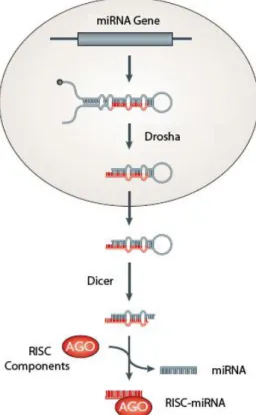
11 Figure 1.1. - miRNA biosynthesis.
Transcripts of miRNA genes fold into a primary structure and are processed by Drosha. pre-miRNAs are exported to the cytoplasm and processed once again before loading onto Ago. Adapted from
Chapman and Carrington (2007).
1. Introduction
1.1. RNA interference
The discovery of RNA interference (RNAi)1 has reshaped the way we think
about gene expression, unveiling hidden layers of genetic regulation where small RNA molecules mediate gene silencing. Moreover, it has opened a wide range of methodological alternatives for functional studies of gene expression. The RNAi process has been implicated in a diverse set of gene-regulatory mechanisms, including the silencing of endogenous genes and containing the spread of parasitic and pathogenic invaders such as transposons and viruses2-3. Following the RNAi
discovery, substantial development has improved our knowledge about small non-coding RNAs. Thanks to the recent development of deep sequencing techniques, we now know of numerous small RNA species and their mechanisms of biosynthesis4.
Small RNAs of the silencing pathway are defined by their reduced length (~20-30 nucleotides (nts)) and their interaction with members from the Argonaute (Ago) protein family. Mammalian genomes encode at least three main classes of small RNAs: microRNAs (miRNAs), small interfering RNAs (siRNAs) and Piwi-interacting RNAs (piRNAs)4-5. This
categorization of small RNAs is based on their biogenesis mechanism and the kind of Ago protein they partner with. The common ground between all these small RNA pathways lies on the involvement of an Ago protein.
12 The biogenesis pathway of miRNAs and siRNAs is well known (Fig. 1.1). The primary precursors of miRNAs (pri-miRNAs) are transcribed from the genome and are made of stem–loop structures that contain the miRNA sequence within the stem. Production of miRNAs depends on the nuclear-localized Drosha, which defines one end of the miRNA duplex and releases a precursor miRNA (pre-miRNA) that is exported to the cytoplasm. Here, Dicer carries out the final processing and one strand is loaded on the RNA-induced silencing complex (RISC)5-6. In contrast with miRNAs, siRNAs do not contain any hairpin structure,
and are provided as a long double-stranded RNA that is directly process by Dicer and loaded onto an Argonaute protein.
The basic RNAi process begins after miRNAs, siRNAs and piRNAs complete their inherent biosynthesis pathway. The resulting single-stranded RNA (ssRNA) is loaded onto an Ago protein located within the RISC. The loaded ssRNA is termed “guide strand” and, in concert with an Argonaute protein, functions as a search engine of the transcriptome, looking for complementary messenger RNAs (mRNAs). Once target moleculesr have been found, Ago proteins direct endonucleolytic activity on its targets, successfully avoiding further translation of that specific mRNA molecule4-7.
1.2. Argonaute proteins
Extensive studies in many different organisms have made increasingly evident that Argonaute proteins display key roles in pathways involving small RNAs. As explained above, it is now known that Ago proteins comprise the central protein component of the RISC, a protein complex that utilizes short guide RNAs to direct specific gene silencing on complementary nucleic acid molecules.
Argonaute proteins consist of four functional domains: the N-terminal, PAZ, Mid and Piwi domains. Eukaryotic Ago proteins that participate in RNAi mechanisms always contain these domains, with the most important being the PAZ and Piwi domains8. The PAZ domain is found in members of the Dicer and
Argonaute proteins, two families with essential roles in RNAi mechanisms. Crystallographic studies have shown that PAZ contains a structural fold that displays an ability for binding single-stranded RNAs9. On the other hand, the Piwi
13 Figure 1.2. – Schematic representation of the domain structure of an
Argonaute protein. The PAZ domain (yellow) involved in small RNA binding
is positioned close to the N-terminal region (grey). The Mid section (light blue) contains the MC domain (dark blue), which is a 5’-cap binding structure. The Piwi domain (orange) contains catalytic residues that possess cleavage activity. Adapted from Hutvagner and Simard (2008).
domain resembles RNase H, an endonuclease that nicks RNA strands in RNA/DNA hybrids, however the Piwi domain directs its activity on RNA/RNA duplexes10.
In animals, phylogenetic analysis indicates that known Argonaute proteins segregate into two groups: the ubiquitously-expressed Ago family, and the germline-restricted Piwi family. Substantial breakthroughs have been made to clarify the biological functions and mechanisms of proteins from the Ago subclade, particularly in RNAi processes. Of the known small RNAs that partner with Ago proteins, the best-characterized are miRNAs and siRNAs, which have been linked to numerous transcriptional and post-transcriptional mechanisms. Although great progress occurred for Ago members the same cannot be said for Piwi proteins, which have lagged behind its counterpart. However, this trend has changed in recent years, as Piwi proteins have now been found to associate with a new class of small RNAs that has fascinating differences from miRNAs and siRNAs.
1.3. Piwi proteins
Piwi proteins are mainly restricted to the germline and have been found to perform critical roles in this context during germ cell development. In flies, mutants in two Piwi-like genes, piwi and aubergine, show severe defects in gametogenesis. piwi has been shown to be essential in the asymmetrical division for the renewal of both male and female germline stem cells (GSCs). piwi mutant larvae contain a normal number of GSCs at the onset of gametogenesis. However, mutant adult gonads lack GSCs and barely contain any gametes11. Additionally, it
was observed that piwi was also expressed in nurse cells surrounding the oocyte. This led to the discovery that piwi expression in the ovary acts to maintain GSC renewal through a somatic signaling mechanism11. Another Piwi family member,
14 with a distinct role in spermatogenesis12. Male flies lacking aub lose the ability to
silence Stellate, an X-linked repetitive locus. In the absence of aub, the hundreds of
Stellate repeats generate a protein that forms crystalline aggregates in
spermatocytes, resulting in sterility12. Wild-type individuals are capable of
silencing Stellate transcripts through a complementary RNA molecule, found to be transcribed from paralogous repeats called suppressor of stellate, Su(Ste)13.
Although Piwi proteins and their germline specificity are conserved among many different organisms, the effects of Piwi mutations seem to be variable but consistently decisive in germ cell survival. Houwing et al. directed their attention to zebrafish, whose genome harbors two Piwi orthologs: ziwi and zili. It was discovered that loss of ziwi, which was found to be expressed only in ovary and testis, strongly affected germ cells yielding sterile adults. The defects were observed early in development, triggering apoptosis in pre-meiotic cells14. In
addition, they found that depletion of germ cells was completed as early as 3 weeks after birth. Similar to ziwi, expression of zili is also specific to the testis and ovary and zili mutant individuals are agametic15. However, the observed loss of
germ cells was not caused by an increase in apoptosis but rather due to an inability to differentiate, probably instigated by defects in meiosis15.
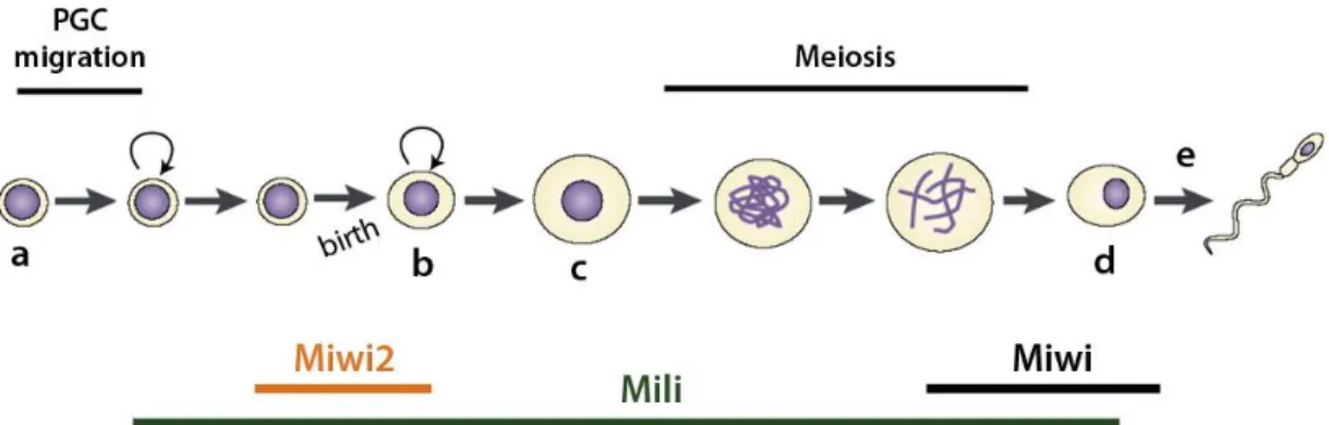
The expression pattern of Piwi proteins remains conserved in mammals. The murine genome encodes three Piwi proteins: Miwi (Piwi-like 1), Mili (Piwi-like 2) and Miwi2 (Piwi-like 4) and all are specifically expressed in the germline during spermatogenesis16-18. Still, they seem to act in a non-redundant fashion since they
are expressed at different developmental time periods (Fig. 1.3.1). Miwi is only found in adult testis, particularly in later stages of spermatogenesis during spermatocyte and spermatid development16. Expression of Mili is considerably
wider, beginning much earlier in primordial germ cells (PGCs) and comprehending most of the spermatogenic process until the spermatid stage17,19. Finally, Miwi2
expression was found to be restricted to a very short window, starting after PGC migration and ending 3 days after birth20.
In recent years we have witnessed abundant progress in the research of mammalian Piwi biology, probably due to the increasing notion that Piwi genes are highly pleiotropic, responsible for many different biological processes. Functional
15 analyses of Piwi in mammalian germ cells have provided outcomes consistent with their expression pattern. miwi knock-out mice exhibit male sterility, characterized by a clear arrest in the first wave of spermatogenesis, at the round-spermatid stage (Fig. 1.3.1, d)16. The authors noticed that the phenotypic effects of the miwi
mutation were extremely similar to CREM-deficient mice (cAMP-responsive element modulator), an essential transcription factor for the onset of spermiogenesis (Fig. 1.3.1, e). A drastic down-regulation of CREM targets was registered in miwi mutants, and this outcome was not caused by Miwi or CREM regulating each other. Strikingly, it was concluded that Miwi is a key regulator of spermiogenesis through coupling with CREM-target mRNAs, thus stabilizing them16. This study revealed an exciting new function for the mammalian piwi
family in maintaining mRNA stability.
Also in congruence with their expression pattern, both mili- and miwi2-deficient individuals were male-sterile17-18. Unlike miwi mutants, earlier defects
were observed in both cases. Neither mili nor miwi2 mutant individuals displayed any post-meiotic germ cells meaning that mutant cells never completed the first round of meiosis. Evidence from mili mutants led to the conclusion that spermatogenesis was blocked during the early stages of meiosis and that subsequent apoptosis was responsible for depletion of the most mature germ cells, deep within the seminiferous tubules17. miwi2-mutant germ cells arrest
Figure 1.3.1 – Schematic representation of spermatogenesis. Before birth, PGCs (a)
migrate and expand before entering the cell-cycle arrest. After birth, germ stem cells (or spermatogonia (b)) resume division and spermatocytes (c) enter meiosis. Following meiosis, cells reach the haploid round spermatid stage (d), after which spermiogenesis (e) takes place. Spermiogenesis is the morphogenetic process that transforms a round spermatid into a mature sperm. The timing of expression for Miwi (black), Mili (green) and Miwi2 (orange) is also represented. Adapted from Aravin et al., (2008).
16 spermatogenesis at the beginning of meiosis as well. However, the number of apoptotic cells among germ cells increased with time, not only in the partially-matured germ cells like mili mutants, but also in the stem cell population18. This
observation suggests a role for Miwi2 in germline stem cell maintenance, much like the Drosophila Piwi. This conclusion was further supported by the expression of Miwi2 in somatic Sertoli cells18. Overall, the studies conducted so far show that
Piwi proteins exhibit a wide range of action and participate in many mechanisms that are essential for germ cell development and survival.
No expression of miwi or miwi2 has been detected in mammalian female gonads. Transcripts of mili have only been recognized in ovaries at the same developmental time point as that of male mice. Yet, the expression began to decline after birth and was completely absent from the adult ovary19. Furthermore,
knock-out mutations for any of the three murine Piwis yields fertile females, with regular gonad development and complete germ cell lineages. It seems that so far the male-specific function of Piwi proteins might be unique to mammals, because other vertebrates such as zebrafish rely on Piwi proteins for germ-cell development in both sexes14-15. Some authors have argued that mammalian Piwi proteins have
male-specific activity because female germ cells do not undergo continuous mitosis and meiosis during adulthood, contrary to the situation in flies and zebrafish21. In
spite of the previous argument, it is possible that the mouse is not the best mammalian model in this case, and that an answer may come from the study of non-model organisms.
1.4. piRNAs, a fortress of genomic integrity
1.4.1. Transposable elements
The integrity of the genetic material contained within our cells is relentlessly threatened by a large variety of environmental or endogenous factors. Ionizing radiation, toxic compounds and DNA replication errors are well known examples of such deleterious agents. However, one of the major hazards that genomes face is DNA itself, particularly transposable elements (TEs). TEs are selfish DNA segments that have the ability to move from one location in the genome to another. This feature makes active TEs highly mutagenic, as the random
17 insertion of their sequences in new locations can disrupt gene function or structure22.
TEs can be divided into two major classes based on the mechanism of transposition: retrotransposons (class I) and DNA transposons (class II)23.
Retrotransposons amplify themselves through a “copy and paste” mechanism, which involves an RNA intermediate that is converted back into DNA via multiple rounds of reverse transcription. Retro-elements can be autonomous, if they encode the necessary components for amplification, or nonautonomous, if they require the necessary proteins to be supplied by the autonomous elements24. On the other
hand, the majority of class II elements exhibit motion at the hand of a “cut and paste” method, and their insertion relies on a transposase enzyme25.
Despite the detrimental consequences of active transposition, these mobile elements have been key players in the shaping and evolution of genomes. In fact, TEs can be found in the genomes of all plants and animals. Furthermore, transposons and remnants of transposon sequences account for nearly half of the human genome (42% for retrotransposons and 3% for DNA transposons)26,
forming what it is usually referred to as “junk DNA”. Whether these iterative sequences are actually “junk” is a complicated matter, however millions of years of evolution have achieved an equilibrium between the deleterious effects of transposition on the individual and the long-term benefits on the species through shaping of the genome27.
1.4.2. Silencing transposable elements
Most transposons remain in a quiescent state, mainly due to defects caused by the accumulation of mutations that interfere with their transposition mechanism. However, intact and competent copies exist within host genomes and, regardless of their importance for genomic evolution, the immediate effect of their transposition is usually deleterious and needs to be controlled. An evolutionary arms-race between the genome and mobile elements stemmed from these interactions and gave rise to several epigenetic mechanisms that cells employ to limit the mobility of the rare, healthy TEs.
18 TE silencing is essentially carried out by two processes: transcriptionally via chromatin modifications, and post-transcriptionally through RNA interference28. Chromatin modifications include changes in histone tails and DNA
methylation. Analyses of TE-associated nucleosomes have shown that they are enriched for methylation of histone H3 at the lysine 9 position (H3K9)29. These
modifications result in heterochromatin that is condensed and thus inaccessible for transcription, keeping TEs in a silent state. Another important signal used for long-term transcription repression lies on DNA methylation of cytosine residues. Mammalian genomes employ this heritable mechanism on genes subject to genomic imprinting, X-chromosome inactivation and retrotransposon silencing. For example, the DNA methyltransferase 3-like (Dnmt3L) is a germ-line specific protein and its deletion handicaps the de novo methylation of retrotransposons, leading to an upregulation of TE transcripts and meiotic collapse of germ cells in mice30. Endogenous siRNAs (endo-siRNAs) have also been implicated in TE control
through RNAi. endo-siRNAs that correspond to transposon and heterochromatic regions were identified in Drosophila somatic cells and transposon transcripts increased dramatically in ago2 mutant flies31. In mice, endo-siRNAs were also
found to repress TEs in oocytes32.
We have seen that animals developed numerous mechanisms to prevent the mobility of TEs and still a significant fraction of their genomes correspond to these selfish agents. Repression of TEs is of the utmost importance in germ cells, since transposition events could lead to a genomic alteration that would be transferred to the offspring, further advancing TE success.
1.5. Piwi proteins and piRNAs
Given the conservation of the main protein motifs between the Ago and Piwi groups, it was expected to some extent that Piwi proteins interacted with small RNAs. In mammals, several studies searching for Piwi-associated small RNAs let to the discovery and characterization of piwi-interacting RNAs (piRNAs)33-36. This
finding widened the spectrum of functional possibilities for Piwi proteins, from post-transcriptional gene silencing and repression of transposable elements to the
19 epigenetic regulation of the genome. Hints of the existence of piRNAs had emerged earlier in how Stellate repeats are silenced in Drosophila13 (see section 1.3).
The piRNAs represent a novel population of small RNAs of approximately 27 nts length. There are several crucial differences between piRNAs and the better known siRNAs or miRNAs. Apart from their distinct lengths (piRNAs are longer and more variable), piRNAs interact exclusively with members of the Piwi subgroup of proteins33,35, while miRNAs and siRNAs associate with Ago proteins.
Furthermore, piRNA sequences are much more diverse. For example, while one miRNA species can have hundreds of its copies in the genome, there are thousands of individual piRNAs, revealing an extremely high complexity of piRNA populations37. Finally, unlike the other small RNA classes, piRNA biogenesis is
independent of Dicer and are not derived from double-stranded precursors38. How
are piRNAs produced then?
Massive sequencing projects using data from cloned piRNAs determined that they are not evenly distributed across the genome, but are rather clustered at specific regions33,35,37. These regions were named piRNA clusters and exhibit
remarkable characteristics. Drosophila piRNAs were mapped to centromeric and telomeric regions of heterochromatin, territories that are densely populated by repetitive elements, such as both intact and defective copies of transposons37.
These clusters contain a bulk of randomly-oriented and nested transposons, and can produce piRNAs from both genomic strands that are ready to be loaded on a Piwi protein. piRNAs that are antisense to transposon sequences can then guide its Piwi partner to direct endonucleolytic activity on complementary mRNAs, preventing transposition.
In accordance, mili- and miwi2-deficient mice display an up-regulation of retrotransposon transcripts18,39. Activation of TEs could be responsible for the
defects in gametogenesis observed in mili and miwi2 mutant mice (see section 1.3). The increased transposition observed in individuals lacking these genes could lead to the generation of double-stranded DNA breaks, activating DNA-damage checkpoints which in turn trigger apoptosis18.
20
1.5.1. piRNA biogenesis
The study of Piwi-associated piRNAs in flies and mice has supported the hypothesis of a two-step biogenesis pathway for piRNAs: a primary processing pathway, thought to be responsible for providing an initial pool of transposon-directed piRNA-Piwi complexes; and the ping-pong amplification loop, a feedback system that shapes the piRNA population in order to target active transposons more efficiently.
Evidence from RT-PCR using primers derived from the same strand of a cluster revealed that transcripts much longer than 30 nts are transcribed from rat piRNA loci40. This line of evidence points to a hypothesis where primary
processing samples from a long single-stranded piRNA transcript, generating an initial set of mature piRNAs20,37. Given the content of these clusters (described
above), these primary piRNAs can recognize and destroy TEs transcripts that originate from dispersed locations in the genome. Exactly how primary processing produces piRNAs from the precursor molecule remains unclear.
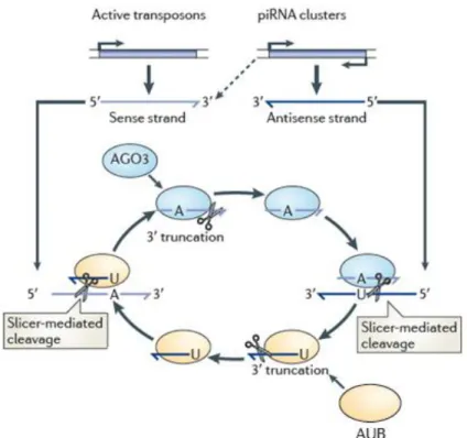
Figure 1.5.1 – The ping-pong amplification loop. Aub associated with antisense piRNAs
cleave complementary targets that originate from active transposons, defining the 5’ end of piRNAs that are loaded onto Ago3. The 3’ end of piRNAs are formed by unknown factors. Ago3-bound piRNAs direct cleavage of antisense piRNAs derived from genomic clusters that are subsequently loaded onto Aub. piRNAs that induce this amplification loop may be maternally inherited. Adapted from Siomi et al., (2011).
21 Primary piRNAs are the first defensive line against TEs in the germline, and they are backed by another mechanism, the ping-pong cycle. Shortly after the discovery of piRNAs, two independent studies published data supporting a Piwi-dependent amplification machinery that boosts piRNA numbers37,41. In Drosophila,
the majority of piRNAs derived from total RNA extractions are antisense to transposon mRNA sequences37. However, sequence analysis of piRNAs bound to
the three fly Piwis (Piwi, Aubergine (Aub), and Ago3) showed that Ago3-associated piRNAs are often complementary to Aub- and Piwi-bound piRNAs. Moreover, piRNAs bound to Piwi and Aub are usually antisense to transposons (76% and 83%, repectively), whereas Ago3 piRNAs are strongly biased for sense transposon strands (75%)37,41. The observed strand bias suggests a model where the antisense
piRNAs (bound to Aub) target and cleave transposon mRNAs. This results in sense fragments that are not merely degraded, but rather loaded onto Ago3. In turn, the Ago3-bound sense transcripts target the large single-stranded precursors that stemmed from piRNA loci, leading to the creation of additional antisense piRNAs that associate with Piwi and Aub (Fig. 1.5.1). This results in a feedback cycle that amplifies the specific fraction of the initial pool of piRNAs that correspond to active transposons. In a way, the ping-pong cycle makes use of the active population of TEs as a scaffold, and directs specific amplification of the piRNA sequences that target those transposons. Compelling evidence supporting a post-transcriptional enhancing of piRNAs has been also found in vertebrates14-15,20. Despite fascinating,
the ping-pong model is a hypothesis that still needs experimental support.
1.5.2. Evolutionary implications of Piwi proteins and piRNAs
Considering these observations, it seems as if piRNA clusters function as repositories of information, accumulating genetic memory of the transposable elements that afflicted that population in the past. Just as if were keeping a
criminal record. Moreover, the cell has also developed a mechanism to access those
records by way of a Piwi protein patrol. If a transposable criminal decides to wander around the cell, Piwi proteins carrying a piRNA mugshot of the perpetrator are able to recognize it and stop it from committing further crimes. For instance, if a new, unseen transposon were to become active in a population, at first it would be extremely successful, able to proliferate without opposition. Deleterious
22 mutations caused by increased transposition would be selected against while individuals with nonsense insertions in gene-poor regions (such as heterochromatic regions where piRNA clusters are located) could most likely escape unharmed. With time, as further transposon copies gather in these areas (where they are prone to accumulate mutations), the genome becomes progressively better protected and has its database updated for the new transposon. Some authors have proposed to explore if the content of piRNA clusters is in fact renewed by random transposition of new TEs, or if a mechanism exists that actively targets new insertions to these piRNA loci42.
1.6. The bovine as an animal model
1.6.1. The mammalian alternative
The mammalian female germline remains a major puzzle in the understanding of Piwi proteins. While a snowball of evidence progressively heightens the importance of Piwi proteins in the male germline, there is an apparent indifference towards Piwi proteins in female mice. As previously discussed, it has been suggested that Piwi function is absent from female mammals because their germ cells are not continuously dividing21. Still, these animals must
employ some mechanism to silence TEs in the germline. Contrary to the case in male gametes, a small RNA profile of mouse oocytes detected very low quantities of the characteristic ~27 nucleotide-long piRNAs34. Instead, it was discovered that
oocytes carry a rich population of retrotransposon-derived siRNAs, which have an average length of 22 nts, are Dicer-dependent and have the ability to post-transcriptionally silence TEs32,34. These observations suggest that mammalian
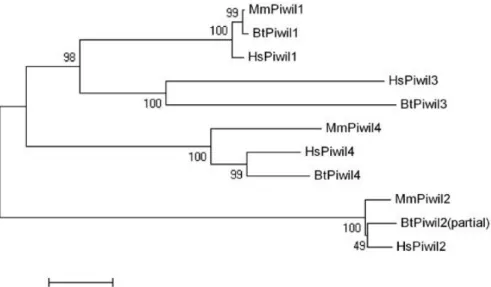
females do not need the effective germline-specific surveillance provided by piRNAs and Piwi for TE silencing. However, some evidence suggests that this situation might not extend to the rest of the mammalian class. Phylogenetic data has shown that many mammalian genomes (including humans and bovine) hold a fourth Piwi-like gene (PIWIL3) that is not present in rodent genomes (Fig. 1.6.1). The high conservation state of this gene means that it cannot be overlooked and a function for Piwi in female mammals should not be ruled out until PIWIL3 and other Piwi genes are examined in other animals.
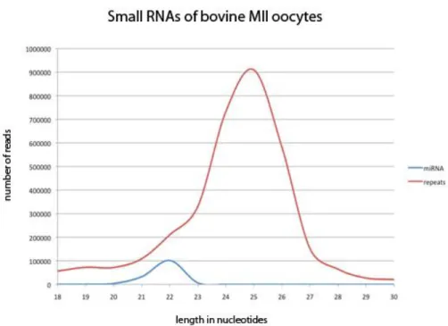
23 Despite the obvious limitations regarding experimental manipulation, the bovine represents a viable alternative for the study of Piwi proteins in the female germline and early embryos. Large amounts of cow ovaries can be obtained as left-over material from local slaughterhouses, and healthy oocytes can be isolated from these ovaries. Collected oocytes can be matured in vitro, fertilized and embryos can be developed until the blastocyst stage. Furthermore, and contrary to the case in mice, the small RNA profile of bovine oocytes contains a pronounced piRNA-like population of 24-28 nts length (Fig. 1.6.2). Sequencing of this population found that they mostly map repetitive elements in the genome, particularly SINE and LINE elements (Ketting, R., unpublished results). This observation reveals a major difference in Piwi-piRNA biology between mammalian species, supporting the notion that rodents might constitute an exception and not the standard in this matter.
1.6.2. Oocyte maturation
Mammalian oogenesis is an intricate process that results in the genesis of the oocyte, a unique cell that, when combined with its male equivalent, is capable of giving rise to a new individual. During oogenesis the egg prepares itself for fertilization and for the beginning of development. While a sperm eliminates all its
Fig. 1.6.1 – Phylogenetic relationships between mammalian Piwi proteins.
Mm = Mus musculus; Hs = Homo sapiens; Bt = Bos taurus. PIWIL3 is lacking in murine genomes, though it is present in many mammals. Roelen, B., Ketting, R.
24 cytoplasm and condenses its nucleus to the smallest possible dimensions, an egg builds up in size gathering reserves to support subsequent growth. Here, we will focus primarily on aspects of oocyte maturation, the process that promotes the structural and molecular changes required to form a developmentally competent gamete43.
Oocyte maturation is a tightly regulated mechanism, and it is carried out at two levels within the cell: nuclear and cytoplasmic maturation. During nuclear maturation, extracellular signals stimulate oocytes that are arrested in the first meiotic division to resume meiosis. This resumption is temporary, as a second arrest takes place at the metaphase II stage. At this point, meiosis will only be completed if a sperm penetrates the cell. On the other hand, cytoplasmic maturation is characterized by the accumulation of factors in the cytoplasm that instill upon the oocyte the capacity to sustain early embryonic development.
Figure 1.6.2 – Small RNA profile of MII stage bovine oocytes. The blue line indicates
reads corresponding to annotated miRNAs. The red line indicates reads mapping to transposons. The shoulder in the transposon curve suggests the presence of siRNAs in addition to the pronounced piRNA-like population. X-axis: length of read in nucleotides. Y-axis: absolute number of obtained reads. From Ketting, R., unpublished results.
25
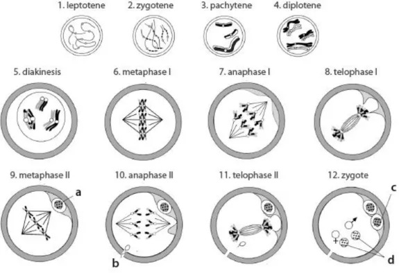
1.6.3. Meiosis
In mammals, oocyte meiosis results in the division of a diploid parental cell into haploid progeny, a cell containing only one member of the pair of homologous chromosomes that were present in the progenitor. This reduction in chromosome number is accomplished by two sequential rounds of nuclear division (meiosis I and meiosis II), which can be divided in the following phases44-45:
Prophase I: starting during fetal development, prophase I is the longest chapter in
meiosis and is characterized by pairing of homologous chromosomes, allowing recombination of genetic material of paternal and maternal origin. Prophase I is divided into five stages: leptotene, zygotene, pachytene, diplotene and diakinesis. At leptotene (Fig. 1.6.3 – 1), base pairing between complementary DNA is thought to mediate the initial association of chromosomes. During zygotene (Fig. 1.6.3 – 2), a protein structure called the synaptonemal complex is formed, and functions to keep homologous chromosomes closely associated and aligned throughout the pachytene stage (Fig. 1.6.2 – 3). Recombination is completed at the pachytene stage, leaving the chromosomes linked at the sites of crossing-over, the chiasmata. The synaptonemal complex disappears at the diplotene stage (Fig. 1.6.3 – 4) and homologous chromosomes separate along their length. However, they remain attached at the chiasmata, which is critical for their alignment at the metaphase. This is the first regulatory point in oocyte meiosis and oocytes can remain arrested at this stage for long periods of time. During the arrest, chromosomes decondense (germinal vesicle (GV)) and are actively transcribed, accumulating the needed materials to support early embryonic development. The prophase chapter ends with diakinesis (Fig. 1.6.3 – 5), which represents the transition to the metaphase. At this point, chromosomes progressively condense and the nuclear envelope is dissolved, in a process called germinal vesicle breakdown (GVBD).
Metaphase I: at this phase, bivalent chromosomes align on the spindle apparatus.
While centromeres of sister chromatids are adjacent to each other and oriented in the same direction, the centromeres of homologous chromosomes face opposite spindle poles. Accordingly, microtubules from the same pole attach to sister chromatids and homologous chromosomes attach to microtubules from opposite poles of the spindle (Fig. 1.6.3 – 6).
26 Figure 1.6.3 – Schematic representation of oocyte meiosis. (a) first polar body; (b)
fertilization; (c) second polar body; (d) pronuclei. Adapted from Jones (1978).
Anaphase I: disruption of the chiasmata that join homologous chromosomes
marks the beginning of anaphase I. This is followed by the separation of homologous chromosomes, but not sister chromatids, towards opposite poles of the spindle (Fig. 1.6.3 – 7).
Telophase I: bringing the first meiotic division to an end, the separated
chromosomes aggregate, culminating with an asymmetric cell division and the extrusion of the first polar body (Fig. 1.6.3 – 8).
Metaphase II: without having re-formed a nucleus, chromosomes align again at
the spindle and microtubules from opposite poles attach to the centromeres of sister chromatids. Most vertebrate oocytes are arrested again at this point, where they remain until fertilization (Fig. 1.6.3 – 9).
Anaphase II: if a sperm penetrates the egg, the link between the centromeres of
sister chromatids is broken, and microtubules pull sister chromatids towards the opposite poles of the spindle (Fig. 1.6.3 – 10).
Telophase II: meiosis in the oocyte is completed with a new asymmetric division
27 Following completion of meiosis, the zygote (Fig. 1.6.3 – 12) contains two pronuclei, one derived from each progenitor. In mammals, the pronuclei subsequently enter the S phase and drive DNA replication as they migrate toward each other. As they converge, the zygote progresses to the M phase of its inaugural mitotic division and both paternal and maternal chromosomes align on a common spindle. Completion of mitosis originates two embryonic cells, each containing a new diploid genome. These cells then initiate the sequence of cell divisions that ultimately leads to the development of a new organism45.
28
2. Materials and Methods
2.1. Oocyte collection, in vitro maturation and fertilization
Bovine oocytes were collected from ovaries donated by a local slaughterhouse. The ovaries were transported in a thermal flask and kept at 30ºC in saline solution supplemented with penicillin/streptomycin (Pen/Strep, 1mL/L). Cumulus-oocyte complexes (COCs) were then obtained by suction of follicular fluid from antral follicules ranging 2-8mm. The collected follicular fluid was analyzed and COCs were selected based on cumulus investment. Only COCs with several layers of compact cumulus were included in experiments. After selection, the COCs were rinsed in B medium (Table 1) thus removing excess of follicular fluid to avoid agglomeration. COCs were then transferred to 4-well plates, each well containing 500uL of maturation medium (Table 1) and incubated at 39ºC with 5% CO2 injection. After 22-24 hours of incubation about 85% of cultured oocytes reached the metaphase II stage of meiosis.
Medium Ingredients
B MilliQ water, M199 with Earle's salts, HEPES and glutamine C LAL water, M199 with Earle's salts and glutamine, NaHCO3
PHE Penicillamine, hypotaurine and epinephrine
Maturation Medium C, Fetal Calf Serum, Pen/Strep, recombinant FSH
Fertilization LAL water, NaCl, KCl, NaHCOMgCl 3, Na2HPO4, Na pyruvate, phenolred, CaCl2.2H2O,
2.6H2O, Pen/Strep, BSA
RD MilliQ waterm NaCl, KCl, NaHCO3, Na2HPO4, Na Lactate, HEPES, phenolred, CaCl2.2H2O,
MgCl2.6H2O, Na Pyruvate, Pen/Strep, BSA
SOF A LAL water, NaCL, KCl, KH2PO4, Na Lactate, MgSO4.7H2O, NaHCO3, CaCl2.2H2O,
phenolred, Non-essential aminoacid solution
SOF B LAL water, Pen/Strep, Na-pyruvate, L-glutamine, BSA SOF 4X SOF A, 1X SOF B
Following maturation the COCs were transferred to fertilization medium containing heparin and PHE (Table 1). At this point, sperm was prepared for in
vitro fertilization. A straw of cryopreserved bull sperm was thawed in a 37ºC
waterbath and layered on a Percoll gradient. This gradient was centrifuged for 30 minutes at 700G for optimal recovery of motile sperm. Before adding to the culture, the appropriate concentration of sperm was determined by sampling a Table 1. Composition of media used in oocyte culture, fertilization and embryo culture
29 small volume of the sperm solution and counting the number of sperm heads in a Bürker Turk chamber. Taking into account which bull was used, the dilution factor was calculated and sperm was added to the fertilization medium (Table 1). The inseminated COCs were again incubated at 39ºC and 5% CO2. The fertilization process takes between 18-22h, after which the putative zygotes were placed in an embryo culture environment. For that purpose, zygotes were rinsed in medium RD (Table 1) and stripped of cumulus cells by physical disaggregation using a vortex mixer. Following denudation, the presumptive zygotes were transferred to a new 4-well plate with synthetic oviductal fluid (SOF medium, Table 1) and incubated at 39ºC, in a 5% CO2 and 7% O2 environment.
2.2. Total RNA extraction and cDNA synthesis
Pools of oocytes and embryos at different developmental time points were collected for transcriptomic analysis using reverse transcriptase-polymerase chain reaction (RT-PCR) and quantitative RT-PCR (qRT-PCR). Samples of 10 individuals were collected for the following developmental time points: germinal vesicle (GV), metaphase I (MI), metaphase II (MII), zygotes, 2-cell, 4-cell, 8-cell, morula and blastocyst. All samples were stored in an eppendorf tube containing 150uL of lysis buffer (RLT buffer (Qiagen) and β-mercaptoethanol) at -80ºC until use.
Extraction of RNA was performed using the RNeasy Micro Kit (Qiagen) according to manufacturer’s instructions. Briefly, lysates were homogenized, diluted 1:1 with 70% ethanol and applied to the micro-column. The process of extraction included an on-column DNAse I treatment to avoid DNA contamination. After washing, the RNA was eluted with 18µL RNAse-free water for a final yield of 16µL total RNA (2µL remain trapped on the column).
Before cDNA synthesis, RNA samples were incubated for 5 min at 70ºC, vortexed and briefly centrifuged. Reverse transcription took place in a total volume of 20uL, containing 10uL of RNA sample and 10uL of the reverse transcription mix (5X first strand buffer, DTT, dNTPs, RNAsin, random primers, and Superscript reverse transcriptase). After combining the RT mix and the RNA, samples were incubated for 1 hr at 50ºC, followed by an enzyme inactivation step of 5 min at 80ºC. For every reaction, a control under the same conditions but
30 without reverse transcriptase (-RT) was created to detect potential genomic DNA contaminations. All cDNA samples were stored at -20ºC until use in polymerase chain reaction (PCR) or quantitative PCR.
2.3. Primer design
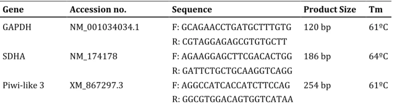
All primers were designed to span an intron using the NCBI Primer Tool. Primers were tested on cDNA of in vitro matured oocytes and the PCR products were run on a 1% agarose gel and sequenced to examine the specificity of the primers. Additionally, the optimal annealing temperature of each primer was determined experimentally with a temperature gradient. The sequences of the primers used are specified in Table 2.
Gene Accession no. Sequence Product Size Tm
GAPDH NM_001034034.1 F: GCAGAACCTGATGCTTTGTG 120 bp 61ºC R: CGTAGGAGAGCGTGTGCTT SDHA NM_174178 F: AGAAGGAGCTTCGACACTGG 186 bp 64ºC R: GATTCTGCTGCAAGGTCAGG Piwi-like 3 XM_867297.3 F: AGGCCATCACCATCTTCCAG 254 bp 61ºC R: GGCGTGGACAGTGGTCATAA
2.4. RT-PCR
All cDNA samples were tested before use for a housekeeping gene to confirm cDNA synthesis. PCR products were then run on a 1% agarose gel to check primer specificity. Amplification of cDNA was carried out by HotStar-Taq polymerase (Qiagen) according to manufacturer’s instructions. Reactions were carried out in a total volume of 25µL: 1µL of sample was added to 24µL of mixture (MgCl2, 10X Buffer, primers, Taq polymerase). Each reaction started with a 15 min
dwell at 95ºC, followed by 40 cycles with 3 steps per cycle: 5 sec of denaturation at 95ºC, 5 sec at the primer-specific annealing temperature and 20 sec of elongation at 72ºC. The reactions ended with a final extension of 7 min at 72ºC. 10µL of the amplified products were run on a 1% agarose gel, and each experiment included a
31 negative control (water blank), a positive control (sequenced product) and a 100- base pair (bp) ladder for reference.
2.5. Quantitative RT-PCR
Quantitative RT-PCR amplifications were performed using iQ-SYBR Green Supermix (Bio-Rad). Reactions for each gene were performed simultaneously for all samples in a 96-well plate and each run was supplemented with a standard curve. All samples and standard curve points were run in technical duplicates. The standard curve used to quantify reference gene SDHA (Succinate dehydrogenase complex subunit A) was made from 10-fold dilutions of PCR product, ranging from 10pg to 1ag. For quantification of the target gene (PIWIL3), the standard curve consisted of 2-fold serial dilutions of cDNA synthesized from approximately 500 oocytes.
qRT-PCR reactions were performed in a total volume of 25µL, where 1µL of cDNA was used for the target gene, and 0.5µL of cDNA for the reference gene. The reaction mix consisted of iQ-SYBR Green Supermix, water and primers. The thermal cycling profile was as follows: 3 min of initial denaturation at 95ºC, followed by 40 cycles of 5 sec at 95ºC, 5 sec at the primer-specific annealing temperature, and 20 sec of elongation at 72ºC; finally, 1 min at 95ºC followed by 10 min at 65ºC.
2.6. Immunocytochemistry
Piwi-like 3 antibodies were produced in rabbits using the N-terminal region (first 20 aminoacids) of the predicted aminoacid sequence of the protein as an antigen (Eurogentec). Prior to its use in immunofluorescence, the specificity of antibodies was tested on a Western blot analysis.
All oocytes were stripped of cumulus cells before staining and fixed for a minimum of 10 min in 4% paraformaldehyde (PFA). After fixation, the oocytes were briefly washed with 0.1% Triton X-100 and 10% FCS in PBS (PBST) and permeabilized for 30 min using 0.5% Triton X-100 and PBST. The oocytes were subsequently blocked for 1h in PBST and incubated with the primary antibody
32 overnight at 4ºC. The primary antibody was washed off three times in PBST (15 min each) followed by incubation in secondary antibody for 1 hr in the dark. After several washes, the oocytes were stained with 4',6-diamidino-2-phenylindole (DAPI) for 15 min and mounted on a slide with Vectashield and isolated with Vaseline.
2.7. Transfection of oocytes
Oocytes were transfected with a fluorescein-labeled dsRNA oligomer (BLOCK-iT Fluorescent Oligo, Invitrogen) by lipofection, using the reagent Lipofectamine 2000 (Invitrogen). Prior to transfection, the oocytes were stripped of cumulus cells by physical disaggregation, washed in PBS and transferred to small droplets of 0,1% pronase for 3-5 min in order to remove the zona pellucida surrounding the oocyte. Enzymatic digestion of the zona was necessary to expose the oocyte plasma membrane to the liposomes. This process was carefully monitored under a stereomicroscope. Once the oocytes were zona-free, they were transferred to a dish containing large volumes of maturation medium to end the enzymatic reaction. The transfection procedure was carried out according to manufacturer’s instructions and published evaluations of the reagent46. First, the
denuded oocytes were placed in a 4-well plate containing maturation medium without antibiotics. At this point, the labeled oligos (LOs) were diluted in Opti-MEM I medium. Subsequently, Lipofectamine 2000 was diluted in Opti-Opti-MEM I for 5 min. Following the 5 min incubation, the diluted LOs were combined with the diluted Lipofectamine 2000 and kept at room temperature for 30 min. This step allowed the LOs-Lipofectamine 2000 complexes to form. Before placing the plate in the incubator, the LOs-Lipofectamine complexes were added to each well containing denuded oocytes and maturation medium.
33
3. Results
3.1. PIWIL3 expression is mainly restricted to germ cells
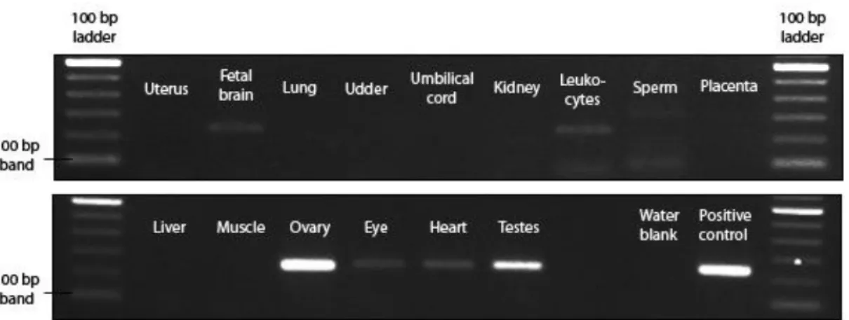
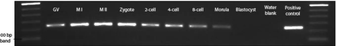
In order to provide context and a thorough description of the expression pattern of PIWIL3, total RNA was extracted from a large variety of bovine tissues and cDNA was made to be used for gene expression analysis using RT-PCR. Amplification of cDNA with PIWIL3 specific primers yielded clear bands of the expected size (254 bp) for both testes and ovary samples (Fig. 3.1). Specific expression was also detected for the following samples: fetal brain, leukocytes, eye and heart. However, in further analysis using quantitative RT-PCR, these samples exhibited a late threshold cycle (data not shown), well below the lower boundary of our standard curve, indicating extremely low starting quantities. A sequenced product of a testes cDNA sample served as a positive control.
3.2. PIWIL3 is expressed throughout meiotic maturation and
early development
Next, we explored the expression pattern of PIWIL3 within the ovary, in particular, during oocyte maturation and early embryo development. Samples ranging from germinal-vesicle (GV) oocytes to blastocysts were collected and, similar to the bovine tissues analysis, total RNA was extracted and cDNA was synthesized by reverse transcription. Tested time points were as follows: GV,
Figure 3.1 - Expression of PIWIL3 in different bovine tissues. cDNA samples from a
large variety of tissues were tested for the presence of PIWIL3 transcripts. Significant expression was only detected in ovary and testes cDNA. -RT controls of each sample yielded no specific bands (not shown).
34 metaphase I (MI), metaphase II (MII), zygote, 2-cell, 4-cell, 8-cell, morula and blastocyst. Specific bands of PIWIL3 transcripts were detected in all developmental time points tested except for the blastocyst stage. The observed expression pattern suggests a role for PIWIL3 throughout meiosis and pre-implantation development, but not during the separation of embryonic and extra-embryonic cell lineages that occurs at the blastocyst stage.
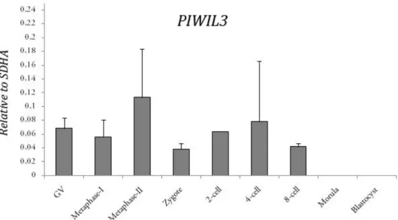
3.3. Relative PIWIL3 expression levels
To determine if the expression of PIWIL3 is under regulation, cDNA made from total RNA extracts was amplified using quantitative RT-PCR (qRT-PCR). The quantitative expression of PIWIL3 was tested throughout maturation and early development, using the same time points as in section 3.2. Expression levels were normalized to the expression of SDHA, a house-keeping gene that is steadily expressed during bovine oocyte maturation and early development, therefore allowing direct comparison with our target gene. For each sample, a corresponding –RT blank was amplified and was found non-detectable or displayed an extremely late threshold cycle, beyond the scope of the standard curve.
No significant up- or down-regulation of PIWIL3 expression was detected throughout the developmental time points tested. For morula and blastocyst samples, expression levels were non-detectable or did not exceed the background amounts of their –RT controls. This result is consistent with the data obtained by RT-PCR and suggests that PIWIL3 does not play a significant role at this point. A moderate (about 1.7-fold), but not significant up-regulation of PIWIL3 transcripts was detected in MII oocytes when compared with GV-stage oocytes and zygotes, indicating a possible role for PIWIL3 in oocyte maturation and meiosis.
Figure 3.2 - Expression of PIWIL3 during oocyte maturation and early development. cDNA
samples ranging from GV-stage oocytes to blastocysts were tested for the presence of PIWIL3 transcripts. Bands of the expected size were present in all time points tested with the exception of blastocysts. -RT controls of each sample yielded no specific bands (not shown).
35
3.4. Piwi-like 3 associates with chromatin during meiosis
In order to identify the expression pattern and sub-cellular localization of Piwi-like 3 protein, whole mount immunofluorescence was performed on in vitro produced oocytes and embryos. In the first experiments, Piwi-like 3 was detected in MII oocytes, but was not found in GV-stage oocytes or 2-cell embryos. Consequently, we decided to pinpoint the exact timing of Piwi-like 3 expression by collecting individuals every 90 min starting at the onset of maturation until the metaphase II stage (after 24h). However, only select time points were included in figure 3.4.1. Piwi-like 3 protein was first detected around the genetic material after 9 hrs of incubation (Fig. 3.4.1, G-I), a moment where the germinal vesicle already exhibits some degree of condensation, but no metaphase plate is yet formed. No specific expression was detected prior to this time point (Fig. 3.4.1, A-F). At 12 hrs into maturation the first metaphase is evident, with individual chromosomes easily identified (Fig. 3.4.1, J-L). Piwi-like 3 was localized in a spindle-like manner, tightly surrounding the paired chromosomes but most likely not binding DNA itself, as gaps matching the position of the chromosomes are visible in the fluorescence pattern (Fig. 3.4.1, K). 15 hrs after incubation, homologous chromosomes were still
Figure 3.3 – Relative mRNA expression levels of PIWIL3 during oocyte maturation and early embryo development. X axis: developmental stages.
Y-axis: relative PIWIL3 amounts normalized to the reference gene SDHA. Error bars represent standard deviation.
36 aligned with the spindle (Fig. 3.4.1, M-O). Apparently, the pattern of Piwi-like 3 expression underwent re-arrangement at these stages. Small clusters of proteins were visible dispersed throughout the cell, giving the impression that pockets of Piwi-like 3 were transiting. This notion was supported by the expression pattern at 16.5 hrs (Fig. 3.4.1, P-R). Here, as homologous chromosomes were being pulled toward opposite poles of the spindle, less of these clusters were detectable and Piwi-like 3 became more localized to one set of the divergent chromosomes.
37 Figure 3.4.1 - Time-lapse immunofluorescence results on in vitro matured oocytes. All rows of three images are
presented in the following order: DAPI stain, Piwi-like 3 stain and overlay; and correspond to the same developmental time point. (A-C) 0 hr, onset of maturation; (D-F) 6 hrs after incubation; (G-I) 9 hrs; (J-L) 12 hrs, metaphase I stage; (M-O) 15 hrs; (P-R) 16,5 hrs, anaphase I stage; (S-U) 18 hrs; (V-X) 21 hrs, metaphase II stage. Scale bar = 10µM. (Y) Amplified section of white rectangle in X. Scale bar = 20 µM
38 Later, at 18 hrs into maturation, Piwi-like 3 was strictly confined to that set of chromosomes, while the other half was condensing in order to be extruded as the first polar body (Fig. 3.4.1, S-U). After 21 hrs in culture, the metaphase II plate had been established and Piwi-like 3 localized around the DNA (Fig. 3.4.1, V-X), similar to the localization at the first metaphase. No Piwi-like 3 was detected in the vicinity of the polar body. At this point, the oocytes arrested meiosis until fertilization.
Following maturation, the oocytes were fertilized and meiotic progression was analyzed in zygotes until the formation of pronuclei. 4 hrs into the resumption of meiosis, sister chromatids were still aligned at the equatorial plate of the
Figure 3.4.2 - Time-lapse immunofluorescence results on in vitro produced zygotes. All
rows of three images are presented in the following order: DAPI stain, Piwi-like 3 stain and overlay; and correspond to the same developmental time point. (A-C) 4 hrs following IVF; (D-F) 6 hrs after incubation; (G-I) 16h after fertilization. Scale bar = 10µM
39 spindle, and Piwi-like 3 remained localized in this context (Fig. 3.4.2, A-C). As sister chromatids separated, a Piwi expression pattern similar to that of the first anaphase emerged (Fig. 3.4.2, D-F). An apparent re-arrangement took place while proteins began to accumulate on one set of chromatids. Piwi-like 3 expression faded after this event, and was last detected when pronuclei had already been formed (Fig. 3.4.2, G-I). Here, Piwi-like 3 can be seen around a chromatin-dense body that is located within the presumptive female pronucleus.
These observations are consistent with the data obtained by PCR analysis. The up-regulation of PIWIL3 mRNA detected at the MII stage corresponds with the protein expression pattern observed by immunofluorescence. However, despite the presence of PIWIL3 mRNA from 2-cell embryos onward, no protein was detected in embryos beyond the 2-cell stage (data not shown).
3.5. Lipofection in bovine oocytes
The next step in our research would include a functional analysis of Piwi proteins in bovine oocytes. One major issue regarding the bovine as a model organism is the difficulty of complete gene inactivation to conduct reverse genetics studies. Fortunately, a large number of techniques are available to manipulate gene expression of mammalian cell lines. In oocytes, microinjection has been predominantly used to insert dsRNA, plasmid constructs and recombinant proteins to facilitate functional studies. One of our goals is to use siRNA-mediated approaches to down-regulate PIWIL3 transcripts and infer on its role during oocyte maturation. However, microinjection is a rather laborious work, and a high number of oocytes (~100) would be necessary to certify protein down-regulation in a western blot analysis. An alternative to this methodology could lie on the transfection of small RNAs via lipofection, a technique that utilizes lipid vesicles to introduce nucleic acids into cells. This method is mostly used in more dense cell cultures and not generally directed for oocyte transfection, also because of the protective layer of glycoproteins that surrounds oocytes, the zona pellucida. Nevertheless, some reports have successfully used lipofection in mouse and rat oocytes47-48.