2017 SSAT PLENARY PRESENTATION
Assessment of the Lymph Node Status in Patients Undergoing
Liver Resection for Intrahepatic Cholangiocarcinoma: the New
Eighth Edition AJCC Staging System
Fabio Bagante1&Gaya Spolverato1&Matthew Weiss2&Sorin Alexandrescu3& Hugo P. Marques4&Luca Aldrighetti5&Shishir K. Maithel6&Carlo Pulitano7& Todd W. Bauer8&Feng Shen9&George A. Poultsides10&Oliver Soubrane11& Guillaume Martel12&B. Groot Koerkamp13&Alfredo Guglielmi1&Endo Itaru14& Timothy M. Pawlik15
Received: 16 February 2017 / Accepted: 6 April 2017 / Published online: 19 April 2017 # 2017 The Society for Surgery of the Alimentary Tract
Abstract
Introduction The role of routine lymphadenectomy for intrahepatic cholangiocarcinoma (ICC) is still controversial. The AJCC eighth edition recommends a minimum of six harvested lymph nodes (HLNs) for adequate nodal staging. We sought to define outcome and risk of death among patients who were staged with≥6 HLNs versus <6 HLNs.
Materials and Methods Patients undergoing hepatectomy for ICC between 1990 and 2015 at 1 of the 14 major hepatobiliary centers were identified.
Results Among 1154 patients undergoing hepatectomy for ICC, 515 (44.6%) had lymphadenectomy. On final pathology, 200 (17.3%) patients had metastatic lymph node (MLN), while 315 (27.3%) had negative lymph node (NLN). Among NLN patients, HLN was associated with 5-year OS (p = 0.098). While HLN did not impact 5-year OS among MLN patients (p = 0.71), the number of MLN was associated with 5-year OS (p = 0.02). Among the 317 (27.5%) patients staged according the AJCC eighth edition staging system, N1 patients had a 3-fold increased risk of death compared with N0 patients (hazard ratio 3.03; p < 0.001).
Gaya Spolverato and Fabio Bagante contributed equally to this work. Electronic supplementary material The online version of this article (doi:10.1007/s11605-017-3426-x) contains supplementary material, which is available to authorized users.
* Timothy M. Pawlik tim.pawlik@osumc.edu 1
Department of Surgery, University of Verona, Verona, Italy 2
Department of Surgery, Johns Hopkins Hospital, Baltimore, MD, USA
3 Department of Surgery, Fundeni Clinical Institute, Bucharest, Romania
4
Department of Surgery, Curry Cabral Hospital, Lisbon, Portugal 5
Department of Surgery, Ospedale San Raffaele, Milan, Italy 6
Department of Surgery, Emory University, Atlanta, GA, USA 7
Department of Surgery, Royal Prince Alfred Hospital, University of Sydney, Sydney, Australia
8 Department of Surgery, University of Virginia, Charlottesville, VA, USA
9 Department of Surgery, Eastern Hepatobiliary Surgery Hospital, Shanghai, China
10
Department of Surgery, Stanford University, Stanford, CA, USA 11
Department of Hepatobiliopancreatic Surgery and Liver Transplantation, AP-HP, Beaujon Hospital, Clichy, France 12
Division of General Surgery, Department of Surgery, University of Ottawa, Ottawa, ON, Canada
13
Department of Surgery, Erasmus University Medical Centre, Rotterdam, Netherlands
14
Gastroenterological Surgery Division, Yokohama City University School of Medicine, Yokohama, Japan
15 Department of Surgery, The Urban Meyer III and Shelley Meyer Chair in Cancer Research, The Ohio State University Wexner Medical Center, 395 W. 12th Ave., Suite 670, Columbus, OH 43210, USA
Conclusion Only one fourth of patients undergoing hepatectomy for ICC had adequate nodal staging according to the AJCC eighth edition. While the six HLN cutoff value impacted prognosis of N0 patients, the number of MLN rather than HLN was associated with long-term survival of N1 patients.
Keywords ICC . Surgery . Staging . Nodal status
Introduction
The American Joint Committee on Cancer (AJCC) staging manual is the most common means to stratify cancer patients with regard to prognosis. In the seventh edition AJCC staging manual, a staging system for ICC was introduced for the first time. The newly released eighth edition AJCC T categories have several modifications for ICC staging, including the ad-dition of tumor size to lesion number and vascular invasion [1–7]. AJCC nodal (N) staging has also been a topic of ongoing debate, as the role for routine lymphadenectomy for ICC has been controversial with no standard approach to assessing regional nodal information [2, 8, 9]. In one study using the Surveillance, Epidemiology, and End Results (SEER) cancer registry, Kim et al. reported on 749 patients who underwent surgical resection of ICC between 1988 and 2011 [10]. In this study, Kim et al. assessed the prognostic performance of AJCC/UICC seventh N stage, lymph node ratio (LNR), and log odds (LODDS) among patients with ICC [1 0]. Interestingly, after curative intent resection of ICC, LODDS and LNR were better predictors of long-term prognosis versus seventh edition AJCC nodal staging. In particular, while LNR performed well among patients who had >3 LNs harvested and examined, LODDS was better at determining prognosis among patients with≤3 LN examined.
While nodal status appears to be an important factor in de-termining prognosis of patients with ICC [11, 12], routine lymphadenectomy is not always performed, especially in Western centers [13, 14]. Data on pathological lymph node status are, therefore, often lacking. Importantly, guidelines of the European Association for the Study of the Liver (EASL) on the management of ICC recommend the removal of lymph nodes defined as suspicious according to preoperative imaging [15]. However, the correlation of radiological lymph node status assessment with pathological N status has yet to be determined. As such, the objective of the current study was to correlate the performance of radiological versus pathological assessment of lymph node status among patients with resectable ICC. Moreover, given that the newly released AJCC eighth edition recommends the recovery of at least six lymph nodes for com-plete pathologic staging, we sought to define the outcome of patients who wereBadequately^ (≥6 nodes harvested) versus Binadequately^ staged (<6 nodes harvested) according to the eighth edition of the AJCC staging manual [16].
Materials and Methods
Patient Demographic and Clinical Data
Patients undergoing liver surgery for histologically confirmed ICC between 1990 and 2015 at one of the following 14 major hepatobiliary centers were identified: Johns Hopkins Hospital, Baltimore, MD; Stanford University, Stanford, CA; University of Virginia, Charlottesville, VA; Emory University, Atlanta, GA; Fundeni Clinical Institute of Digestive Disease, Bucharest, Romania; Curry Cabral Hospital, Lisbon, Portugal; Ospedale San Raffaele, Milan, Italy; Royal Prince Alfred Hospital, University of Sydney, Sydney, Australia; Eastern Hepatobiliary Surgery Hospital, Shanghai, China; Beaujon Hospital, Clichy, France; University of Ottawa, Ottawa, Ontario, Canada; Erasmus University Medical Centre, Rotterdam, Netherlands; Yokohama City University School of Medicine, Yokohama, Japan; and University of Verona, School of Medicine, Verona, Italy. The Institutional Review Board of each institution ap-proved the study. Only patients who underwent curative intent surgery were included, while patients with metastatic disease and patients who underwent an R2 resection were excluded.
Standard patient demographic and clinicopathologic data were collected including age, sex, American Society of Anesthesiologists (ASA) physical status classification, and presence of underlying liver disease, such as cirrhosis, chronic hepatitis B infection, and chronic hepatitis C infection. Serum level of carcinoembryonic antigen (CEA) and cancer antigen (CA) 19-9 was also included. Data regarding treatment details were collected including type of surgery and receipt of adju-vant chemotherapy and radiotherapy. The type of surgery was classified as wedge liver resection, minor resection (removal of≤2 Couinaud segment), and major resection (removal of ≥3 Couinaud segment) [17]. Resection margin status was record-ed and classifirecord-ed as microscopically negative (R0) and micro-scopically positive (R1). Tumor-specific characteristics in-cluding tumor size, number, grade, number of lymph nodes harvested, number of metastatic nodes, presence of vascular invasion (macroscopic and microscopic), perineural invasion, biliary invasion, and direct invasion of contiguous organs were obtained. Lymphadenectomy was performed such that the regional lymph nodes including the nodes in the hepatoduodenal ligament (station 12), the nodes along the common hepatic artery (station 8), and the nodes on the pos-terior surface of the head of the pancreas (station 13) were harvested, in addition, for left side ICC, also the nodes along the trunk of left gastric artery (station 7). Data on tumor stage
were also collected according to both the seventh and the eighth edition AJCC staging system [1, 16]. Nodal status was assessed preoperatively by endoscopic ultrasound echography (EUS), computer tomography (CT) scan, magnetic resonance imaging (MRI), and positron emission tomography (PET) scan; nodes were classified on preoperative imaging as posi-tive, negaposi-tive, or suspicious. Date of last follow-up and vital status were collected on all patients.
Statistical Analysis
Continuous variables were reported as medians with inter-quartile ranges (IQRs), while categorical variables were re-ported as whole numbers and percentages. The endpoint for the survival analysis was overall survival (OS). OS was de-fined as the time interval between the date of surgery and the date of death. Time was censored at the date of the last follow-up assessment for patients who were still alive at the time of analysis. Survival curves were estimated using the Kaplan-Meier method, and differences between the curves were com-pared using the log-rank test. Univariate Cox proportional hazard models were used to evaluate associations between variables and OS. The coefficients from the Cox models were reported as hazard ratios (HRs) with corresponding 95% con-fidence intervals (CIs). A Bayesian model was developed to analyze the prognosis of patients with or without metastatic nodes (N1 versus N0) based on the number of harvested nodes [18]. The results of the Bayesian model were presented as 5-year OS and HR with associated 95% credible intervals (Cr Is). All analyses were performed using the statistical software programs STATA (v. 12.0, StataCorp, College Station, TX), OpenBugs (v.2011), and R CRAN (v. 3.2.2, 2015) with the packages Bsurvival,^ BHmisc,^ and BR2OpenBUGS.^ A p value <0.10 was considered statistically significant.
Results
Baseline Characteristics of the Study Group
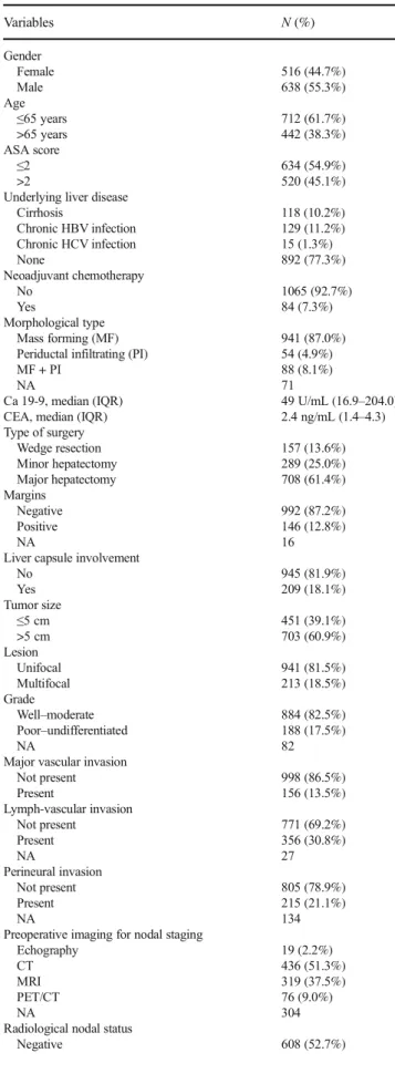
Among 1154 patients who underwent hepatectomy for ICC, most patients were male (n = 638, 55.3%) and younger than 65 years (n = 712, 61.7%; Table1). Based on the ASA phys-ical status classification, 634 (54.9%) patients were ASA≤2 and 520 (45.1%) patients were ASA 3 or 4. Preoperatively, a minority of patients (n = 84, 7.3%) underwent neoadjuvant chemotherapy. About two thirds of patients (n = 708, 61.4%) underwent a major hepatectomy, while 157 (13.6%) underwent a wedge resection and 289 (25.0%) a minor hepa-tectomy. A resection with negative margins (R0) was per-formed in the majority of patients (n = 992, 87.2%), while 146 (12.8%) patients had R1 resections. Mass forming
Table 1 Baseline characteristics of the study group (n = 1154)
Variables N (%) Gender Female 516 (44.7%) Male 638 (55.3%) Age ≤65 years 712 (61.7%) >65 years 442 (38.3%) ASA score ≤2 >2 634 (54.9%) 520 (45.1%) Underlying liver disease
Cirrhosis 118 (10.2%) Chronic HBV infection 129 (11.2%) Chronic HCV infection 15 (1.3%) None 892 (77.3%) Neoadjuvant chemotherapy No 1065 (92.7%) Yes 84 (7.3%) Morphological type Mass forming (MF) 941 (87.0%)
Periductal infiltrating (PI) 54 (4.9%) MF + PI
NA
88 (8.1%) 71
Ca 19-9, median (IQR) 49 U/mL (16.9–204.0)
CEA, median (IQR) 2.4 ng/mL (1.4–4.3)
Type of surgery Wedge resection 157 (13.6%) Minor hepatectomy 289 (25.0%) Major hepatectomy 708 (61.4%) Margins Negative 992 (87.2%) Positive 146 (12.8%) NA 16
Liver capsule involvement
No 945 (81.9%) Yes 209 (18.1%) Tumor size ≤5 cm 451 (39.1%) >5 cm 703 (60.9%) Lesion Unifocal 941 (81.5%) Multifocal 213 (18.5%) Grade Well–moderate 884 (82.5%) Poor–undifferentiated 188 (17.5%) NA 82
Major vascular invasion
Not present 998 (86.5%) Present 156 (13.5%) Lymph-vascular invasion Not present 771 (69.2%) Present 356 (30.8%) NA 27 Perineural invasion Not present 805 (78.9%) Present 215 (21.1%) NA 134
Preoperative imaging for nodal staging
Echography 19 (2.2%)
CT 436 (51.3%)
MRI 319 (37.5%)
PET/CT 76 (9.0%)
NA 304
Radiological nodal status
(n = 941, 87.0%) was the most common morphological sub-type of ICC. Overall, 941 (81.5%) patients had a single tumor, while 213 (18.5%) patients had multifocal disease. According to the AJCC seventh edition T staging system, 487 (42.2%) patients were classified as stage T1, 207 (17.9%) as stage T2a, 123 (10.7%) as stage T2b, 195 (19.9%) as stage T3, and 142 (12.3%) as stage T4. Using the AJCC eighth edition T staging system, 249 (21.6%) patients were classified as stage T1a, 270 (23.4%) as T1b, 402 (34.8%) as T2, 167 (14.5%) as T3, and 66 (5.7%) as T4.
Radiological Nodal Status
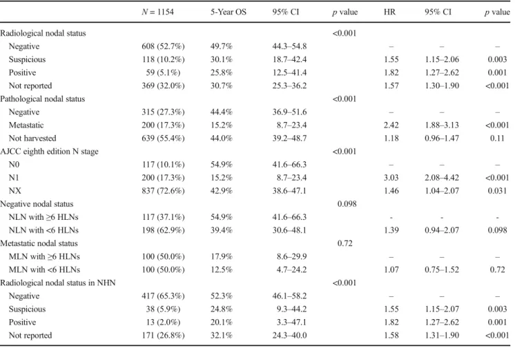
A total of 785 (68.0%) patients had data on preoperative ra-diological nodal staging. EUS, CT, MRI, and PET were used to preoperatively assess nodal status in 19 (2.4% of 785), 383 (48.9% of 785), 307 (39.1% of 785), and 76 (9.7% of 785) patients, respectively. Among radiological-staged patients, nodal status was negative (R-NLN) in 608 (77.5% of 785) patients, suspicious (R-SLN) in 118 (15.0% of 785), and met-astatic (MLN) in 59 (7.5% of 785) patients. Patients with R-NLN had a 5-year OS of 49.7% (IQR, 44.3–54.8) compared with a 5-year OS of 30.1% (IQR, 18.7–42.4) for patients with
R-SLN and 25.8% (IQR, 12.5–41.4) for patients with R-MLN (p < 0.001; Table2, Fig.S1). Compared with patients with R-NLN, patients with R-SLN (HR 1.55, 95% CI, 1.15–2.06; p = 0.003) and R-MLN (HR 1.82, 95% CI, 1.27–2.62; p = 0.001) were at higher risk of death (Table2).
Among 317 (27.5%) patients who had data on both radio-logical and pathoradio-logical nodal evaluation, the incidence of NLN was 66.5% (n = 127) among patients initially deemed R-NLN compared with 42.5% (n = 34) among patients who were preoperatively staged R-SLN; in contrast, the incidence of NLN was only 34.8% (n = 16) among patients deemed preop-eratively to be R-MLN. The incidence of MLN increased from 33.5% (n = 64) among patients who were R-NLN to 57.5% (n = 46) and 65.2% (n = 30) among patients who were R-SLN or R-MLN, respectively (p < 0.001; Table 3). Radiological nodal status was associated with advanced disease (T3/T4 AJCC seventh ed. and eighth ed. stages; both p ≤ 0.003; Table3). Of note, the area under the receiver operating charac-teristic (ROC) curve comparing nodes deemed preoperatively as positive or suspicious versus documented metastatic nodal disease on final pathology was 0.63.
Nodal Status
At the time of hepatectomy, nodes were harvested in 515 (44.6%) patients, while 639 (55.4%) patients did not undergo lymphadenectomy. When lymphadenectomy was performed, the median number of harvested lymph node (HLN) was 4 (IQR, 2–8). Overall, 200 (17.3%) patients had metastatic lymph nodes (MLNs), and 315 (27.3%) patients had no evi-dence of lymph node metastasis (NLN). Among the 200 (17.3%) patients with MLN, 110 (55.0% of MLN group) pa-tients had 1 MLN, 65 (32.5% of MLN group) 2–5 MLNs, and 25 (12.5% of MLN group)≥6 MLNs. Among the 315 (27.3%) patients with NLN, 67 (21.3% of NLN group) patients had 1 HLN, 131 (41.6% of NLN group) 2–5 HLNs, and 117 (37.1% of NLN group) ≥6 HLNs. The 5-year OS of patients with NLN was 44.4% (IQR, 36.9–51.6) versus 15.2% (IQR, 8.7– 23.4) for patients with MLN (HR 2.42, 95% CI 1.88–3.13; p < 0.001; Fig.1).
To verify the prognostic role of six HLNs as the minimum number recommended by the AJCC eighth edition staging system for an adequate nodal staging, MLN and NLN patients were dichotomized in two groups with a cutoff of six HLNs. Among the 200 (17.3%) patients with MLN, 100 (50.0% of MLN group) patients had ≥6 HLNs with a 5-year OS of 17.9% (IQR, 8.6–29.9) versus a 5-year OS of 12.5% among the 100 (50.0% of MLN group) patients who had <6 HLNs (IQR, 4.7–24.2; p = 0.71; Fig.S2). Patients with MLN had a similar risk of death when stratified by <6 versus≥6 HLNs (HR 1.07, 95% CI 0.75–1.52, p = 0.72). Among the 315 (27.3%) patients with NLN, 117 (37.1% of NLN group) pa-tients had≥6 HLNs with a 5-year OS of 54.9% (IQR, 41.6–
Table 1 (continued)
Variables N (%)
Suspicious 118 (10.2%)
Positive 59 (5.1%)
Not reported 369 (32.0%)
Pathological nodal status
Negative 315 (27.3%)
Metastatic 200 (17.3%)
Not harvested 639 (55.4%)
Lymph nodes harvesteda, median (IQR) 4 (2–8) Metastatic lymph nodesb
1 110 (55.0%)
2–5 65 (32.5%)
≥6 25 (12.5%)
AJCC eighth edition N status
N0 117 (10.1%)
N1 200 (17.3%)
NX 837 (72.6%)
AJCC seventh edition T status
T1 487 (42.2%)
T2a 207 (17.9%)
T2b 123 (10.7%)
T3 195 (16.9%)
T4 142 (12.3%)
AJCC eighth edition T status
T1a 249 (21.6%)
T1b 270 (23.4%)
T2 402 (34.8%)
T3 167 (14.5%)
T4 66 (5.7%)
NA not available, IQR interquartile range a
Of 515 patients who underwent lymphadenectomy bOf 200 patients with metastatic nodes;
66.3) compared with a 5-year OS of 39.4% (IQR, 30.6–48.1; p = 0.098; Fig.S3) among the 198 (62.9% of NLN group) patients who had <6 HLNs. Interestingly, patients with NLN who had <6 nodes harvested tended to have an increased risk of death compared with patients who had≥6 HLNs (HR 1.39, 95% CI 0.94–2.07, p = 0.098).
Patients Without Nodes Harvested
A total of 639 (55.4%) patients did not undergo lymphadenec-tomy (Nx); Nx patients had a 5-year OS of 44.0% (IQR, 39.2– 48.7) (HR 1.18, 95% CI 0.96–1.47; p = 0.11) (Table2). When pathological Nx patients were stratified according to
Table 2 Comparison of nodal status—Kaplan-Meier analysis and risk of death
N = 1154 5-Year OS 95% CI p value HR 95% CI p value
Radiological nodal status <0.001
Negative 608 (52.7%) 49.7% 44.3–54.8 – – –
Suspicious 118 (10.2%) 30.1% 18.7–42.4 1.55 1.15–2.06 0.003
Positive 59 (5.1%) 25.8% 12.5–41.4 1.82 1.27–2.62 0.001
Not reported 369 (32.0%) 30.7% 25.3–36.2 1.57 1.30–1.90 <0.001
Pathological nodal status <0.001
Negative 315 (27.3%) 44.4% 36.9–51.6 – – –
Metastatic 200 (17.3%) 15.2% 8.7–23.4 2.42 1.88–3.13 <0.001
Not harvested 639 (55.4%) 44.0% 39.2–48.7 1.18 0.96–1.47 0.11
AJCC eighth edition N stage <0.001
N0 117 (10.1%) 54.9% 41.6–66.3 – – –
N1 200 (17.3%) 15.2% 8.7–23.4 3.03 2.08–4.42 <0.001
NX 837 (72.6%) 42.9% 38.6–47.1 1.46 1.04–2.07 0.031
Negative nodal status 0.098
NLN with≥6 HLNs 117 (37.1%) 54.9% 41.6–66.3 - -
-NLN with <6 HLNs 198 (62.9%) 39.4% 30.6–48.1 1.39 0.94–2.07 0.098
Metastatic nodal status 0.72
MLN with≥6 HLNs 100 (50.0%) 17.9% 8.6–29.9 – – –
MLN with <6 HLNs 100 (50.0%) 12.5% 4.7–24.2 1.07 0.75–1.52 0.72
Radiological nodal status in NHN <0.001
Negative 417 (65.3%) 52.3% 46.1–58.2 – – –
Suspicious 38 (5.9%) 24.8% 9.3–44.2 1.55 1.15–2.07 0.003
Positive 13 (2.0%) 20.1% 3.3–47.1 1.82 1.27–2.62 0.001
Not reported 171 (26.8%) 32.1% 24.3–40.0 1.58 1.31–1.90 <0.001 OS overall survival, CI confidence interval, HLN harvested lymph node, MLN metastatic lymph node
Table 3 Association between radiological nodal status, pathological nodal status, AJCC seventh ed., and AJCC eighth ed. T stages
Radiological modal status p value Negative Suspicious Positive
Pathological nodal statusa <0.001
Negative 127 (66.5%) 34 (42.5%) 16 (34.8%) Positive 64 (33.5%) 46 (57.5%) 30 (65.2%)
AJCC seventh edition T stagesb <0.001
T1/T2a/T2b 471 (77.5%) 74 (62.7%) 31 (52.5%)
T3/T4 137 (22.5%) 44 (37.3%) 28 (47.5%)
AJCC eighth edition T stagesb 0.003
T1a/T1b/T2 406 (66.8%) 70 (59.3%) 27 (45.8%) T3/T4 202 (33.2%) 48 (40.7%) 32 (54.2%) a n = 317 patients b n = 785 patients
preoperative radiological nodal status, 417 (65.3%) were in the R-NLN group, 38 (5.9%) in the R-SLN, and 13 (2.0%) in the R-MLN group; 171 (26.8%) did not have any informa-tion on radiological node status (R-Nx). Among pathological Nx patients, 5-year OS among patients with R-NLN was 52.3% (IQR, 46.1–58.2) versus 24.8% (IQR, 9.3–44.2) for R-SLN, 20.1% (IQR, 3.3–47.1) for R-MLN and 32.1% (IQR, 24.3–40.0) for R-Nx (Table2). Compared with patients who had R-NLN, patients with R-SLN (HR 1.55, 95% CI 1.15–2.07), MLN (HR 1.82, 95% CI 1.27–2.62), and R-Nx (HR 1.58, 95% CI 1.31–1.90) had an increased hazard of death (all p≤ 0.003; Table2).
AJCC Eighth Nodal Staging
Among the 317 (27.5%) patients with MLN or NLN with≥6 HLNs—the cutoff recommended by the AJCC eighth edition staging system—117 (36.9% of 317) patients were defined as N0 and 200 (63.1% of 317) as N1. The 5-year OS of N0 patients was 54.9% (IQR, 41.6–66.3; Table2) versus 15.2% (IQR, 8.7–23.4) for N1 patients (p < 0.001; Fig.2). In turn, N1 patients had an increased hazard of death compared with N0 patients (HR 3.03, 95% CI 2.08–4.42; p < 0.001).
To further investigate the effect of the number of HLN on the prognosis of N0 and N1 patients, a Bayesian Weibull model was developed. N1 patients had an increased risk of death (HR 2.42, 95% Cr Int, 1.69–3.38) compared with N0 patients who had one HLN. Of note, when the number of HLN was increased to 3, 6, 8, and 10, the risk of death among N1 patients versus N0 patients increased incrementally to 2.61, 2.89, 3.11, and 3.34, respectively. Specifically, 5-year OS among N0 patients increased with higher numbers of HLN from 38.3% with 1 HLN to 42.7, 49.2, 53.4, and 57.3% with 3, 6, 8, and 10 HLNs, respectively. Conversely, 5-year OS among N1 patients did not varying considerably when HLN increased, ranging from 9.7% with 1 HLN to only 15.5% with 10 HLNs (TableS1).
Discussion
Several studies have investigated a variety of clinicopatholog-ical factors and long-term survival, identifying tumor size, tumor number, vascular invasion, and lymph node metastasis as independent predictors of OS and recurrence-free survival (RFS) among patients undergoing surgery for ICC [2, 15, 19, 20
]. In particular, the role of lymphadenectomy for ICC has been extensively debated [2, 8–10]. In a recent meta-analysis that evaluated the management of lymph node basin during liver resection for ICC, the authors suggested that surgeons should strongly consider lymph node dissection at the time of surgery although there was insufficient data to support a strong recommendation for routine lymphadenectomy [21]. The newly released eighth edition AJCC staging manual ad-vocates, however, for recovery of at least six lymph nodes during the time of surgery for ICC [16]. Given the large vari-ability in lymphadenectomy among Eastern and Western cen-ters, little data exist regarding the optimal number of lymph nodes to harvest at the time of surgery for ICC. In addition, no study has explicitly sought to evaluate the prognostic rele-vance of preoperative radiological versus pathological lymph node status. While several studies have evaluated the sensitiv-ity, specificsensitiv-ity, and predictive value of ultrasound, CT, MRI, and PET [22–24], no study had assessed the prognostic ability of preoperatively determine N status and long-term outcomes following resection of ICC. The current study was important, therefore, as we determined the prognostic impact of preoper-ative radiologic nodal status. In addition, we assessed the eighth AJCC edition’s recommendation for a minimum recov-ery of six lymph nodes and its association with long-term outcome among a large multiinstitutional cohort of patients with ICC.
In addition to pathological staging, preoperative radiologi-cal lymph node status, as assessed by EUS, CT, MRI, or PET, was correlated with long-term prognosis among patients un-dergoing resection of ICC (Table2). Specifically, patients who
Fig. 1 Kaplan-Meier overall survival curves stratified by pathological nodal status
Fig. 2 Kaplan-Meier overall survival curves stratified by AJCC eighth edition nodal stages
had metastatic lymph node disease on preoperative imaging had a 5-year OS of only 25.8%, which was almost one half the 5-year OS (49.7%) among patients who had no lymph node disease suspected on preoperative imaging. Interestingly, pa-tients who had nodes deemed as suspicious on preoperative imaging had a comparable 5-year outcome (30.1%) as patients who had metastatic nodes. In fact, patients who had nodes deemed as positive or suspicious on preoperative imaging had an 82 and 55% increased hazard of death long-term com-pared with patients who had no nodal disease.
Perhaps not surprisingly, pathological nodal status also was associated with prognosis and, in fact, was much more strong-ly correlated with long-term outcome than preoperative strong-lymph node assessment. Specifically, patients with pathologic N1 disease according to the AJCC eighth edition staging system had almost a 2.5-fold increased risk of death at 5 years com-pared with N0 patients. The superiority of pathological versus radiological nodal status to predict long-term survival might be expected. While preoperative imaging can often accurately predictBtrue^ nodal status, some inaccuracy and lack of cor-relation of preoperative imaging with final pathology can oc-cur. Specifically, in the current study, the area under the ROC curve comparing nodes that wereBpositive^ or Bsuspicious^ on preoperative imaging with true nodal disease on final pa-thology was 0.63. These data suggest that the correlation of preoperative imaging to detect nodal disease was good to fair. Moreover, radiological node status was also correlated with both AJCC seventh and eighth editions’ T stages, as patients with advanced T stage disease were more likely to have pos-itive or suspicious nodes on the preoperative imaging (Table3).
Recently, the AJCC eighth edition proposed a cutoff of six lymph nodes to N stage patients adequately. The impact of number of nodes examined has not been examined among patients undergoing resection of ICC. As such, we investigat-ed the effect of a cutoff of six nodes on long-term outcome. Of note, OS among patients with N0 disease who had≥6 HLNs was 54.9%, which was markedly better than the 15.2% sur-vival noted among patient with N1 disease. Perhaps of more interest, when assessed using a Bayesian Weibull model, the 5-year OS of N0 patients improved with increasing numbers of HLN, while the 5-year OS of N1 patients did not change with a higher number of HLN (TableS1). In addition, while the 5-year OS of patients with N1 disease who had≥6 HLNs was no different than patients who had <6 HLNs, the 5-year OS of patients with N0 disease with <6 HLNs was somewhat worse versus N0 patients who had a more thorough lymph node harvest of≥6 nodes.
The study had several limitations that should be considered when interpreting the results. While one strength of the current report was that it involved multiple centers, the multiinstitutional nature likely led to different radiological imaging techniques. In turn, the sensitivity and specificity of these different techniques
in staging the nodal basin may vary [23–25]. The multicenter nature of the study also did not allow for standardization of operative or perioperative approach, especially in terms of per-formance and extent of lymphadenectomy. Finally, the long du-ration and the multiinstitutional nature of the study likely caused some heterogeneity in ICC treatment approach. However, given the rarity of ICC, obtaining data from multiple centers increased the sample size and made the data more generalizable.
Conclusion
In conclusion, while pathological nodal status was strongly associated with long-term outcome, only one fourth of pa-tients undergoing liver resection for ICC had adequate nodal staging according to the newly released AJCC eighth edition staging system. Moreover, our results suggested that radiolog-ical lymph node staging could be inaccurate in up to 40% of patients and should not be considered a valid alternative to lymphadenectomy. Furthermore, the best ability to discrimi-nate between patients with favorable prognosis and patients with poor prognosis based on the lymph node status was reached when≥6 lymph nodes were harvested. In other words, the quality of the lymph node staging in terms of hazard of death of metastatic patients compared with non-metastatic pa-tients increased from 1.6 to 1.8 with radiological assessment to 2.4 with pathology assessment; of note, the hazard of death increases to 3-fold when the AJCC eighth ed. staging system recommendations were fulfilled. While the six HLN cutoff value was associated with prognosis among patients staged as N0, the number of HLN was not associated with long-term survival among patients with N1 disease. These data serve to emphasize the important prognostic role of patholog-ical staging of nodal disease among patients undergoing re-section of ICC.
Compliance with Ethical Standards
Conflict of Interest The authors declare that they have no conflict of interest.
References
1. Edge SB BD, Compton CC, Fritz AG, Greene FL, Trotti A. AJCC Cancer Staging Manual (7th edn). 2010.
2. de Jong MC, Nathan H, Sotiropoulos GC, et al. Intrahepatic chol-angiocarcinoma: an international multi-institutional analysis of prognostic factors and lymph node assessment. Journal of clinical oncology : official journal of the American Society of Clinical Oncology. 2011;29(23):3140–3145.
3. Farges O, Fuks D, Le Treut YP, et al. AJCC 7th edition of TNM staging accurately discriminates outcomes of patients with resect-able intrahepatic cholangiocarcinoma: By the AFC-IHCC-2009 study group. Cancer. 2011;117(10):2170–2177.
4. Igami T, Ebata T, Yokoyama Y, Sugawara G, Takahashi Y, Nagino M. Staging of peripheral-type intrahepatic cholangiocarcinoma: ap-praisal of the new TNM classification and its modifications. World journal of surgery. 2011;35(11):2501–2509.
5. Ali SM, Clark CJ, Mounajjed T, et al. Model to predict survival after surgical resection of intrahepatic cholangiocarcinoma: the Mayo Clinic experience. HPB : the official journal of the International Hepato Pancreato Biliary Association. 2015;17(3): 244–250.
6. Jiang W, Zeng ZC, Tang ZY, et al. A prognostic scoring system based on clinical features of intrahepatic cholangiocarcinoma: the Fudan score. Annals of oncology : official journal of the European Society for Medical Oncology / ESMO. 2011;22(7):1644–1652. 7. Hyder O, Marques H, Pulitano C, et al. A Nomogram to Predict
Lon g-term Survival A fter Rese cti on for Intrah epat i c Cholangiocarcinoma: An Eastern and Western Experience. JAMA surgery. Mar 5 2014.
8. Clark CJ, Wood-Wentz CM, Reid-Lombardo KM, Kendrick ML, Huebner M, Que FG. Lymphadenectomy in the staging and treat-ment of intrahepatic cholangiocarcinoma: a population-based study using the National Cancer Institute SEER database. HPB : the offi-cial journal of the International Hepato Pancreato Biliary Association. 2011;13(9):612–620.
9. Mavros MN, Economopoulos KP, Alexiou VG, Pawlik TM. Treatment and Prognosis for Patients With Intrahepatic Cholangiocarcinoma: Systematic Review and Meta-analysis. JAMA surgery. Apr 9 2014.
10. Kim Y, Spolverato G, Amini N, et al. Surgical Management of Intrahepatic Cholangiocarcinoma: Defining an Optimal Prognostic Lymph Node Stratification Schema. Annals of surgical oncology. 2015;22(8):2772–2778.
11. Vitale A, Moustafa M, Spolverato G, Gani F, Cillo U, Pawlik TM. Defining the possible therapeutic benefit of lymphadenectomy among patients undergoing hepatic resection for intrahepatic cholan-giocarcinoma. Journal of surgical oncology. 2016;113(6):685–691. 12. Nuzzo G, Giuliante F, Ardito F, et al. Improvement in perioperative
and long-term outcome after surgical treatment of hilar cholangio-carcinoma: results of an Italian multicenter analysis of 440 patients. Archives of surgery. 2012;147(1):26–34.
13. Shimada M, Yamashita Y, Aishima S, Shirabe K, Takenaka K, Sugimachi K. Value of lymph node dissection during resection of intrahepatic cholangiocarcinoma. The British journal of surgery. 2001;88(11):1463–1466.
14. Bagante F, Gani F, Spolverato G, et al. Intrahepatic Cholangiocarcinoma: Prognosis of Patients Who Did Not Undergo Lymphadenectomy.
Journal of the American College of Surgeons. 2015;221(6):1031–1040 e1034.
15. Bridgewater J, Galle PR, Khan SA, et al. Guidelines for the diag-nosis and management of intrahepatic cholangiocarcinoma. Journal of hepatology. 2014;60(6):1268–1289.
16. Amin MB EiC, American Joint Committee on Cancer. Springer. 2017.
17. Couinaud C. Liver anatomy: portal (and suprahepatic) or biliary segmentation. Digestive surgery. 1999;16(6):459–467.
18. Bagante F, Tran T, Spolverato G, et al. Perihilar Cholangiocarcinoma: Number of Nodes Examined and Optimal Lymph Node Prognostic Scheme. Journal of the American College of Surgeons. 2016;222(5): 750–759 e752.
19. Hyder O, Marques H, Pulitano C, et al. A nomogram to predict long-term survival after resection for intrahepatic cholangiocarcino-ma: an Eastern and Western experience. JAMA surgery. 2014;149(5):432–438.
20. Doussot A, Gonen M, Wiggers JK, et al. Recurrence Patterns and Disease-Free Survival after Resection of Intrahepatic Cholangiocarcinoma: Preoperative and Postoperative Prognostic Models. Journal of the American College of Surgeons. 2016;223(3):493–505 e492.
21. Amini N, Ejaz A, Spolverato G, Maithel SK, Kim Y, Pawlik TM. Management of lymph nodes during resection of hepatocellular carcinoma and intrahepatic cholangiocarcinoma: a systematic re-view. Journal of gastrointestinal surgery : official journal of the Society for Surgery of the Alimentary Tract. 2014;18(12):2136– 2148.
22. Holzapfel K, Gaa J, Schubert EC, et al. Value of diffusion-weighted MR imaging in the diagnosis of lymph node metastases in patients with cholangiocarcinoma. Abdominal radiology. 2016;41(10): 1937–1941.
23. Hanninen EL, Pech M, Jonas S, et al. Magnetic resonance imaging including magnetic resonance cholangiopancreatography for tumor localization and therapy planning in malignant hilar obstructions. Acta radiologica. 2005;46(5):462–470.
24. Kluge R, Schmidt F, Caca K, et al. Positron emission tomography with [(18)F]fluoro-2-deoxy-D-glucose for diagnosis and staging of bile duct cancer. Hepatology. 2001;33(5):1029–1035.
25. Roche CJ, Hughes ML, Garvey CJ, et al. CT and pathologic assess-ment of prospective nodal staging in patients with ductal adenocar-cinoma of the head of the pancreas. AJR. American journal of roentgenology. 2003;180(2):475–480.