Relationship between the presence of liver
metastases with histological grading, depth of
invasion and nodal involvement in sporadic
adenocarcinoma of the large intestine
Relação entre presença de metástases hepáticas com grau histológico, profundidade de
invasão e envolvimento nodal no adenocarcinoma esporádico de intestino grosso
Eduardo Cambruzzi1, 2; Larissa R. Roman2; Andressa A. Noschang2; Fernanda B. Pacheco2; Luana R. Gassen2; Luciana H. Miranda2; Karla Lais Pêgas3
1. Instituto de Cardiologia do Rio Grande do Sul. 2. Universidade Luterana do Brasil. 3. Santa Casa de Misericórdia.
First submission on 06/06/15; last submission on 06/06/15; accepted for publication on 24/06/15; published on 20/08/15
ABSTRACT
Introduction: Large intestine adenocarcinoma (LIA) is the most common cancer of the gastrointestinal tract, and corresponds to the ifth most common malignancy in Brazil. The main prognostic factors related to LIA are depth of tumor invasion and perivisceral
lymph nodes status. Objective: To estimate the relationship between pathological indings and the presence of liver metastases (LM)
in LIA cases. Method: We evaluated 51 cases of LIA, previously submitted to surgical resection, in order to determine the following
variables: topography, tumor size, macroscopic appearance, degree of differentiation, depth of invasion, nodal status, and presence
of LM. Results: The average age was 64.8 years, with predominance of men (n = 26/51.0%) and lesions in the sigmoid colon
(n = 18/35.3%). The main general characteristics of the sample were ulcerative-vegetative lesions (n = 20/39.2%), no annular
tumors (n = 3/64.7%), moderately differentiated tumor (n = 44/86.3%), absence of mucinous areas (n = 40/78.4%), and mesocolon
invasion (n = 29/56.9%). LM were found in 14 cases (27.5%), and is associated with presence of nodal metastases (p = 0.005). Tumor
size (p = 0.72), macroscopic appearance (p = 0.362), histological grade (p = 0.147), and depth of invasion (p = 0.195) showed no
association with LM presence. Conclusion: LIA has a wide anatomical and pathological heterogeneity. In this study, the presence of
LM associated with LIA was related to perivisceral lymph nodes status, with no relation to tumor size, degree of differentiation, and depth of invasion, which suggests that identifying neoplastic angiolymphatic invasion is a possible predictor of liver involvement.
Key words: adenocarcinoma; neoplastic metastasis; colorectal cancer; pathology; prognosis.
INTRODUCTION
The primary adenocarcinoma of the large intestine is among the ten most common types of cancer worldwide, and it is also the most common form of malignancy of the gastrointestinal tract. It corresponds to the third leading cause of death in women and the ifth among men in Brazil, although these rates are higher in North America and Europe(1-3).A
progressively higher incidence over the last 15 years have been observed; however, the distribution of colorectal cancer in relation to gender and age has remained constant(1-4). The risk
for developing this cancer increases after the age of 40 for both men and women, and this risk doubles with each subsequent decade. Family history is a risk factor for colorectal carcinoma, especially in patients younger than 50 years, the prevalence of adenomas of the colon in relatives of patients with colorectal carcinoma is 39%(1-4).
Colorectal adenocarcinoma may present polypoid, vegetative, ulcerative, ulcerative-vegetative, and iniltrative macroscopic forms. Histologically, it can be classiied into grades based on tubular architectural pattern of neoplastic cells(2, 3, 5).
Although poorly differentiated and mucinous neoplasms are
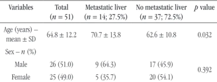
TABLE 1 –LIA: sample characterization
Variables Total (n = 51)
Metastatic liver (n = 14; 27.5%)
No metastatic liver (n = 37; 72.5%)
p value
Age (years) –
mean ± SD 64.8 ± 12.2 70.7 ± 13.8 62.6 ± 10.8 0.032
Sex – n (%)
Male 26 (51.0) 9 (64.3) 17 (45.9)
0.392
Female 25 (49.0) 5 (35.7) 20 (54.1)
LIA: large intestine adenocarcinoma; SD: standard deviation.
associated with lower disease-free survival rates, the two most important prognostic factors are the depth of invasion and the presence of metastases in regional lymph nodes. Besides these latter, the metastases may involve the lungs and bones, but as a consequence of the venous drainage of the colon, the liver is the most common location for metastatic lesions(2, 3, 5). About 25%
of patients with colorectal cancer may present liver metastasis at the time of primary tumor diagnosis, and 50% of patients may show liver metastases during the course of the disease(2, 3, 5).
In this study, the authors assessed the relationship between liver metastases and anatomopathological indings of colorectal adenocarcinoma, as topography, tumor size, macroscopic appearance, degree of differentiation, presence of mucinous areas, depth of invasion, and in periviscerais lymph
nodes metastasis.
METHOD
This is a cross-sectional retrospective study involving patients with large intestine adenocarcinoma (LIA), who had undergone previous surgical resection and staging. Initially, 51 different LIA cases were selected for the study, previously analyzed in the Pathology Laboratory of the Grupo Hospitalar Conceição (GHC) in Porto Alegre (RS). Data collection was carried out between March 2014 and October 2014, and was approved by the Ethics Research Committee (ERC) of GHC, project nº 14-001. We included in the sample only those cases histologically classiied as primary large intestine adenocarcinoma according to World Health Organization criteria. All other histological types of primary benign or malignant neoplasm of the large intestine and cases of metastasis or secondary involvement of the organ were excluded.
The demographic characteristics (age and sex) were collected from the medical records. Each case was previously ixed in 10% formalin, stained with hematoxylin-eosin (HE), and the following anatomopathological data were reviewed and described:
1) anatomical position: cecum, ascending colon, sigmoid colon, transverse colon, and rectum;
2) tumor size: is in centimeters, in the longer axis of the
lesion;
3)macroscopic appearance: iniltrative, ulcerative, ulcerative-iniltrative, ulcerative-vegetative and vegetative;
4) presence of annular or no annular lesion;
5) degree of differentiation: well-differentiated, moderately-differentiated and poorly-differentiate;
6) presence of mucous producing area;
7) depth of invasion: mucosa, submucosa, muscle, perivisceral tissue, serous and adjacent organ invasion;
8) surgical margins status: free or positive; 9) presence of lymph nodes metastases.
The evaluated cases were also grouped by TNM staging system; T: tumor, N: lymph node, M: metastasis. Data were recorded and analyzed in a Microsoft Excel 2007 spreadsheet. Quantitative variables were expressed as mean and standard deviation (DP) or median and interquartile range. Categorical variables were described by absolute and relative frequencies. To compare means between groups we used the Student-t test; in case of asymmetrical distribution, we used the Mann-Whitney test; Pearson chi-squared tests or Fisher’s exact test were used for the difference between proportions; in case of signiicance, the adjusted residual test was applied. The signiicance level was 5% (p ≤ 0.05), and analyzes were performed in SPSS software version 21.0.
RESULTS
The group of patients analyzed had a mean age of 64.8 years, predominantly male. The prevalence of liver metastasis was 27.5% (conidence interval [CI] 95%: 15.2%-39.8%).
Tables 1 and 2 show the results obtained by the research.
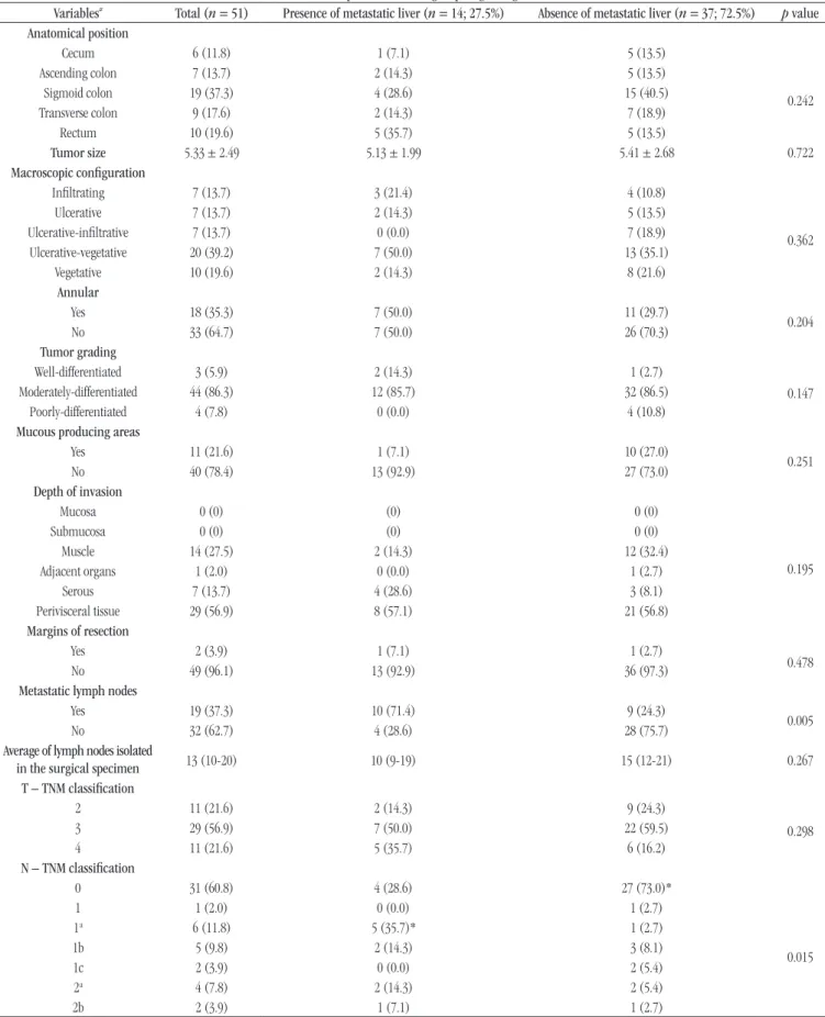
The number of lymph node metastasis (p = 0.005) and group
N of TNM system (p = 0.015) were associated with the presence of
liver metastases. The pathological variables, such as anatomical position (p = 0.242), tumor size (p = 0.722), macroscopic appearance (p = 0.362), degree of differentiation (p = 0.147),
presence of mucous producing area (p = 0.251), depth of invasion
(p = 0.195), and positive resection margins (p = 0.478), had no
TABLE 2 – LIA: comparison between groups regarding the variables
Variables# Total (n = 51) Presence of metastatic liver (n = 14; 27.5%) Absence of metastatic liver (n = 37; 72.5%) p value
Anatomical position
Cecum 6 (11.8) 1 (7.1) 5 (13.5)
0.242
Ascending colon 7 (13.7) 2 (14.3) 5 (13.5)
Sigmoid colon 19 (37.3) 4 (28.6) 15 (40.5)
Transverse colon 9 (17.6) 2 (14.3) 7 (18.9)
Rectum 10 (19.6) 5 (35.7) 5 (13.5)
Tumor size 5.33 ± 2.49 5.13 ± 1.99 5.41 ± 2.68 0.722
Macroscopic coniguration
Iniltrating 7 (13.7) 3 (21.4) 4 (10.8)
0.362
Ulcerative 7 (13.7) 2 (14.3) 5 (13.5)
Ulcerative-iniltrative 7 (13.7) 0 (0.0) 7 (18.9)
Ulcerative-vegetative 20 (39.2) 7 (50.0) 13 (35.1)
Vegetative 10 (19.6) 2 (14.3) 8 (21.6)
Annular
Yes 18 (35.3) 7 (50.0) 11 (29.7)
0.204
No 33 (64.7) 7 (50.0) 26 (70.3)
Tumor grading
Well-differentiated 3 (5.9) 2 (14.3) 1 (2.7)
0.147
Moderately-differentiated 44 (86.3) 12 (85.7) 32 (86.5)
Poorly-differentiated 4 (7.8) 0 (0.0) 4 (10.8)
Mucous producing areas
Yes 11 (21.6) 1 (7.1) 10 (27.0)
0.251
No 40 (78.4) 13 (92.9) 27 (73.0)
Depth of invasion
Mucosa 0 (0) (0) 0 (0)
0.195
Submucosa 0 (0) (0) 0 (0)
Muscle 14 (27.5) 2 (14.3) 12 (32.4)
Adjacent organs 1 (2.0) 0 (0.0) 1 (2.7)
Serous 7 (13.7) 4 (28.6) 3 (8.1)
Perivisceral tissue 29 (56.9) 8 (57.1) 21 (56.8)
Margins of resection
Yes 2 (3.9) 1 (7.1) 1 (2.7)
0.478
No 49 (96.1) 13 (92.9) 36 (97.3)
Metastatic lymph nodes
Yes 19 (37.3) 10 (71.4) 9 (24.3)
0.005
No 32 (62.7) 4 (28.6) 28 (75.7)
Average of lymph nodes isolated
in the surgical specimen 13 (10-20) 10 (9-19) 15 (12-21) 0.267
T – TNM classiication
2 11 (21.6) 2 (14.3) 9 (24.3)
0.298
3 29 (56.9) 7 (50.0) 22 (59.5)
4 11 (21.6) 5 (35.7) 6 (16.2)
N – TNM classiication
0 31 (60.8) 4 (28.6) 27 (73.0)*
0.015
1 1 (2.0) 0 (0.0) 1 (2.7)
1a 6 (11.8) 5 (35.7)* 1 (2.7)
1b 5 (9.8) 2 (14.3) 3 (8.1)
1c 2 (3.9) 0 (0.0) 2 (5.4)
2a 4 (7.8) 2 (14.3) 2 (5.4)
2b 2 (3.9) 1 (7.1) 1 (2.7)
# variables expressed as mean ± SD, median (25-75 percentile) or n(%); * statistically significant association by adjusted residual testing to 5% of significance; LIA: large intestine
DISCUSSION
Colorectal carcinoma is among the ten main types cancers in Brazil, affecting both sexes in similar prevalence. The age proile of colorectal carcinoma has dramatic increase in incidence from 40 years for women and 50 years for men. It is estimated that, in 2014, in Brazil, 32,600 cases of colorectal carcinoma were found,
15,070 men and 17,530 women(6). In this study, data of age group
was equivalent to those from the literature. The study population consisted of an age average of 64.8 years, ranging 12.2 years, in
26 male (51%) and 25 female (49%). Oliveira et al. analyzed 129
cases of colorectal carcinoma and determined that the mean age of patients was 56.9 years, ranging 25-87 years, of which 51.2% were female patients (51.2%, p > 0.05)(7). Torres Neto et al. evaluated
355 cases of colorectal cancer, which was more frequent in women,
the age ranged from 5-98 years, mean age of 60.2 years(8).
Colorectal adenocarcinoma may grade in well-differentiated, moderately-differentiated, poorly-differentiated and undifferentiated lesions. The iniltrating edge of adenocarcinomas may have rounded or spiculated tumor growth and, in its progression, the adenocarcinoma gradually invades the intestine layers. During this mural progression of the cancer, there may be angiolymphatic invasion in cancer and potential spread and tumor implantation in regional lymph nodes and distant organs(1, 2, 4, 9, 10). In this study,
the anatomopathological variables, such as anatomical position
(p = 0.242), tumor size (p = 0.722), macroscopic appearance
(p = 0.362), the presence or absence of annular lesion (p = 0.204),
degree of differentiation (p = 0.147), presence of mucous producing
area (p = 0.251), depth of invasion (p = 0.195), and presence of
positive surgical margins (p = 0.478), were not associated with the
presence of liver metastases. Madeira et al. analyzed 50 LIA cases
and reported that there was no association between the presence of nodal metastases and age, sex, microscopic edge, presence of mucous producing areas, degree of differentiation, and tumor
size(2). Fujimoto et al. reported that the presence of liver metastases
on 122 cases with clinical ive years follow-up, was signiicantly associated with tumor size, degree of differentiation, depth of invasion, presence of neoplastic angiolymphatic invasion and presence of metastases in regional lymph(11). Kitajima et al.
investigated the depth of submucosa invasion in 865 cases of invasive colorectal carcinoma and concluded that nodal
metastasis rates are associated with depth of submucosa lesion(12).
Inoue et al. compared the depth of submucosa lesion in 118 cases
of colorectal carcinomas sessile and supericial type, which were resected endoscopically, and total depth was signiicantly higher in sessile type(13).
The liver is the most common site of distant metastases of LIA, found in up to 25% patients at diagnosis(1-3, 5-7, 11). The
prevalence of liver metastases in our study was 27.5%. The average age of the group with liver metastasis was signiicantly
higher than the group without metastasis. Fernandes et al.
analyzed 749 LIA cases and determined that the main sites of metastases were liver (64.51%), peritoneum (19.35%), and lungs (9.67%). Among metastases associated with colorectal cancer,
the liver was also the most affected organ (54.11%)(14). Murad et
al. reported that in a sample of 49 patients with liver metastases
from colorectal adenocarcinoma, 31 cases (63.2%) had already had hepatic involvement in the initial surgical procedure or presence of metastasis within six months of initial diagnosis. In 18 cases (36.8%), the appearance of metastasis occurred six
months after colorectal surgery(9). Ambiru et al. determined that
the survival of 168 patients with liver metastases associated with colorectal carcinoma was 42% in three years and 26% in ive years. The main prognostic factors included nodal status of the initial resection of the primary tumor, surgical margin status,
number of liver metastases, and adjuvant chemotherapy(15). Arru
et al., among the 297 cases of resected liver metastases associated with colorectal carcinoma, determined that the time period of disease-free survival was associated with the degree of differentiation, serum level of carcinoembryonic antigen, tumor size greater than 5 cm and time period of metastasis detection after initial surgical treatment(16). Ohji et al. selected 120 cases of
advanced colorectal carcinoma, including liver metastases in 60 cases and determined that the presence of nodal metastasis, and the immunohistochemical expression of neprilysin (CD10) and vascular endothelial growth factor (VEGF) are associated with the development of metastatic lesions(17).Ueno et al. reported that the
degree of differentiation was associated with groups T and N of the TNM system, the serum levels of carcinoembryonic antigen, the presence of liver, lung and peritoneal metastases, the number
of liver metastases and survival time after hepatectomy(18).Lin
et al. determined that in cases of colorectal adenocarcinoma, the presence of tumor deposits in perivisceral tissues was
associated with liver and nodal metastasis, perineural invasion
and disease-free interval after resection of the primary tumor(19).
Adachi et al. reported that the presence of liver metastases in adenocarcinoma colorectal cancer was associated with primary tumors with size equal or greater than 6 cm, presence of serosal invasion, neoplastic angiolymphatic invasion, and metastasis to
regional lymph nodes(20).
In our study, the presence of regional lymph node metastasis
(p = 0.005) was associated with the presence of liver metastasis.
RESUMO
Introdução: O adenocarcinoma de intestino grosso (AIG) é o tumor maligno mais frequente do trato digestivo e corresponde à quinta
neoplasia maligna mais comum no Brasil. Os principais fatores prognósticos do AIG são profundidade de invasão neoplásica e
status dos linfonodos periviscerais. Objetivo: Estimar a relação entre achados anatomopatológicos e presença de metástases hepáticas
(MH) em casos de AIG. Método: Foram avaliados 51 casos de AIG, previamente submetidos à ressecção cirúrgica, e determinadas as
seguintes variáveis: topografia, tamanho tumoral, conformação macroscópica, grau histológico, profundidade de invasão, status
nodal e presença de MH. Resultados: A média de idade correspondeu a 64,8 anos, com predomínio de homens (n = 26/51,0%) e
lesões do cólon sigmoide (n = 18/35,3%). Lesões ulcerovegetantes (n = 20/39,2%), tumores não anelares (n = 3/64,7%), neoplasias
moderadamente diferenciadas (n = 44/86,3%), ausência de áreas mucoprodutoras (n = 40/78,4%) e invasão do mesocólon (n =
29/56,9%) foram as principais características gerais da amostra. MH foram encontradas em 14 casos (27,5%), estando associadas à presença de metástases nodais (p = 0,005). Tamanho tumoral (p = 0,72), configuração macroscópica (p = 0,362), grau histológico
(p = 0,147) e profundidade de invasão (p = 0,195) não apresentaram associação com a presença de MH. Conclusão: O AIG
apresenta heterogeneidade anatomopatológica ampla. No presente estudo, a presença de MH associadas ao AIG esteve relacionada
com o status dos linfonodos periviscerais, não havendo relação com tamanho tumoral, grau de diferenciação e profundidade de
invasão, sugerindo que a identificação de invasão neoplásica angiolinfática é possível fator preditivo do envolvimento hepático.
Unitermos: adenocarcinoma; metástase neoplásica; neoplasias colorretais; patologia; prognóstico.
for colorectal cancer resection, and the presence of nodal metastases was associated with the degree of differentiation(21).
Fonseca et al. assessed 521 surgical specimens of patients operated
for colorectal cancer and the average of lymph nodes affected
by metastases corresponds to 2.57 ± 5.34 lymph nodes(22), while
Santo et al. found an average of 3.4 ± 3.3 metastatic lymph node
from 51 specimens examined (42.8%/n = 119)(23). Homma et al.
analyzed 65 patients with colorectal adenocarcinoma invading the submucosa or the muscle propria, and concluded that the resection of the affected regional lymph nodes by metastases reduced the local recurrence rate of these carcinomas(24). In another study
Homma et al. determined that sex, histologic grade, neoplastic angiolymphatic invasion or nodal status were not signiicantly
associated with tumor recurrence(25). Ahmadi et al. analyzed 824
patients with colorectal carcinoma and found that the right colon tumors are more often associated with nodal metastases, and were
also associated with age(26). Toh et al. investigated 207 cases of
adenocarcinoma colorectal and demonstrated that the radial extent of the tumor and submucosal invasion area can predict the
regional nodal status(27).
CONCLUSION
LIA has good anatomopathological heterogeneity, and the depth of invasion and lymph node status are considered important prognostic factors. In this study, the presence of liver metastases determined by LIA was associated with the status of perivisceral lymph nodes, with no relation to tumor size, degree of differentiation, and depth of invasion, suggesting that identifying neoplastic angiolymphatic invasion by microscopic examination is a potential predictor of liver involvement.
REFERENCES
1. Nadal LRM, Adachi CT, Nunes MAA, et al. Evolução do carcinoma colorretal, comparando doentes com idades acima e abaixo de 40 anos, quanto à diferenciação tumoral e ao estádio do tumor. Rev Bras Colo-proctol. 2009; 29(3): 351-7.
2. Madeira B, Pêgas K, Zettler C, Cambruzzi E. A relação entre metástases em linfonodos regionais e fatores prognósticos no adenocarcinoma esporádico de intestino grosso. Rev Bras Colo-proctol. 2009; 29(4): 472-8.
3. Costa SRP, Lima OAT, Cunha TMR, Soares AF. Hepatectomia direita ampliada com ressecção parcial da veia cava para metástase colorretal: relato de caso. Rev Bras Colo-proctol. 2010; 30(2): 232-6.
4. Loy TS, Kaplan PA. Villous adenocarcinoma of the colon and rectum: a clinicopathologic study of 36 cases. Am J Surg Pathol. 2004, 28: 1460-5.
MAILING ADDRESS
Eduardo Cambruzzi
Universidade Luterana do Brasil (ULBRA); Av. Farroupilha, 8001; São José; CEP: 92425-900; Canoas-RS, Brazil; e-mail: [email protected]. 6. Instituto Nacional de Câncer. Tipos de câncer [Internet]. [citado em
18 maio 2015]. Available at: http://www.inca.gov.br/wps/wcm/connect/ tiposdecancer/site/home/colorretal/deinicao.
7. Oliveira RG, Faria FF, Lima Junior ACB. Cirurgias êntero-colorretais: abordagem cirúrgica de 129 pacientes do SUS no Programa de Pós-Graduação Sensu Lato em coloproctologia. Rev Bras Colo-proctol. 2010; 30(3): 333-43.
8. Torres Neto JR, Teixeira FR, Prudente ACL, Silvino CJ, Arciere JS, Vieira Filho MC. Estudo demográico do câncer de cólon e reto no estado de Sergipe. Rev Bras Colo-proctol. 2008; 28(2): 215-22.
9. Murad JC, Ribeiro-Jr U, Corbetti CE, et al. Análise de fatores clínicos e histopatológicos em metástases hepáticas de adenocarcinoma colorretal. ABCD, Arq Bras Cir Dig. 2007; 20(4): 241-4.
10. Henrique-Filho C, Bromberg SH, Barreto E, Godoy AC, Mattosinho-França LC. Valor prognóstico do grau de diferenciação celular, da presença de muco e do padrão de crescimento da margem invasiva em adenocarcinomas colorretais Dukes B. Arq Gastroenterol. 2004 Set; 41(3): 185-9.
11. Fujimoto Y, Nakanishi Y, Sekine S, et al. CD10 expression in colorectal carcinoma correlates with liver metastasis. Dis Colon Rectum. 2005 Oct; 48(10): 1883-9.
12. Kitajima K, Fujimori T, Fujii S, et al. Correlations between lymph node metastasis and depth of submucosal invasion in submucosal invasive colorectal carcinoma: a Japanese collaborative study. J Gastroenterol. 2004 Jun; 39(6): 534-43.
13. Inoue Y, Ohki T, Nakagawa R, Yamamoto M. Depth of submucosal invasion in the sessile ans supericial types of submucosal invasive colorectal carcinoma: objective versus subjective measurement. Hepatogastroenterology. 2013 Nov-Dec; 60(128): 1916-21.
14. Fernandes GMM, Leme CVD, Ruiz-Cintra MT, Pavarino EC, Netinho JG, Goloni-Bertollo EM. Clinical and epidemiological evaluation of patients with sporadic colorectal cancer. J Coloproctol. 2014; 34(4): 216-23. 15. Ambiru S, Miyazaki M, Isono T, et al. Hepatic resection for colorectal metastases: analysis of prognostic factors. Dis Colon Rectum. 1999 May; 42(5): 632-9.
16. Arru M, Aldrighetti L, Castoldi R, et al. Analysis of prognostic factors inluencing long-term survival after hepatic resection for metastatic colorectal cancer. World J Surg. 2008 Jan; 32(1): 93-103.
17. Ohji Y, Yao T, Eguchi T, et al. Evaluation of risk of liver metastasis in colorectal adenocarcinoma based on the combination of risk factors including CD10 expression: multivariate analysys of clinicopathological and immunohistochemical factors. Oncol Rep. 2007 Mar; 17(3): 525-30. 18. Ueno H, Konishi T, Ishikawa Y, et al. Prognostic value of poorly differentiated clusters in the primary tumor in patients undergoing hepatectomy forcolorectal liver metastasis. Surgery. 2015 May; 157(5): 899-908.
19. Lin Q, Wei Y, Ren L, et al. Tumor deposit is a poor prognostic indicator in patients who underwent simultaneous resection for synchronous colorectal liver metastases. Onco Targets Ther. 2015 Jan; 8: 233-40. 20. Adachi Y, Inomata M, Kakisako K, et al. Histopathologic characteristics of colorectal cancer with liver metastasis. Dis Colon Rectum. 1999 Aug; 42(8): 1053-6.
21. Kuhnen RB, Kock KS. Número de gânglios dissecados em peças operatórias de pacientes submetidos à ressecção cirúrgica de câncer colorretal: retrospectiva de 10 anos HNSC – Tubarão, SC. Rev Bras Colo-proctol. 2007; 27(4): 417-22.
22. Fonseca LM, Quites LV, Cabral MMD, Silva RG, Luz MMP, Lacerda Filho A. Câncer colorretal: resultados da avaliação patológica padronizada de 521 casos operados no Hospital das Clínicas da UFMG. Rev Bras Colo-proctol. 2011; 31(1): 17-25.
23. Santo GFE, Aguilar-Nascimento JE, Kishima MO, Takiuchi A. Correlação de fatores anatomopatológicos com a sobrevida de pacientes operados por adenocarcinoma colorretal. Rev Col Bras Cir. 2008; 35(3): 82-187.
24. Homma Y, Hamano t, Otsuki Y, et al. Severe tumor budding is a risk factor for lateral lymph node metastasis in early rectal cancers. J Surg Oncol. 2010 Sep; 102(3): 230-4.
25. Homma Y, Humano T, Otsuki Y, et al. The total number of lymph node metastases is a more signiicant risk factor for poor prognosis than positive lateral lymph node metastasis. Surg Today. 2015 Feb; 45(2): 168-74.
26. Ahmadi O, Stringer MD, Black MA, McCall JL. Inluence of age and site of disease on lymph node yield in colorectal cancer. N Z Med J. 2014 Jun; 127(1395): 31-40.