Faculdade de Medicina de Lisboa
The role of A
2A
receptors in cognitive
decline – decoding the molecular shift
towards neurodegeneration
Mariana Temido Mendes Ferreira
Orientadores:
Doutora Luísa Maria Vaqueiro Lopes
Professor Doutor Tiago Fleming de Oliveira Outeiro
Tese especialmente elaborada para obtenção do grau de
Doutor em Ciências Biomédicas, Especialidade em
Neurociências
Faculdade de Medicina de Lisboa
The role of A
2Areceptors in cognitive decline – decoding
the molecular shift towards neurodegeneration
Mariana Temido Mendes Ferreira
Orientadores: Doutora Luísa Maria Vaqueiro Lopes
Professor Doutor Tiago Fleming de Oliveira Outeiro
Tese especialmente elaborada para obtenção do grau de Doutor em Ciências Biomédicas, Especialidade em Neurociências
Júri: Presidente:
Doutor José Luís Bliebernicht Ducla Soares, Professor Catedrático em regime de tenure e membro do Conselho Científico da Faculdade de Medicina da Universidade de Lisboa
Vogais:
Doutor Christophe Bernard, Investigador Coordenador do Institut de Neurosciences des Systèmes, Aix-Marseille Université
Doutora Paula Isabel Antunes Pousinha, Professora Auxiliar do Institut de Pharmacologie Moléculaire et Cellulaire, Côte d’Azur Université
Doutora Ana Luísa Monteiro de Carvalho, Professora Auxiliar da Faculdade de Ciências e Tecnologia da Universidade de Coimbra
Doutora Ana Maria Ferreira de Sousa Sebastião, Professora Catedrática da Faculdade de Medicina da Universidade de Lisboa
Doutor Alexandre Valério de Mendonça, Investigador Coordenador da Faculdade de Medicina da Universidade de Lisboa
Doutora Luísa Maria Vaqueiro Lopes, Professora Associada Convidada da Faculdade de Medicina da Universidade de Lisboa (Orientadora)
Fundação para a Ciência e Tecnologia, referência SFRH/BD/52228/2013
i
As opiniões expressas nesta publicação são da exclusiva responsabilidade do seu autor, não cabendo qualquer responsabilidade à Faculdade de Medicina da Universidade de Lisboa pelos conteúdos nele apresentados.
A impressão desta dissertação foi aprovada pelo
Conselho Científico da Faculdade de Medicina de Lisboa
em reunião de 22 de Maio de 2018.
iii
The experimental work herein described was performed at Instituto de Medicina Molecular - Faculdade de Medicina da Universidade de Lisboa except when otherwise stated, under the supervision of Luísa V. Lopes, PhD and Tiago F. Outeiro, PhD.
v
“It always seems impossible until it´s done”
vii
Aging is associated with cognitive decline both in humans and animals. Importantly, aging is the main risk factor for nerurodegenerative diseases, namely Alzheimer’s disease (AD), which primarily affects synapses in the temporal lobe and hippocampal formation. In fact, synaptic dysfunction plays a central role in AD, since it drives cognitive decline. Indeed, in age-related neurodegeneration, cognitive decline has a stronger correlation to early synapse loss than neuronal loss in patients. Despite the many clinical trials conducted to identify drug targets that could reduce protein toxicity in AD, such targets and strategies have proven unsuccessful. Therefore, efforts focused on identifying the early mechanisms of disease pathogenesis, driven or exacerbated by the aging process, may prove more relevant to slow the progression rather than the current disease-based models.
A recent genetic study discovered a significant association of the adenosine A2A receptor encoding gene (ADORA2A) with hippocampal
volume in mild cognitive impairment and Alzheimer’s disease. There is compelling evidence from animal models of a cortical and hippocampal upsurge of adenosine A2A receptors (A2AR) in
glutamatergic synapses upon aging and AD. Importantly, the blockade of A2AR prevents hippocampus-dependent memory deficits and
synaptic impairments in aged animals and in several AD models. Accordingly, in humans, several epidemiological studies have shown that regular caffeine consumption attenuates memory disruption during aging and decreases the risk of developing memory impairments in AD
viii
patients. Together, these data suggest that A2AR might be a good
candidate as trigger to synaptic dysfunction in aging and AD. The main goal of this dissertation was then to explore the synaptic function of A2AR in age-related conditions.
We have assessed the A2AR expression in human hippocampal slices
and found a significant upsurge of A2AR in hippocampal neurons of
aged humans, a phenotype aggravated in AD patients. Increased selective expression of A2AR driven by the CaMKII promoter in rat
forebrain neurons was sufficient to mimic aging-like memory impairments, assessed by the Morris water maze task, and to uncover an LTD-to-LTP shift in the Schaffer collaterals-CA1 synapse of hippocampus. This shift was due to an increased NMDA receptor gating and associated to increased Ca2+ influx. The mGluR5-NMDAR
interplay was identified as a key event in A2AR-induced synaptic
dysfunction. Moreover, chronic treatment with an A2AR selective
antagonist, orally delivered for one month, rescued the aberrant NMDAR overactivation and the plasticity shift. Importantly, the same LTD-to-LTP shift was observed in memory-impaired aged rats and APP/PS1 mice modeling AD, a phenotype rescued upon A2AR
blockade.
These data support a key role for over-active hippocampal A2AR in
aging and AD-dependent synaptic and cognitive dysfunction and may underlie the significant genetic association of ADORA2A with AD. Importantly, this newly found interaction might prove a suitable alternative for regulating aberrant mGluR5/NMDAR signaling in AD without disrupting their constitutive activity.
ix
O envelhecimento está associado a défices cognitivos tanto em humanos como em animais. Para além disso, o envelhecimento é o principal fator de risco para as doenças neurodegenerativas, nomeadamente doença de Alzheimer (DA), a qual afecta de uma forma muito significativa as sinapses do lobo temporal e da formação hipocampal. Estes défices cognitivos estão associados a alterações estruturais e funcionais no hipocampo, nomeadamente disfunção sináptica.
A disfunção sináptica apresenta um papel central na DA, uma vez que leva a défices cognitivos. De facto, na neurodegeneração associada à idade, o declínio cognitivo apresenta uma correlação mais forte com a perda sináptica precoce do que com morte neuronal. Apesar do largo número de ensaios clínicos conduzidos no sentido de identificar alvos para fármacos que possam reduzir a toxicidade proteica em DA, tais alvos e estratégias terapêuticas têm-se revelado infrutíferos. Por conseguinte, esforços no sentido de identificar os mecanismos iniciais subjacentes à doença, os quais podem ser provocados ou exacerbados pelo processo de envelhecimento, poderão mostrar-se mais relevantes para retardar a progressão da doença do que os modelos actuais baseados na própria doença.
A gama de proteínas sinápticas é complexa e os mecanismos subjacentes à transmissão sináptica excitatória são rigorosamente regulados pela actividade sináptica. A activação dos receptores NMDA tem um papel fundamental uma vez que pode tanto induzir potenciação de longa duração (LTP) como depressão de longa duração (LTD),
x
sendo que alterações nestes receptores foram previamente reportadas no contexto de envelhecimento e doença de Alzheimer. Para além disso, foi também reportado um envolvimento dos receptores metabotrópicos de glutamato (mGluR) na disfunção sináptica mediada por Aβ, de tal forma que a proteína Aβ facilita LTD mediada por mGluR e inibe LTP, levando a uma diminuição na densidade das espinhas dendríticas.
O papel da LTP foi intensivamente estudado no contexto da aprendizagem e memória. Pelo contrário, a relação entre LTD e memória está muito menos estudada, tanto em condições fisiológicas como patológicas.
A LTD pode ser definida como um enfraquecimento duradouro de uma sinapse em resposta a um estímulo de baixa frequência. O estímulo desencadeador para a LTD dependente da actividade e induzida sinapticamente é predominantemente um aumento no cálcio pós-sináptico (Ca2+). Uma vez que aumentos pós-sinápticos de Ca2+ estão
implicados na indução tanto de LTP como de LTD, o conceito de que um aumento elevado de Ca2+ intracelular resulta em LTP e um
aumento modesto resulta em LTD encontra-se largamente aceite na comunidade científica. Alguns estudos científicos reportaram um aumento da susceptibilidade para LTD associado ao envelhecimento, enquanto outros não observaram diferenças na magnitude de LTD em animais envelhecidos. Estas discrepâncias em termos de resultados podem ser explicadas por diferenças em termos de estirpe de ratos utilizada, padrão de estimulação e rácio Ca2+/Mg2+ no meio de
perfusão das fatias de hipocampo. De facto, as diferenças relacionadas com a idade em termos de indução de LTD foram revertidas com a
xi
manipulação do rácio Ca2+/Mg2+, consistente com a ideia de que
alterações na regulação de Ca2+ com o envelhecimento desencadeiam
este aumento na susceptibilidade para LTD. Contudo, os mecanismos que levam a alterações na regulação do Ca2+ no processo de LTD
durante o envelhecimento fisiológico e também em patologias associadas ao envelhecimento continuam por esclarecer.
Recentemente, um estudo genético observou uma associação significativa entre o gene que codifica para os receptores da adenosina A2A (A2AR) (ADORA2A) e o volume hipocampal em défice cognitivo
ligeiro e doença de Alzheimer. Existe evidência robusta de um aumento da expressão de A2AR em sinapses glutamatérgicas a nível
cortical e hipocampal no envelhecimento e DA. Para além disso, o bloqueio de A2AR previne défices sinápticos e de memória
dependentes do hipocampo em animais envelhecidos e modelos de DA. Em humanos, vários estudos epidemiológicos demonstraram que o consumo regular de cafeína atenua os défices de memória no envelhecimento e diminui o risco de desenvolver défices de memória em doentes de Alzheimer. Consequentemente, estes resultados sugerem que os A2AR são um potencial estímulo desencadeador de
disfunção sináptica no envelhecimento e DA. Desta forma, o objectivo da presente dissertação foi explorar a função sináptica dos receptores A2AR em patologias associadas ao envelhecimento.
Avaliou-se a expressão de A2AR em secções humanas de hipocampo e
observou-se um aumento significativo na expressão de A2AR em
neurónios hipocampais de idosos, um fenótipo agravado em doentes de Alzheimer. Através de um modelo transgénico de rato com sobreexpressão dos receptores A2A controlada através do promotor
xii
CaMKII nos neurónios do prosencéfalo, observou-se que o aumento da expressão de A2AR, maioritariamente pre-sinápticos, foi suficiente para
desencadear défices de memória semelhantes aos observados no envelhecimento, os quais foram avaliados através do teste do labirinto de Morris. Para além disso, a sobreexpressão neuronal de A2AR leva a
um aumento da probabilidade de libertação de glutamato e a uma sobreactivação dos receptores NMDAR. Em termos de plasticidade sináptica, estes animais apresentam uma potenciação da sinapse fibras de Schaffer-CA1 do hipocampo quando é aplicado um estímulo de baixa frequência que habitualmente induz LTD. Esta inversão de LTD para LTP encontra-se associada ao aumento da ativação do receptor NMDA, com um consequente aumento da entrada de Ca2+.
Identificou-se a interacção mGluR5-NMDAR como tendo um papel chave na disfunção sináptica induzida por A2AR. O tratamento agudo quer com
o antagonista selectivo SCH 58261 quer com o antagonista não selectivo cafeína reverte esta inversão na plasticidade. Para além disto, e de forma mais relevante, o tratamento crónico com um antagonista seletivo dos A2AR, administrado por via oral durante um mês, reverteu
a sobre-activação dos receptores NMDA e a inversão na plasticidade sináptica.
Foram também usados ratos envelhecidos (aproximadamente 18 meses de idade) com défices de memória e em ratinhos APP/PS1 com cerca de 12 meses de idade, um modelo de Doença de Alzheimer. Foram já previamente reportados défices de memória nestes ratinhos nesta idade. De forma muito relevante, esta mesma inversão sináptica foi observada em animais envelhecidos e em ratinhos APP/PS1. Para além disso, observou-se um aumento da expressão de A2AR no hipocampo
xiii
dos ratinhos APP/PS1, em comparação com ratinhos de estirpe selvagem. Consistente com o papel chave dos receptores A2AR, o
bloqueio agudo destes receptores reverte por completo estes défices sinápticos.
Em suma, a interacção NMDAR/mGluR5/A2AR descrita nesta
dissertação pode constituir uma alternativa viável para regular a sinalização aberrante mGluR5/NMDAR em Doença de Alzheimer sem afetar a sua actividade constitutiva. Para além disso, todos estes resultados apontam para um papel chave da sobre-activação de A2AR a
nível hipocampal na disfunção sináptica e cognitiva observada no envelhecimento e na DA e poderá estar subjacente à associação genética significativa do gene ADORA2A com DA.
xv
Temido-Ferreira M, Ferreira DG, Batalha VL, Marques-Morgado I,
Coelho JE, Pereira P, Gomes R, Pinto A, Carvalho S, Canas PM, Cuvelier L, Buée-Scherrer V, Faivre E, Baqi Y, Müller CE, Pimentel J, Schiffmann SE, Buée L, Bader M, Outeiro TF, Blum D, Cunha RA, Marie H, Pousinha PA and Lopes LV (2018) Age-related shift in LTD is dependent on neuronal adenosine A2A receptors interplay with
mGluR5 and NMDA receptors; Molecular Psychiatry; doi: 10.1038/s41380-018-0110-9.
Temido-Ferreira M, Pousinha PA and Lopes LV; The synaptic
pathophysiology of aging: implications for cognitive defects; Review;
in preparation.
Other publications
Ferreira DG, Temido-Ferreira M, Miranda HV, Batalha VL, Coelho JE, Szegö ÉM, Marques-Morgado I, Vaz SH, Rhee JS, Schmitz M, Zerr I, Lopes LV, Outeiro TF (2017) α-synuclein interacts with PrPC to induce cognitive impairment through mGluR5 and NMDAR2B.
Nature Neuroscience 20(11):1569-1579.
Batalha VL, Ferreira DG, Coelho JE, Valadas JS, Gomes RA,
Temido-Ferreira M, Shmidt T, Baqi Y, Buée L, Müller CE, Hamdane
M, Outeiro TF, Bader M, Meijsing SH, Sadri-Vakili G, Blum D, Lopes LV. (2016) The caffeine-binding adenosine A2A receptor induces
age-xvi
like HPA-axis dysfunction by targeting glucocorticoid receptor function. Scientific Reports 11;6:31493.
xvii
ABBREVIATION LIST ... xxi
CHAPTER I - Introduction ... 1
1. State of the art ... 3
1.1. Memory and the hippocampus ... 3
1.2. Synaptic Plasticity ... 9
1.3. Aging and Synaptic dysfunction ... 15
1.4. Physiopathological role of A2AR in the hippocampus ... 28
2. Aims ... 43 3. Technical approaches ... 45 3.1. Electrophysiology... 45 CHAPTER II - Methods ... 51 1. Human samples ... 53 2. Animals ... 53
3. Generation and maintenance of transgenic animals ... 54
4. Oral administration of the drug ... 55
5. RNA extraction and quantitative real time PCR analysis (RT-qPCR) ... 56
6. In situ hibridization ... 57
7. Behavioral assessments ... 59
7.1. Morris water maze ... 59
7.2. Y-maze behavior test ... 60
8. Electrophysiology experiments ... 61
8.1. Field potential recordings ... 61
8.2. Patch-clamp recordings ... 62
xviii
10. Transfection of primary neuronal cultures ... 67 11. Construct generation ... 67 12. Ca2+ imaging ... 68 13. Immunocytochemistry ... 69 14. Immunohistochemistry ... 69 15. Electron microscopy ... 71 16. Fractionation ... 72 17. Western blotting ... 73 18. Drugs ... 74 19. Statistics ... 75 CHAPTER III - Results ... 77
1. Increased levels of A2AR in human aged and Alzheimer’s
disease brain ... 81 2. Physiopathological levels of A2AR in neurons impair
hippocampus-dependent spatial memory ... 83 3. Increased levels of A2AR enhance glutamate release probability
... 93 4. A2AR increase NMDAR-mediated currents in CA1 pyramidal
neurons ... 97 5. Physiological levels of A2AR lead to a NMDAR-mediated
LTD-to-LTP shift ... 100 6. Blockade of A2AR in vivo rescues the LTD-to-LTP shift in
Tg(CaMKII-hA2AR) animals ... 108
7. Increased levels of A2AR impair calcium homeostasis ... 110
8. LTD-to-LTP shift in aged and APP/PS1 animals is rescued by A2AR blockade ... 113
CHAPTER IV - Conclusions ... 119 1. Discussion ... 121 2. Future Perspectives ... 133
xix
3. Acknowledgements ... 137 CHAPTER IV - References ... 139
xxi A1R – Adenosine A1 receptors
A2AR – Adenosine A2A receptors
Act-β – Actin-β
AD – Alzheimer’s disease
AMPA – α-Amino-3-hydroxy-5-methyl-4isoxazolepropionic acid ANOVA – Analysis of variance
AP5 - 2-Amino-5-phosphonopentanoic acid ATP – Adenosine triphosphate
BDNF – Brain derived neurotrophic factor
BiFC – Bimolecular fluorescence complementation BSA – Bovine serum albumin
C57BL6/J – “Black Six” or B6 Strain Mice CA1 – Cornu ammonis 1
CA2 – Cornu ammonis 2 CA3 – Cornu ammonis 3
CaMKII – Calcium calmodulin dependent protein kinase II cAMP – 3’-5’-cyclic adenosine monophosphate
CaN - Calcineurin
CGS 21680 - 2-(4-(2-Carboxyethyl)phenethylamino)-5'-N-ethylcarboxamidoadenosine
xxii CNS – Central nervous system
CREB – cAMP response element-binding protein CTR – Control
CypA – Cyclophilin A DG – Dentate gyrus
DHPG – (S)-3,5-Dihydroxyphenylglycine DMEM – Dulbecco’s modified Eagle’s medium DMSO – Dimethylsulfoxide
DNA – Deoxyribonucleic acid
DNQX - 6,7-Dinitro-1,4-dihydroquinoxaline-2,3-dione DTT – Ditiotreitol
EC – Entorhinal cortex
EDTA – Ethylenediaminetetraacetic acid EPM – Elevated plus maze
fEPSP – field Excitatory postsynaptic potential FBS – Fetal bovine serum
GABA – Gamma-aminobutyric acid GFAP – Glial fibrillary acidic protein GPCR – G-Protein coupled receptor HBSS – Hank’s balanced salt solution HRP – Horseradish peroxidase
xxiii ICC – Immunocytochemistry IHC – Immunohistochemistry IP3 - Inositol-1,4,5-triphosphate KW6002 – 8-[(E)-2-(3,4-Dimethoxyphenyl)ethenyl]-1,3-diethyl-7-methyl-purine-2,6-dione
LFS - Low frequency stimulation LTD – Long-term depression LTP – Long-term potentiation
MAP2 – Microtubule-associated protein 2 MCI – Mild cognitive impairment
mGluR5 – Metabotropic glutamate receptor 5 MPEP – 2-Methyl-6-(phenylethynyl)pyridine MTL – Medial temporal lobe
MWM – Morris water maze NaCl – Sodium chloride
NMDA - N-Methyl-D-aspartate OF – Open field test
PI - Phosphatidylinositol-4,5-biphosphate PBS – Phosphate buffered saline
xxiv PFA – Paraformaldehyde
PFC – Pre-frontal cortex PKA – Protein kinase A PKC – Protein kinase C PSD – Postsynaptic density
RIPA – Radioimmunoprecipitation assay buffer RT – Room temperature
RT-qPCR – Real-time quantitative polymerase chain reaction SEM – Standard error of mean
SCH 58261 – 2-(Furan-2-yl)-7-phenethyl-7H-pyrazolo[4,3-e][1,2,4]triazolo[1,5-c]pyrimidin-5-amine
SDS – Sodium dodecyl sulfate TBS – Tris buffered saline
Tg(CaMKII-hA2AR) – Transgenic rats overexpressing human
adenosine A2AR Receptor under the control of the CaMKII promoter
VDCC – Voltage-dependent calcium channels WHO – World health organization
1
CHAPTER I
3
1.
State of the art
1.1.
Memory and the hippocampus
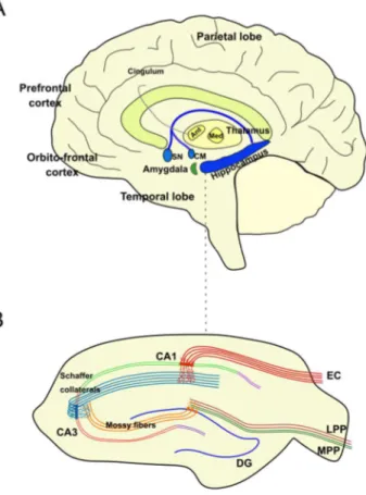
Encoding, consolidation and retrieval of mnemonic information is critically dependent on a large reciprocal network of regions that includes neocortical association regions, subcortical nuclei, the medial temporal lobe (MTL), parahippocampal areas and the hippocampal formation. The hippocampus is considered the central node in this circuit. It receives input from almost all neocortical association areas via perirhinal and parahippocampal cortices and finally through the entorhinal cortex (EC) (Bartsch and Wulff, 2015) (Figure 1.1).
The gross anatomical analysis of the hippocampal formation dates back to Arantius who first described the appearance of human hippocampal formation and gave it the name hippocampus (derived from the Greek word for sea horse). The term cornu ammonis was introduced by the neuroanatomist Lorente de Nó (Lorente de No, 1934).
The hippocampal formation comprises four relatively simple cortical regions. These include the dentate gyrus, the hippocampus proper (which can be divided into three sub-fields, namely CA3, CA2 and CA1), the subicular complex (which can also be divided into three subdivisions: the subiculum, presubiculum and parasubiculum) and the entorhinal cortex which, particularly in the rodents, is generally divided into medial and lateral subdivisions (Amaral and Witter, 1989).
4
Figure 1.1. Hippocampal anatomy. (A) Principal anatomy of the hippocampal memory systems and the brain regions involved in learning and memory. (B) Hippocampal slice. Abbreviations: Ant, anterior thalamic nuclei; CA, cornu ammonis; CM, corpus mammillaris; DG, dentate gyrus; EC, entorhinal cortex; LPP, lateral performant pathway; Med, medial thalamic nuclei; MPP, medial performant pathway; Mtt, mamillothalamic tract; SN: septal nucleus.
1.1.1. Structural composition
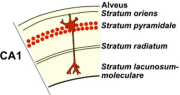
The human hippocampus can be divided into the fields CA3, CA2 and CA1. These hippocampal fields have essentially one cellular layer,
5
called the pyramidal cell layer – stratum pyramidale. The limiting surface with the ventricular lumen is formed by axons of the pyramidal cells – the alveus. Between it and the pyramidal cell layer, the stratum
oriens mainly contains the basal dendrites of the pyramidal cells as
well as several types of interneurons. The region superficial to the pyramidal cell layer (toward the hippocampal fissure) contains the apical dendrites of the pyramidal cells and interneurons. This region has historically been divided into (1) the stratum lucidum, (2) the
stratum radiatum and (3) the stratum lacunosum-moleculare
corresponding to the most superficial portion of this region, adjacent to the hippocampal fissure. The apical dendrites of pyramidal neurons make up the stratum radiatum (Figure 1.2) (Schultz and Engelhardt, 2014).
In the stratum lucidum of CA3, the mossy fibers travel and form synapses with proximal dendrites just above the pyramidal cell layer of CA3. In the human hippocampus a fraction of the mossy fibers also travels within the CA3 pyramidal cell layer. Stratum lucidum is absent in CA2 and CA1 regions, which do not receive mossy fiber input (Schultz and Engelhardt, 2014).
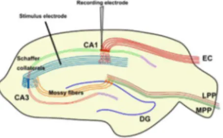
The hallmark of CA3 neurons is that their proximal dendrites are contacted by mossy fibers which correspond to the dentate granule cell axons. The mossy fibers traverse the stratum lucidum immediately above the CA3 pyramidal cell layer. In CA1, the projections from CA3 and CA2 – the so-called Schaffer collaterals – terminate in the stratum
radiatum and stratum oriens (Figure 1.2) (Schultz and Engelhardt,
6
The CA2 region has a relatively compact and narrow pyramidal cell layer. The borders of this region are difficult to establish in routine preparations of the human hippocampus (Schultz and Engelhardt, 2014).
The pyramidal cell layer of CA1 becomes thicker and more heterogeneous in monkeys and humans as compared to rodents. Stereological studies estimate the total number of CA1 neurons as approximately 14 x 106. The appearance of CA1 varies along its
transverse and rostrocaudal axes. Based on pigmentoarchitectonic studies, the human pyramidal cell layer can be subdivided into an outer and inner pyramidal cell layer. Close to the CA2 border, the CA1 cells of both sublayers appear most tightly packed and the pyramidal cell layer is at its thinnest. The border of CA1 with CA2 is very difficult to define because some CA2 pyramidal cells appear to extend over the emerging CA1 pyramidal cell layer. The pyramidal layer of CA1 overlaps that of the subiculum in an oblique manner (Schultz and Engelhardt, 2014).
Figure 1.2. Section of CA1 with different layers. Abbreviations: CA1, cornu ammonis 1. (Adapted from (Amaral and Witter, 1989)).
7 1.1.2. Connectivity
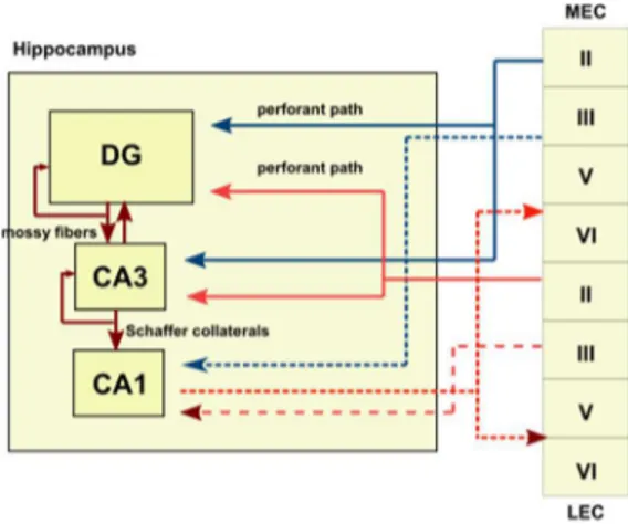
The hippocampus is a three-layered structure that is reciprocally connected to other cortical and subcortical areas. The principal neurons of the hippocampus are organized in layers and receive unidirectional input from the EC, where layer II neurons project to granule cells in the dentate gyrus (DG) via the perforant path (Bartsch and Wulff, 2015; Strange et al., 2014). The trisynaptic pathway from the DG to the CA3 via mossy fibers and onward to CA1 via Schaffer collaterals is the principal feed-forward circuit involved in the process of information through the hippocampus (Bartsch and Wulff, 2015). Additionally, layer III neurons from the EC directly project to CA1 neurons via the temporoammonic path (perforant path to CA1) (Bartsch and Wulff, 2015). CA1 pyramidal cells – the major output neurons – project via the subicular complex back to deep layers of the EC and to various subcortical and cortical areas (Bartsch and Wulff, 2015b; Murray et al., 2011). Importantly, the structure of this feed-forward circuit with its limited redundancy may be critical for learning and memory but may also contribute to its vulnerability during insults (Bartsch and Wulff, 2015; Lavenex and Amaral, 2000).
The DG, with three distinct layers (molecular, granular, and polymorphic), consists mainly of granule cells and receives input from the EC. The axons of the DG granule cells form the mossy fiber system and project to the CA3 (Amaral et al., 2007; Bartsch and Wulff, 2015). Mossy fibers also project back onto granule cells, thus forming a recurrent network. Additionally, the DG receives information from the contralateral hippocampus via commissural projections (Amaral et
8
al., 2007; Bartsch and Wulff, 2015). Axon collaterals of CA3 pyramidal neurons synapse onto other CA3 neurons, forming a recurrent autoassociative network, whereas CA3 neurons projecting back to the dentate network form a heteroassociative network (Bartsch and Wulff, 2015; Lisman, 1999). CA1 pyramidal neurons receive information which has been pre-processed in the subnetworks of the DG and CA3, but also receives direct projections from the EC, suggesting that the function of the CA1 neurons includes comparing new information from the EC with stored information via CA3 in terms of mismatch, error and novelty detection (Figure 1.3) (Bartsch and Wulff, 2015; Lisman and Otmakhova, 2001).
Figure 1.3. Schematic representation of the hippocampal trisynaptic circuit.
Firstly, granule neurons in the hippocampal dentate gyrus receive afferent inputs, via the performant path, from the layer II of the lateral and medial entorhinal cortex. Next, granule neurons project to the CA3 pyramidal neurons via mossy fibers and, ultimately, CA1 neurons receive inputs from the CA3 by the Schaffer collaterals, by the contralateral hippocampus through associational/commissural fibers or direct inputs from the performant path. To close the hippocampal synaptic loop, CA1 pyramidal neurons project back to the entorhinal cortex. Abbreviations: CA1, CA2
9
and CA3, cornu ammonis 1, 2 and 3; DG, dentate gyrus; LEC, lateral entorhinal cortex; MEC, medial entorhinal cortex (Adapted from (Alves, 2017)).
1.2.
Synaptic Plasticity
Neural plasticity is the ability of the brain or brain structures to adapt in response to intrinsic or extrinsic stimuli such as changes in the environment, development or lesions (Bartsch and Wulff, 2015; Kolb and Muhammad, 2014). Neural plasticity can occur on various functional and structural levels, such as changes in membrane excitability, synaptic plasticity and changes in dendritic and axonal structure (Bartsch and Wulff, 2015; Kolb and Gibb, 2014). Neural plasticity can also occur as structural and functional adaptations and reorganization of neuronal populations, which is reflected in modifications of recruitment and strength of connectivity of networks and circuits. Furthermore, neural plasticity can show a time- and age-dependency and can result in positive or negative adaptive effects (Bartsch and Wulff, 2015; Kolb and Gibb, 2014).
The hippocampus has been for long considered a classic example for the study of neural plasticity as many paradigms of synaptic plasticity such as Long-term potentiation (LTP) and Long-term depression (LTD), excitatory postsynaptic potential (EPSP)-spike potentiation and spike-timing dependent plasticity, have been identified and demonstrated in hippocampal circuits (Malenka, 1994).
Synaptic plasticity can be defined as activity-dependent modifications in the efficacy and strength of synaptic transmission of pre-existing
10
synapses (Citri and Malenka, 2008). Long-term synaptic plasticity can last from minutes to several days and even years (Abraham et al., 1994, 2002; Staubli and Scafidi, 1997) and therefore may be associated with the formation of long-term memories (Dong et al., 2013; Ge et al., 2010). Long-term potentiation (LTP) and long-term depression (LTD) are the two main forms of long-term synaptic plasticity thought to play a role in hippocampal functioning (Pinar et al., 2017). LTP is defined as the long-lasting enhancement in signal transmission between two neurons after continuous stimulation and has been for long considered the cellular and molecular basis of memory (Citri and Malenka, 2008). However, much less is known about LTD and its role on learning and memory, either in physiological or pathological conditions, although some reports have already described an association between LTD impairments and cognitive deficits, namely in animal models of stress and Alzheimer’s Disease (AD) (Lanté et al., 2015; Wong et al., 2007). One of the earliest reports of lasting activity-dependent depression of transmission at central synapses concerned the phenomenon of heterosynaptic depression (Lynch et al., 1977). This study demonstrated that in the CA1 region of the hippocampus in vitro, stimuli delivered to one pathway that induced LTP resulted in a reversible depression in the non-tetanised pathway (Lynch et al., 1977). However, this phenomenon was ignored for many years until the 1990s, when it was demonstrated that low-frequency stimulation (LFS) was effective at inducing LTD without the requirement for the induction of LTP, a process called homosynaptic de novo LTD (Dudek and Bear, 1992; Mulkey and Malenka, 1992).
11
Initial reports demonstrated that the induction of LTD in the CA1 region of the hippocampus was dependent on the synaptic activation of NMDAR, NMDAR-dependent LTD, which is usually induced by LFS (Dudek and Bear, 1992; Fujii et al., 1991; Kemp et al., 2000; Mulkey and Malenka, 1992). Furthermore, application of NMDA by itself has also been shown to induce lasting synaptic depression, a form of chemical-LTD, which appears to share common mechanisms with LFS-induced LTD (Lee et al., 1998). However, these two forms of LTD also have important differences (Kameyama et al., 1998; Morishita et al., 2001).
A second major form of LTD requires the activation of mGluRs (Bashir and Collingridge, 1994; Bashir et al., 1993). Additionally, LTD can be obtained by brief application of mGluR agonist DHPG, designated DHPG-LTD (Huber et al., 2001; Palmer et al., 1997). The patterns of activation that are required to induce mGluR-LTD are generally similar to those required to induce NMDAR-LTD, although there are differences depending on the synapse type. For example, NMDAR-LTD is usually induced at CA1 synapses by single-shock LFS, whereas mGluR-LTD is usually induced by paired-pulse LFS (Collingridge et al., 2010; Massey and Bashir, 2007).
However, it should be noted that with very few exceptions (Cho et al., 2000; Palmer et al., 1997; Wang and Gean, 1999) the requirement of NMDA and mGlu receptor activation in LTD is mutually exclusive; thus in virtually all cases of LTD, where there is a requirement for NMDAR activation, there is no role for mGluR activation and vice versa (Kemp and Bashir, 2001).
12
The trigger for postsynaptically induced, activity-dependent LTD is predominantly an increase in calcium (Ca2+). Since postsynaptic rises
in Ca2+ are implicated in the induction of both LTD and LTP (Bliss
and Collingridge, 1993; Lynch et al., 1983), particular properties of the Ca2+ signal (temporal, spatial or magnitude) may determine whether
LTP or LTD is induced. The suggestion of Lisman et al (Lisman, 1989) that large increases in intracellular Ca2+ result in LTP induction
and modest increases in intracellular Ca2+ result in LTD induction
extended the theory of Bienenstock et al., who had proposed that some function of postsynaptic activity controlled the direction (increase or decrease) of synaptic plasticity (Bienenstock et al., 1982).
The increase in postsynaptic Ca2+ that results in LTD can arise from a
variety of sources. For example, several reports describe Ca2+ influx
through NMDAR as fundamental for LTD induction (Ahmed et al., 2011; Dudek and Bear, 1992). Moreover, there is evidence that calcium entry through voltage-dependent Ca2+ channels (VDCC) can
also be important in LTD induction (Christie et al., 1997; Cummings et al., 1996; Oliet et al., 1997; Otani and Connor, 1998; Wang et al., 1997). However, this is still controversial, since other studies showed no effect of nifedipine, a VDCC blocker, in LTD magnitude (Udagawa et al., 2006). mGlu receptor-mediated release of Ca2+ from intracellular
stores also contributes to LTD. Group I mGlu receptors are coupled to phosphatidylinositol-4,5-biphosphate (PI) hydrolysis. One of the products of PI hydrolysis, inositol-1,4,5-triphosphate (IP3), causes
release of calcium from intracellular stores (Pin and Duvoisin, 1995) (Figure 1.4).
13
Despite NMDAR, VDCC and group I mGluR drive Ca2+ influx to
induce LTD, their relative contribution depends significantly on the pattern of stimulation, Ca2+/Mg2+ ratio in the bath medium and age of
the animals (Kumar and Foster, 2014; Norris et al., 1996).
How does the increase in Ca2+ mediate long-lasting changes in
synaptic transmission? One important Ca2+-dependent cascade
involves the binding of Ca2+ to calmodulin, thus forming a
calcium-calmodulin complex. This complex subsequently activates both calmodulin dependent protein kinase (CaMKII) and calcium-calmodulin dependent protein phosphatase, calcineurin (CaN; also known as PP2B). Moderate increases in postsynaptic Ca2+, such as
those expected from LFS, activates calcineurin, which will then lead to the activation of protein phosphatase 1 and/or phosphatase 2 (PP1/2) and subsequent LTD via dephosphorylation of AMPA receptors and other substrates (Lee et al., 2000; Mulkey et al., 1993, 1994; O’Dell and Kandel, 1994) (Figure 1.4). Protein phosphatases can also dephosphorylate and inactivate CaMKII, thereby facilitating LTD. Consistent with this hypothesis, a CaMKII antagonist facilitated DHPG-induced LTD in the CA1 region (Schnabel et al., 1999). However, Mockett et al. showed that mGluR-LTD, induced by DHPG or paired-pulse synaptic stimulation, was dependent on CaMKII activation (Mockett et al., 2011). Given these contradictory results, the role of CaMKII in regulating LTD is not completely understood. Additionally, group I mGluR-dependent activation of phospholipase C (PLC) leads not only to Ca2+ release from intracellular stores but also
to PKC activation. The activation of PKC can lead to phosphorylation of the GluA2 subunit at the serine 880 residue and lead to AMPAR
14
lateral diffusion and subsequent internalization (Gladding et al., 2009; Lüscher and Huber, 2010) (Figure 1.4).
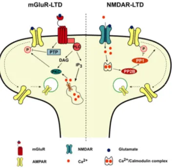
Figure 1.4. mGluR and NMDAR-LTD exhibit different underlying mechanisms.
mGluR activation results in Ca2+ release from intracellular stores and a
PKC-dependent AMPAR internalization. NMDAR activation drives Ca2+ influx, which
activates protein phosphatases, leading to subsequent AMPAR dephosphorylation and internalization. Abbreviations: DAG: Diacylglycerol; IP3: Inositol-1,4,5-triphosphate; PKC: Protein kinase C; PLC: Phospholipase C; PP1: Protein phosphatase 1; PP2B: Protein phosphatase 2B; PTP: Protein tyrosine phosphatase.
LTD can also be mediated by persistent presynaptic changes, and the proportion of presynaptic versus postsynaptic alterations probably depends on various factors, including the brain region, the animal and its developmental stage. There is evidence that LTD (Enoki et al.,
15
2009), including NMDAR-LTD (Stanton et al., 2003) and mGlu-LTD (Foy et al., 1987), can involve a reduction in the probability of glutamate release, which can be triggered by changes in the presynaptic terminal or by postsynaptic changes that are communicated across the synapse via a retrograde messenger (Feinmark et al., 2003; Stanton et al., 2003).
1.3.
Aging and Synaptic dysfunction
Nowadays, the world’s population is aging: the number and proportion of older persons in the societies is growing significantly. According to the World Health Organization (WHO), by 2020, the number of people aged 60 years and older will outnumber children younger than 5 years. Moreover, between 2015 and 2050, the proportion of the world’s population over 60 years will nearly double from 12% to 22%. Importantly, aging is the main risk factor for Alzheimer’s disease (AD) (Reitz et al., 2011). These profound demographic changes place understanding the aging process as one of the big challenges for scientific research nowadays. Although aging affects the entire body, its impact on brain and cognition has a profound effect on the life quality of the individuals. Given the critical role of the hippocampus in learning and memory, it is very important to comprehend the aging-related alterations in this brain structure (Burke and Barnes, 2010). Importantly, memory capabilities that depend on proper hippocampal function appear to be of particular vulnerability to the aging process (McEwen, 1997). Much work has been focused on the hippocampus,
16
as age-related decline in performance dependent on this region is consistently found across species and tasks (Barnes, 1979; Colombo et al., 1997; Erickson and Barnes, 2003; Mabry et al., 1996; Oler and Markus, 1998; Tanila et al., 1997). These age-related memory impairments can be explained in part by changes in neural plasticity or cellular alterations that directly affect mechanisms of plasticity (Burke and Barnes, 2006). Although several age-related neurological changes have been identified during normal aging, these tend to be subtle compared with the ones observed in age-associated disorders, such as Alzheimer’s and Parkinson’s disease (Burke and Barnes, 2006). Consequently, understanding age-related changes in cognition sets a background against which it is possible to assess the effects of the disease (Burke and Barnes, 2006).
1.3.1. Neuronal loss
For a long time, aging had been associated with neuronal loss independently of brain region (Ball, 1977; Brizzee et al., 1980; Brody, 1955; Coleman and Flood, 1987). However, the methods used in those studies question the accuracy of such results (Burke and Barnes, 2006; Morrison and Hof, 1997) and subsequent studies have conclusively shown that the cell number is preserved in aging in several brain areas including the hippocampus (Gazzaley et al., 1997; Keuker et al., 2003; Merrill et al., 2000; Pakkenberg and Gundersen, 1997; Peters et al., 1994; West, 1993). Rather than neuronal loss, alterations in dendritic branching and spine density seem to be a better correlate of aging in
17
terms of neuronal morphology (Foster et al., 1991; Pyapali and Turner, 1996; Scheibel et al., 1976). In fact, dendritic branching is greater in the dentate gyrus and in the layer II of the parahippocampal gyrus of aged individuals (Buell and Coleman, 1979, 1981, Flood et al., 1985, 1987a), while no age-associated changes were observed in CA3 (Flood et al., 1987b) and CA1 (Hanks and Flood, 1991). Further non-human studies confirmed the results observed in the CA1 (Markham et al., 2005; Pyapali and Turner, 1996; Turner and Deupree, 1991). Unlike hippocampus, the morphology of neurons from pre-frontal cortex (PFC) seems to be vulnerable to the aging process (Burke and Barnes, 2006), where a decrease of dendritic branching was reported in aged rats (Grill and Riddle, 2002) and humans (de Brabander et al., 1998; Uylings and de Brabander, 2002). Similar to the dendritic branching results, no alterations were found in synaptic density in DG and CA1 across species (Curcio and Hinds, 1983; Markham et al., 2005; Williams and Matthysse, 1986). There is, however, one study that observed a reduction in spine density in the subiculum of aged monkeys (Uemura, 1985). Loss of CA1 synapses is characteristic of AD and, to a lesser extent, mild cognitive impairment in humans (Scheff et al., 2007). Altogether this data highlight the differences between normal aging, characterized by structural preservation in the MTL, and AD, which is associated with neuronal and synaptic loss in the MTL and in the hippocampus in particular (Morrison and Baxter, 2012).
18 1.3.2. Synaptic dysfunction
Since cell number is not affected in aging, there must be other alterations that are in the genesis of age-associated memory deficits. Deficits in neurogenesis could account for these age-related synaptic impairments. Although aged rats exhibit decreased neurogenesis (Kuhn et al., 1996), this alteration does not correlate with memory performance, since old rats with lower neurogenesis rates actually perform better in spatial memory tasks (Bizon and Gallagher, 2005; Bizon et al., 2004).
Age-associated impairments can be due to synaptic changes that would then lead to altered mechanisms of plasticity (Burke and Barnes, 2006, 2010). Although the total number of Schaffer collaterals-CA1 synapses is preserved across different age groups (Bear et al., 1987), the amplitude of the Schaffer collaterals-induced field EPSP (fEPSP) recorded in CA1 was reduced in aged memory-impaired animals (Barnes et al., 1992; Landfield et al., 1986; Rosenzweig et al., 1997). Furthermore, the postsynaptic density (PSD) area of axospinous synapses was significantly reduced in aged learning-impaired rats (Nicholson et al., 2004). However, at the CA3-CA1 synapse, the size of the unitary EPSP remains constant during aging (Barnes et al., 1997). Together, these data suggest that aging might not be associated with alterations in the strength of individual synaptic connections but instead with an increase in non-functional or silent synapses in the hippocampus (Burke and Barnes, 2006, 2010).
Furthermore, biophysical properties of hippocampal neurons such as the resting membrane potential, membrane time constant, input
19
resistance, threshold to action potential are largely preserved over the lifespan (Burke and Barnes, 2010; Rosenzweig and Barnes, 2003). A notable exception is age-related changes in calcium regulation in aged hippocampal neurons, which have an increased Ca2+ influx through
L-type Ca2+ channels. The pathophysiological role of L-type Ca2+
channels in age-related synaptic dysfunction will be further explored below.
1.3.3. Synaptic plasticity
The alterations observed at individual synapses have a significant impact on synaptic plasticity. Long-term synaptic plasticity processes (LTP and LTD) have been correlated with memory performance for a long time and are proposed as a main neurophysiological correlate of memory (Citri and Malenka, 2008; Lynch, 2004). Importantly, age-associated memory deficits correlate with impairments in either LTP or LTD (Ge et al., 2010; Tombaugh et al., 2002).
In aged animals, LTP has been found to be reduced (Barnes, 1979; Deupree et al., 1993, 1993; Rex et al., 2005; Sankar et al., 2000), not altered (Costenla et al., 1999; Kumar et al., 2007; Landfield and Lynch, 1977; Landfield et al., 1978; Norris et al., 1996; Rex et al., 2005) or even strengthened (Costenla et al., 1999; Diógenes et al., 2011; Huang and Kandel, 2006; Kumar and Foster, 2004; Pinho et al., 2017). This is inconsistent with the classical correlation between increased LTP magnitude and better performance on hippocampal-dependent memory tasks. The synaptic circuit that is being potentiated
20
(Rex et al., 2005) and the stimulation protocol (Burke and Barnes, 2006; Costenla et al., 1999) may then account for the differences observed in LTP magnitude. Generally, age-associated alterations in LTP are only observed when weaker stimulus protocols are used, either an increase (Costenla et al., 1999; Diógenes et al., 2011; Huang and Kandel, 2006; Pinho et al., 2017) or decrease (Tombaugh et al., 2002) in LTP.
Some authors report increased susceptibility to LTD during aging (Norris et al., 1996), whereas others fail to observe alterations in LTD magnitude in aged animals (Foster and Kumar, 2007; Kumar et al., 2007). These discrepancies can be explained by differences in animal strain, stimulation pattern or Ca2+/Mg2+ ratio. Accordingly,
paired-pulse LFS (PP-LFS) does not induce changes in LTD between young and aged animals (Foster and Kumar, 2007), suggesting that different mechanisms may be involved in the induction of LTD by LFS and PP-LFS. Also, age-related differences in LTD induction could be rescued by manipulating the extracellular Ca2+/Mg2+ ratio. Indeed, the fact that
induction of LTD is a function of age and the level of Ca2+ in the
recording medium strongly support an age-related Ca2+ dysregulation
and a shift in Ca2+-dependent induction mechanisms rather than in the
LTD intrinsic capacity (Foster and Kumar, 2007; Kumar et al., 2007). It had been hypothesized that postsynaptic intracellular levels of Ca2+
are involved in setting the synaptic modification curve, which determines the probability that a synapse will be depressed or potentiated for a given pattern of input (Bear et al., 1987; Foster and Kumar, 2002). Accordingly, since Ca2+ homeostasis is disrupted in
21
Thibault and Landfield, 1996), we can expect alterations in the probability for a given synapse to undergo potentiation or depression. All these observations support the calcium hypothesis of aging, which implicates raised intracellular Ca2+ as the major source of functional
impairment and degeneration in aged neurons (Khachaturian, 1989, 1994; Verkhratsky and Toescu, 1998).
1.3.4. Calcium homeostasis
To avoid excessive intracellular levels of calcium ([Ca2+]
i)elevations,
neurons are equipped with complex machinery that permanently modulates the temporal and spatial patterns of Ca2+ signaling
(Arundine and Tymianski, 2003; Delorenzo et al., 2005). Brief elevations of [Ca2+]
i are essential in controlling membrane excitability
and modulating synaptic plasticity mechanisms, gene transcription and other major cellular functions (Arundine and Tymianski, 2003; Delorenzo et al., 2005). However, long-lasting elevation of [Ca2+]
i
triggers neurotoxic signaling pathways that ultimately drive cell death (Arundine and Tymianski, 2003; Delorenzo et al., 2005).
Several studies reported an age-associated increase in basal [Ca2+] i
levels (Hajieva et al., 2009; Raza et al., 2007) and action potential-evoked calcium influx (Oh et al., 2013). Also, hippocampal slice neurons from aged rats display an increase in calcium spike duration (Pitler and Landfield, 1990). The fact that this increase in calcium spike duration is accompanied by a prolongation of Ca2+-dependent
22
due to increased voltage-dependent Ca2+ influx (discussed further
below) (Pitler and Landfield, 1990). Furthermore, another study reported an age-associated increase in [Ca2+]
i levels upon synaptic
activation, although resting free Ca2+ ions were unaltered (Thibault et
al., 2001). Consistent with an upsurge of [Ca2+]
i levels, BAPTA
treatment, a calcium chelator, improves spatial learning in aged animals (Tonkikh et al., 2006) and enhances fEPSP in aged slices, an effect that is lost when Ca2+ influx is partially blocked (Ouanounou et
al., 1999).
However, there are also reports of no change in resting free Ca2+ ions
in aging subjects (Gant et al., 2006; Gibson et al., 1986; Murchison and Griffith, 1998). Hence, there are conflicting reports regarding which elements of calcium homeostasis change with aging and the direction of these changes. In some cases, the contradictory results may result from different rodent species or strain, tissue preparations, duration for which neurons/slices are kept in vitro before Ca2+
determination, cell type, Ca2+ indicator, method of loading indicator,
and calibration (Oh et al., 2013; Raza et al., 2007). Alterations in mechanisms that tightly control [Ca2+]
i levels, such as
buffering (de Jong et al., 1996; Satrústegui et al., 1996; Villa and Meldolesi, 1994), extrusion (Martinez-Serrano et al., 1992; Michaelis et al., 1984, 1992; Raza et al., 2007) and uptake (Gibson et al., 1986) may also account for the age-related disruption of Ca2+ homeostasis.
Reductions in the expression of the calcium-buffering proteins calbindin D-28K and parvalbumin have been observed in the aging brain (Armbrecht et al., 1999; Bu et al., 2003; Geula et al., 2003; de Jong et al., 1996; Kishimoto et al., 1998) and such impairments were
23
associated with faster age-related decline in hippocampal metabolism (Moreno et al., 2012). Also, endogenous buffer capacity was shown to be reduced in intact hippocampal neurons from aged rats (Tonkikh et al., 2006). However, there are also reports of an enhanced endogenous buffer capacity in dissociated basal forebrain neurons of aged rats (Murchison and Griffith, 1998) and in the proximal apical dendrite of CA1 pyramidal neurons in acute slices of aged rats (Oh et al., 2013). These results indicate that increasing calcium buffering blunts the enhanced calcium accumulation in aged rats, but fails to prevent the aging-related increase in calcium influx following a burst of action potentials (Oh et al., 2013).
Alterations in voltage-dependent calcium channels may also underlie the increased Ca2+ influx. There is compelling evidence of an
age-associated increase in L-type Ca2+ channels expression
(Núñez-Santana et al., 2014; Thibault and Landfield, 1996; Veng and Browning, 2002) that can be further correlated with performance in hippocampus-dependent memory tests (Thibault and Landfield, 1996). Furthermore, a significant increase in voltage-gated Ca2+ currents in
CA1 hippocampal neurons was found in aged rats (Campbell et al., 1996). Consistent with the enhanced role of VDCC in Ca2+
dysregulation, L-type Ca2+ channel blocker MEM 1003 rescued
deficits in trace eye-blink conditioning in aged rabbits, an hippocampal-dependent task (Rose et al., 2007). Plus, treatment with nimodipine, an L-type Ca2+ channel blocker, improved spatial working
24
Given the key role of NMDAR in synaptic plasticity and memory (Tsien et al., 1996), putative alterations in NMDAR may account for Ca2+ dysregulation.
In aged CA1 pyramidal neurons there is an increased duration of NMDAR-mediated responses (Jouvenceau et al., 1998). Consistent with this hypothesis, aged animals display an NMDAR overactivation upon glutamate or glycine stimulation, despite a decrease in the density of these receptors (Serra et al., 1994). Accordingly, several studies report a decrease in NMDAR expression in the hippocampus, by western blotting analysis and binding of both NMDAR agonists and antagonists (Clayton et al., 2002; Kito et al., 1990; Magnusson et al., 2002; Pittaluga et al., 1993; Tamaru et al., 1991; Wenk et al., 1991). Furthermore, physiological studies indicate the NMDAR-mediated excitatory postsynaptic potentials in the Schaffer collateral pathway of the hippocampus are reduced by approximately 50-60% in spatial memory-impaired aged animals (Barnes et al., 1997; Billard and Rouaud, 2007; Brim et al., 2013). However, such decrease in NMDAR-mediated responses may be due to the previously described increase in non-functional synapses with aging. There is also an increased MK-801 binding in animals with learning and retention deficits (Ingram et al., 1992; Topic et al., 2007). Since MK-801 only labels open channels, an increase in NMDAR channel open time may act as a compensatory mechanism for the apparent decrease in receptor number (Kumar, 2015).
These discrepant results can also be explained in part by strain effects and differences in technical approaches. NMDAR channels are blocked by extracellular Mg2+ (Mayer et al., 1984; Nowak et al.,
25
1984), and recent data have suggested an age-related increase in NMDAR sensitivity to Mg2+ blockade (Eckles and Browning, 1997).
While in one study, NMDAR-mediated fEPSPs were recorded in a Mg2+-free medium (Jouvenceau et al., 1998), another measured
fEPSPs in the presence of 0.2 mM of Mg2+ (Barnes et al., 1997).
Accordingly, Barnes et al. did not find impairments of NMDAR activation in aged animals when the pyramidal cells were directly depolarized by current injection, thus releasing the NMDAR-related channels of their Mg2+ blockade (Barnes et al., 1996). Furthermore,
differences in protein levels can be due to subregions specificities. For example, Liu et al. showed that a decrease in NR1 (constitutive NMDAR subunit) protein levels was only present in CA2/3 regions of the hippocampus, while no changes were observed for CA1 and DG (Liu et al., 2008). On the other hand, differences in NMDAR density were more significative in the intermediate hippocampus, rather than in the dorsal portion (Magnusson et al., 2006).
Furthermore, NMDAR antagonist treatment enhances neurogenesis in aged rat hippocampus, which could have an impact in cognition (Nacher et al., 2003). Accordingly, an increased activation of NMDAR in other pathological conditions has been already described (Auffret et al., 2010a, 2010b; Ferreira et al., 2017a; Pousinha et al., 2017). Altogether, these age-related alterations in NMDAR expression and activation further strengthens an instrumental role of NMDAR in this age-associated Ca2+ dysregulation.
Besides age-associated alterations in sources of Ca2+ dysregulation,
26
pathways that translate changes in Ca2+ regulation into altered neuronal
function and cognition (Foster et al., 2001). Concretely, several studies described alterations in the activity of protein kinases and phosphatases involved in the expression of synaptic plasticity (Davis et al., 2000; Foster et al., 2001; Karege et al., 2001). These kinases and phosphatases are thought to act on AMPAR to mediate the expression of LTP and LTD, respectively. Although in most cases the expression of a particular kinase or phosphatase is unaltered, their basal activity is decreased, localization is shifted or stimulation induced activation is impaired upon aging (Foster, 2004).
CaMKII is an enzyme crucial for synaptic plasticity (Lisman et al., 2002). Aged rats display impairments in activity-dependent regulation of CaMKII transcription (Davis et al., 2000). Furthermore, an age-associated decrease in the activation/translocation of CaMKII was also described (Eckles et al., 1997; Mullany et al., 1996; Parfitt et al., 1991).
Postsynaptic inhibition of protein phosphatase 1 (PP1) increased synaptic transmission in aged animals, supporting the hypothesis that increase PP1 activity may underlie the age-related decrease in synaptic transmission (Norris et al., 1998). PP1 activity is not directly regulated by Ca2+ but by calcineurin (CaN), the unique phosphatase that is
directly regulated by the level of intracellular Ca2+ (Foster et al., 2001).
Hippocampal CaN and PP1 activity increase with aging and is associated with L-type calcium channels function (Foster et al., 2001). Furthermore, this impaired CaN activity drives increased dephosphorylation of CaN substrate proteins, such as bcl-2 family member protein (BAD) and cAMP response element binding protein
27
(CREB), which is associated with decreased cell viability, susceptibility to neurotoxicity (Walton and Dragunow, 2000) and impairments in terms of maintenance of LTP and memory (Impey et al., 1998; Silva et al., 1998). Consistently, performance in hippocampus-dependent memory tasks can be correlated with CaN activity in aged animals, further stressing hippocampal CaN activity as a useful marker of cognitive decline, since CaN activity can link Ca2+
homeostasis disruption with memory impairments through mechanisms controlling synaptic modification (Foster et al., 2001). Altogether, these data support a shift in the balance of activity of Ca2+
-dependent kinase/phosphatase enzymes as an important marker for age-associated changes in neuronal function and cognition (Figure 1.5).
28
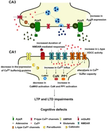
Figure 1.5. Aging is associated with increased intracellular levels of calcium caused by an aberrant contribution of NMDAR, Voltage-dependent calcium channels and impairments in calcium buffering mechanisms. This increase in
Ca2+ leads to the disruption of the balance kinases/phosphatases and thus to
alterations in LTP and LTD, which can be further correlated with cognitive deficits. Abbreviations: CaMKII: Ca2+/calmodulin-dependent protein kinase II; CaN:
calcineurin; PP1: Protein phosphatase 1; VDCC: Voltage-dependent calcium channels.
1.4.
Physiopathological role of A
2AR in the hippocampus
1.4.1. Adenosine
Adenosine is an endogenous nucleotide mainly produced by the degradation of ATP, being present in all cells as a metabolite (Stone et al., 1985). In situations of compromised energy charge or when cells demand an increased consume of ATP, the intracellular concentration of adenosine, estimated to be in the nanomolar range, increases up to micromolar concentrations (Bardenheuer and Schrader, 1986; Nordström et al., 1977) and this metabolite acts with a homeostatic role in the control of cellular metabolism (Arch and Newsholme, 1978). Adenosine has been shown to play a role in the regulation of physiological activity in several organs and tissues, such as purinergic nucleic acid base synthesis, amino acid metabolism and modulation of cellular metabolic status (Daly, 1982; Stone et al., 1985; Williams, 1989).
29
In the central nervous system, adenosine has important neuromodulatory actions and acts as a fine-tuner of synaptic communication, since it is a relevant player in neuron-glia communication and can affect the release and action of many neurotransmitters and other neuromodulators (Ribeiro and Sebastião, 2010). In fact, adenosine can exert its actions either presynaptically - inhibiting or facilitating neurotransmitter release - or postsynaptically - modulating neurotransmitter receptors activity, through the activation of different membrane receptors with opposite actions (Ribeiro and Sebastião, 2010). Consequently, the effects of adenosine depend on the expression pattern and signaling of such receptors, the brain region and the pathophysiological condition. Accordingly, the neuromodulatory role of adenosine is mediated by a balance between the inhibitory and excitatory actions via A1 and A2A receptors (A1R and A2AR),
respectively. Adenosine can also activate adenosine A2B and A3
receptors. However, those receptors are mainly involved in the peripheral effects of adenosine (Cunha, 2001; Ribeiro and Sebastião, 2010).
1.4.2. Adenosine A2A receptors
Adenosine mediates its effects mainly through activation of A1 and
A2AR receptors, metabotropic G-protein coupled receptors (GPCR).
A1R are usually coupled to adenylate cyclase inhibitory proteins
(Gi/Go) and A2AR to adenylate cyclase excitatory proteins (Gs)
30
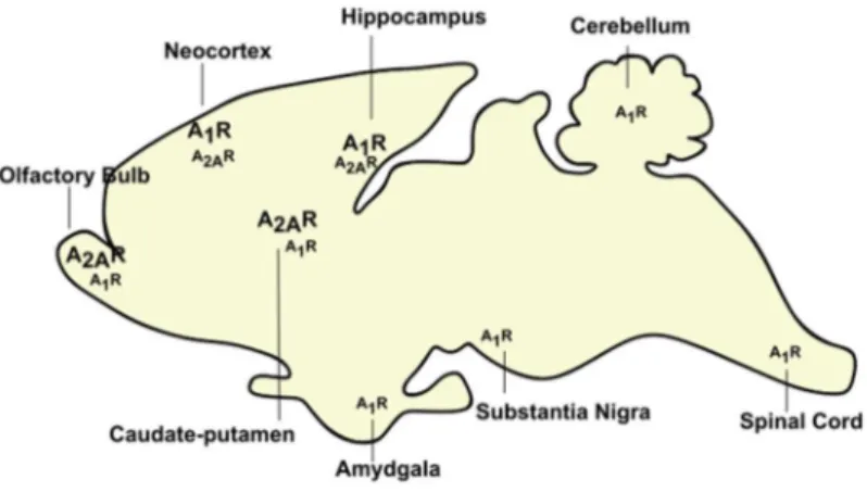
brain has a direct impact on adenosine differential modulation in the brain. A1R are widely distributed, being more abundant in the cortex,
cerebellum and hippocampus (Reppert et al., 1991). On the opposite, A2AR display a more restricted expression pattern: A2AR is highly
expressed in the olfactory bulb and striatum (Jarvis and Williams, 1989), whereas in the neocortex and hippocampus they are present at residual levels (Cunha et al., 1994a; Kirk and Richardson, 1995) (Figure 1.6). Both A1R and A2AR are mostly located in synapses
(Rebola et al., 2003a, 2005a), in particular in excitatory (glutamatergic) synapses (Rebola et al., 2005b; Tetzlaff et al., 1987), although both receptors were shown to be also present in other synapses, such as GABAergic (Cunha and Ribeiro, 2000; Rombo et al., 2015; Shindou et al., 2002) and dopaminergic (Borycz et al., 2007; Garção et al., 2013).
Figure 1.6. Distribution of adenosine receptors A1 and A2A in the brain. The
hippocampus displays high levels of A1R and low levels of A2AR. (Adapted from
31
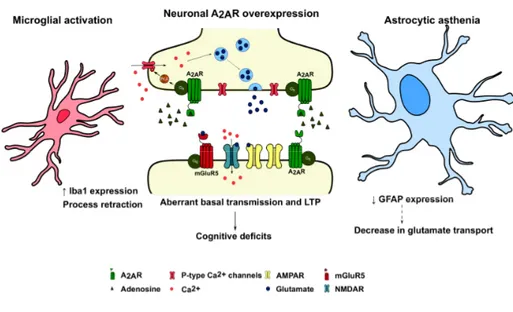
A2AR have an important role on astrocytes, microglia and neurons.
A2AR were shown to be localized in astrocytes (Brambilla et al., 2003;
Cristóvão-Ferreira et al., 2013; Matos et al., 2012a; Orr et al., 2015), where they control uptake of glutamate (Matos et al., 2012a, 2012b) and mediate glucose metabolism, astrogliosis, proliferation, cellular volume and the release of neurotrophic factors and interleukins (Boison et al., 2010; Daré et al., 2007). Plus, different brain insults cause an upregulation of the expression and density of A2AR in
activated microglia (Yu et al., 2008). Both in vitro and in vivo studies further support a role for A2AR in controlling several important
functions operated by microglia, such as process retraction (Gyoneva et al., 2014; Orr et al., 2009), synthesis and release of inflammatory mediators, levels of inflammatory enzymes and proliferation (Santiago et al., 2014).
In neurons, under basal conditions, adenosine preferentially stimulates A1R, which shows a higher affinity for adenosine than A2AR
(Fredholm et al., 2001), leading to inhibition of glutamatergic synaptic transmission in the hippocampus (Sebastião et al., 1990). Adenosine can also activate A2AR, which decreases A1R binding and thus an
inhibition of A1R actions (Lopes et al., 2002). Accordingly, A2AR,
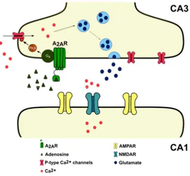
predominantly presynaptic (Rebola et al., 2005a), increase the release of glutamate in the hippocampus (Cunha et al., 1994b; Lopes et al., 2002), possibly by inducing Ca2+ uptake (Gonçalves et al., 1997) and
PKA-dependent Ca2+ currents through P-type Ca2+ channels in the
presynaptic CA3 neurons (Mogul et al., 1993) that project onto the CA1 pyramidal cells (Figure 1.7).
32
Figure 1.7. Presynaptic effects of A2AR in the glutamatergic synapses of the
hippocampus. A2AR increase the release of glutamate in the hippocampus, possibly
by inducing Ca2+ uptake and PKA-dependent Ca2+ currents through P-type Ca2+
channels in the presynaptic CA3 neurons that project onto the CA1 pyramidal cells. Abbreviations: CA1 and CA3: Cornu ammonis 1 and 3; PKA: Protein kinase A.
Postsynaptically, A2AR promote the function of AMPAR (Dias et al.,
2012) and NMDAR (Ferreira et al., 2017a; Rebola et al., 2008; Sarantis et al., 2015). Concretely, previous studies hinted at a possible A2AR-NMDAR interaction, since A2AR can control expression
(Ferreira et al., 2017b, 2017a), recruitment (Rebola et al., 2008) and the rate of desensitization (Sarantis et al., 2015) of NMDAR. Group I metabotropic glutamate receptors, namely mGluR5, are postsynaptic and tightly coupled to NMDA receptors (Ferreira et al., 2017a; Jia et al., 1998; Takagi et al., 2010), conferring them the ability to either
