Sara Carina Duarte da Silva
Março de 2011 U M in ho |2 01 1 S ar a C ar in a D ua rt e da S ilv aInsights into pathology and
neurodegeneration features in a transgenic
mouse model of Machado-Joseph disease
In si g h ts i n to p a th o lo g y a n d n e u ro d e g e n e ra ti o n f e a tu re s in a t ra n sg e n ic m o u se m o d e l o f M a ch a d o -J o se p h d is e a se
Sara Carina Duarte da Silva
Trabalho efectuado sob a orientação de:
Orientador: Professora Doutora Patrícia Maciel
Co-orientador: Dr.ª Anabela Fernandes
Supervisora: Professora Doutora Dorit Schuller
Dissertação de Mestrado
Mestrado em Genética Molecular
Insights into pathology and
neurodegeneration features in a transgenic
mouse model of Machado-Joseph disease
NOME: Sara Carina Duarte da Silva
ENDEREÇO ELECTRÓNICO: sarasilva@ecsaude.uminho.pt TELEFONE: 969809120
NÚMERO DO BILHETE DE IDENTIDADE: 12551200
TÍTULO DA TESE: Insights into pathology and neurodegeneration features in a transgenic mouse model of Machado-Joseph disease
ORIENTADOR: Professora Doutora Patrícia Maciel
CO-ORIENTADOR: Dr.ª Anabela Fernandes
SUPERVISORA: Professora Doutora Dorit Schuller
TESE DE MESTRADO EM: Genética Molecular
E AUTORIZADA A REPRODUÇAO INTEGRAL DESTA TESE APEN AS PARA EFEITOS DE INVESTIGAÇAO, MEDIANTE DECLARAÇAO ESCRITA DO INTERESSADO, QUE A TAL SE COMPROMETE;
Universidade do Minho, ___/___/______
NOME: Sara Carina Duarte da Silva
ENDEREÇO ELECTRÓNICO: sarasilva@ecsaude.uminho.pt TELEFONE: 969809120
NÚMERO DO BILHETE DE IDENTIDADE: 12551200
TÍTULO DA TESE: Insights into pathology and neurodegeneration features in a transgenic mouse model of Machado-Joseph disease
ORIENTADOR: Professora Doutora Patrícia Maciel
CO-ORIENTADOR: Dr.ª Anabela Fernandes
SUPERVISORA: Professora Doutora Dorit Schuller
TESE DE MESTRADO EM: Genética Molecular
E AUTORIZADA A REPRODUÇAO INTEGRAL DESTA TESE APENAS PARA EFEITOS DE INVESTIGAÇAO, MEDIANTE DECLARAÇAO ESCRITA DO INTERESSADO, QUE A TAL SE COMPROMETE;
Universidade do Minho, ___/___/______
Por me fazerem feliz e estarem sempre
comigo
Papá, mamã, Zé e Peck
Durante a escrita desta tese, a conquista e o desânimo são dois sentimentos que se aliam, mas estas últimas palavras que vou escrever, fazem com que a conquista prevaleça o desânimo, já que muitas foram as pessoas que me ajudaram a ultrapassar todas as dificuldades e me deram força para continuar; a todas as pessoas que contribuíram de alguma forma para a escrita desta tese um MUITO OBRIGADA!
À Professora Patrícia Maciel por me ter dado a oportunidade de permanecer no seu laboratório após o estágio curricular e me ter incentivado a ingressar num projecto de mestrado; por tudo muito obrigada…
À Professora Maria João Sousa, coordenadora do mestrado em Genética Molecular, por estar sempre disponível para me ouvir e elucidar sobre muitos aspectos do mestrado;
A todos os professores que estiveram envolvidos neste curso, obrigada pela organização das aulas e pelo incentivo a tarefas inovadoras, em especial à minha Supervisora, professora Dorit Elisabeth Schuller.
A todos os NERD’s pela boa disposição, pelas discussões científicas, pelo apoio e interajuda, pelos petiscos da terça-feira à tardinha…
A todos os elementos do ICVS que sempre que nos cruzámos nos corredores, um “olá” e um sorriso nunca são demais…
Ao Luís “da histologia”, por estar sempre disponível, por fazer os H&E clarinhos como eu gosto, por me ajudar na interpretação de muitos resultados histológicos, por tudo!
À Fernanda Marques (Nanditi), pela constante preocupação e boa disposição (mesmo que aches que não), pelas músicas que pões para eu ouvir, pela amizade, obrigada Nanda, pela pessoa fantástica que és (mesmo que aches que não);
vi
apoio;
Às minhas colegas de grupo: Anabela, Ana João, Teresa, Andreia de Castro, Fátima, Cláudia Botelho.
Anabela: por me teres ajudado nos meus primeiros passos, por me teres orientado nas etapas da minha curta vida científica, mas principalmente por não seres apenas uma colega de trabalho mas sim uma amiga, por todas as conversas que nos ajudam a desviar o pensamento dos nossos “jurandis”, pela companhia, pela amizade. Obrigada pelas conversas dentro do biotério, onde nos sentimos protegidas, eu, tu e os nossos ratinhos. Obrigada.
Ani Joni: por me ouvires sempre quando me irrito, por te irritares juntamente comigo, por teres a capacidade de ouvir as minhas lamentações, pelos conselhos científicos, por te teres tornado uma amiga e não uma colega de trabalho, pelas conversas no bar acompanhadas pelos panikes gordurosos, obrigada pela tua transparência.
Teresa: a tua curta passagem pelo grupo deixou saudades; obrigada pela simplicidade da tua maneira de ser, obrigada pelas palavras de força e pelas tuas gargalhadas. Ah! Já me esquecia de agradecer os pastéis de Belém…
De Castro: pela tua capacidade incrível de me ajudares a resolver problemas, por te conhecer apenas pelo tom de voz, por saber que quando dizes: “Sara vou-te matar!!!” já sei que o problema é de fácil resolução, pelas tuas gargalhadas tão contagiantes, pelo teu jeito de ser tão diferente do meu, por seres uma investigadora crítica com o teu trabalho e por me ajudares a criticar o meu, obrigada por tudo.
Fátima: pelas tuas piadas sempre no timing oportuno e pela tua sensatez;
Aos meus amigos Andreia Carvalho, Loirinha, Ju, Poke, Ti, Zé Manel. Por estarem sempre, sempre lá.
Carvalho: não existem palavras suficientes para descrever o que tens sido para mim ao longo destes 7 anos de convivência diária; acho que os nicknames que nos colocaram “tico e teco” se encaixam perfeitamente no que somos uma para a outra. Têm sido muitas lágrimas e muitos sorrisos ao longo desta nossa amizade, muitas derrotas e muitas conquistas, e tu, tu estás sempre lá. É principalmente a ti que dedico esta tese, pois és tu que estás sempre comigo, pois és tu que compreendes sempre as minhas dúvidas e festejas as minhas vitórias. Simplesmente esta tese é tua.
Loirinha: amiga de todos os dias. Conheces-me como a palma da tua mão. Apesar de nem sempre entenderes o que significa “escrever uma tese” e fazer “investigação”, sempre estives-te comigo e agradeço-te por tudo que és para mim.
Ju: obrigada pelas palavras de força e por sempre me dizeres: “tu vais conseguir”.
Poke, Ti, Zé Manel: apesar de ser difícil para vocês compreender o meu tipo de “trabalho” e nem sempre compreenderes porque é que eu não tenho horários, eu sei que sois os meus amigos do coração e tudo que eu precisei/preciso posso contar convosco.
À minha segunda família: Sr.Joaquim, D.Mena, Bela, Carla, Lando, Jorge, Céu. Sois um porto seguro e sei que posso contar convosco para tudo. Sr. Joaquim, sei que mesmo ausente fisicamente, me acompanhou sempre ao longo da escrita desta tese e ao longo desta etapa da minha vida, e me deu sempre força para continuar, obrigada.
Aos meus meninos: Zé Lando, Catarina e Francisca, o vosso sorriso e a vossas brincadeiras fazem-me tão bem!! Olhar simplesmente para vocês dá-me ânimo, força, alegria, coragem.
Ao Xico Zé, pelo amor durante estes 6 anos, pela compreensão, pela atenção, pela paciência. Por saberes que o cerebelo é uma das áreas afectadas na doença de Machado-Joseph,
viii
me amares da maneira que amas. Obrigada.
A toda a minha família.
Ao meu avô Cristiano. Fazes-me muita falta e sei que estás sempre comigo.
A minha avó Mimi, à Nhi e ao mano, por serem tudo para mim, por serem a família mais compreensiva do mundo. À minha avó por estar sempre preocupada com a comidinha para trazer para o trabalho. À Nhi por fazer tudo por mim. Ao mano pelas palavras de incentivo e por sentires o orgulho que sentes por mim. Obrigada por tudo.
Por fim, às pessoas mais importantes da minha vida, que me deram a oportunidade de viver, que me deram todas as oportunidades e fizeram de mim tudo aquilo que sou hoje. Por todos os esforços que fizeram por mim, por estarem sempre lá; por ficarem (mesmo durante a noite) a ouvir tudo aquilo que preciso de desabafar, por me tratarem como uma princesa, pelo amor e carinho diários, por tudo, tudo. Aos meus pais dedico o fruto do meu trabalho, esta tese.
Machado-Joseph disease (MJD), also known as Spinocerebellar Ataxia 3 (SCA3), is the most common autosomal dominant ataxia worldwide, and is caused by a CAG repeat expansion within the coding region of the ATXN3 gene. The clinical variability of the disease phenotype as well as the age of onset depend on the length of the expanded repeat. The anticipation phenomenon is most frequently associated with repeat expansions in paternal transmission. MJD patients with a repeat expansion above 44 CAGs in the ATXN3 gene present cytoplasmic and/or intranuclear ataxin-3 aggregates and neuronal cell loss in specific areas of the brain. However, some questions remains unanswered in this disease: why only some subpopulations of neurons are affected, although ataxin-3 is everywhere; what underlies this selective neuronal vulnerability; are these neurons dysfunctional or dying?
In an attempt to address these issues, we took advantage of studying a cDNA transgenic mouse model (CMVMJD) expressing the mutant human ataxin-3 under the regulation of the CMV promoter (pCMV), previously generated in our lab. This transgenic mouse model shows an important overlap with genetic and clinical features of MJD, namely genetic instability of the expanded CAG repeat and a motor impairment phenotype.
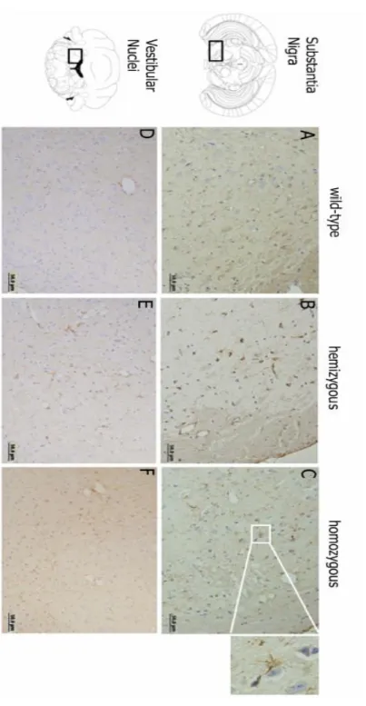
In this work, we performed an extensive pathological analysis of MJD mouse brains, that revealed a significant atrophy in the thalamus and in the dentate neurons. Increased GFAP immunostaining with reactive astrocytes was observed in the vestibular nuclei and substantia nigra of transgenic mice. Regarding cell death, we have searched for evidence of different cell death types (apoptosis and necrosis) by TUNEL assay, caspase-3 analysis and Fluoro-Jade B staining. We did not find any differences between wild-type and MJD transgenic mice, suggesting that probably the affected neurons are not dying, at least by apoptosis or necrosis, instead, they might just be dysfunctional. We also analysed of the somatic mosaicism in neuronal and non-neuronal tissues through aging revealed a significant increase in the mosaicism index of specific brain regions such as the pons, substantia nigra, cerebellar cortex, hipocampus, striatum, deep cerebellar nuclei and hypothalamus with age. However, there was no correlation between the extent of the mosaicism and the pathological involvement of a given region.
The results allow us to conclude that the pCMVMJD94 mouse is a good model to study the pathogenic mechanisms of MJD, mimicking an early stage of the disease.
A doença de Machado-Joseph, também conhecida por Ataxia Espinocerebelosa tipo 3 (SCA3), é a ataxia autossómica dominante mais comum em todo o mundo, causada por uma repetição de CAGs na região codificante do gene ATXN3. A variabilidade no fenótipo da doença assim como a idade de surgimento dos sintomas depende no tamanho da repetição expandida. O fenómeno de antecipação está mais frequentemente associado a transmissões paternas. Pacientes com DMJ com expansões acima de 44 CAGs revelam agregados citoplasmáticos e /ou nucleares e perda neuronal em áreas do cérebro específicas. Algumas questões chave permanecem por esclarecer. Por exemplo, nesta doença apenas algumas subpopulações de neurónios são afectadas, embora a ataxina-3 seja expressa em todas as áreas. O que está por detrás desta vulnerabilidade neuronal selectiva? Estarão estes neurónios a morrer ou estarão disfuncionais? Numa tentativa de responder a estas perguntas, utilizámos um modelo em ratinho que expressa o cDNA da ataxina-3 humana mutada, sob a regulação do promotor CMV (pCMV), previamente gerado no nosso laboratório. Neste trabalho, fizemos uma extensa análise patológica de cérebros de ratinho DMJ que revelaram uma atrofia relevante no tálamo e núcleos denteados. Também se observou um aumento da proteína GFAP, revelando um aumento da reactividade dos astrócitos, nos núcleos vestibular e substantia nigra dos ratinhos transgénicos. Também fizemos um rastreio de diferentes tipos de morte celular (apoptose e necrose) por TUNEL, análise da activação da caspase-3 (imunohistoquímica e western-blot) e coloração com Fluoro-Jade B. Não encontrámos diferenças significativas entre ratinhos do tipo selvagem e transgénicos, sugerindo que provavelmente estes neurónios não estarão a morrer, pelo menos por apoptose ou necrose, estando possivelmente disfuncionais. Para além disso, a repetição de CAG expandida, variou em mais de 50% das transmissões nos ratinhos transgénicos, com expansões típica transmitidas em meioses paternas e contracções em maternas. A análise do mosaicismo somático em tecidos neuronais e periféricos, durante o envelhecimento, revelou um aumento no índex de mosaicismo em regiões específicas do cérebro e mostrou ser dependente da idade. Este ratinho transgénico apresenta características genéticas e clínicas importantes que se sobrepõem às da DMJ, nomeadamente instabilidade intergeracional da expansão de CAGs, características patológicas da doença (astrogliose e neurónios atrofiados, nas regiões relevantes) e um fenótipo de descoordenação motora.
Estes resultados permitiram-nos concluir que o ratinho pCMVMJD94 é um bom modelo para estudar os mecanismos patogénicos da DMJ, mimetizando estadios precoces da doença.
Dedication ... iii
Agradecimentos/Acknowledgments ...v
Abstract ... ix
Resumo ... xi
Abbreviations ... xv
Chapter 1. General Introduction ... 3
1.1 Polyglutamine disorders: an overview... 3
1.2 Instability of the CAG repeat ... 5
1.3 Polyglutamine pathogenic mechanisms ... 7
1.3.1 Misfolded proteins and aggregation ... 7
1.3.2 Transcriptional deregulation ... 8
1.3.3 Mitochondrial impairment ... 9
1.4 Neuropathology and neuron-specific toxicity in PolyQ diseases ... 10
1.5 Mechanisms of cell death ... 12
1.6 Machado-Joseph disease ... 16
1.6.1 Clinical definition ... 16
1.6.2 Pathology ... 17
1.6.3 Genetics ... 18
1.6.4 ATXN3 gene product ... 19
1.7 Different mouse models for the same disease ... 20
Chapter 2. Objectives ... 27
Chapter 3. Brain pathology in MJD transgenic animals ... 31
3.1 Background ... 31
3.2 Materials and methods ... 32
3.3 Results and discussion ... 33
3.4 Conclusion ... 38
Chapter 4. Mechanisms of cell death ... 41
4.1 Background ... 41
4.2 Materials and methods ... 42
xiv
Chapter 5. CAG repeat instability ... 55
5.1 Background ... 55
5.2 Materials and methods ... 57
5.3 Results and discussion ... 59
5.4 Conclusion ... 64
Chapter 6. General discussion ... 67
Chapter 7. Main conclusions ... 73
Chapter 8. Future perspectives ... 77
xv
List of Abbreviations
μL – Microliter μm – Micrometer AD – Alzheimer's Disease AH – Anterior HornALS – Amyotrophic Lateral Sclerosis AR – Androgen receptor
AT3 – Ataxin-3 protein
AT3Q(n) – Ataxin-3 protein containing n glutamine residues
ATXN3 – Ataxin-3 gene ATP – Adenosine triphosphate AIF – Apoptosis Inducing Factor Apaf-1 – Apoptotic peptidase activating factor
AMPA – Agonist of AMPA receptor a.a. –Aminoacid
bp – Base pairs
C. elegans – Caenorhabditis elegans C/P – Caudate/Putamen
CAG – Trinucleotide codon for glutamine
CBP – CREB binding protein
cDNA – complementary DNA cys – Cysteine
cyt c – Cytochrome c
CREB – Cyclic AMP-response Element Binding protein
CNS – Central Nervous System CMVp – Cytomegalovirus promoter DN – Dentate Nucleus
DNA – Deoxyribonucleic acid
DRPLA – Dentatorubral-Pallidoluysian Atrophy
DAB – 3, 34-biiaminobenzidine DM – Myotonic Dystrophy
ECL- Enhanced GChemiLuminescenc ER – Endoplasmic reticulum
E.coli – Escherichia coli
FADD – Cytosolic adaptors proteins GABA – Gamma-Amino Butyric Acid GFAP – Glial Fribillary Acidic Protein HD – Huntington's Disease
HDAC – Histone Deacetylase Hdj-1 – 40-kDa heat-shock protein
xvi
H&E – Hematoxylin&Eosin
Hprt – Hypoxanthine Phosphoribosyl Transferase
Hsp – Heat shock protein HAT – Histone Acetylase Htt – Huntingtin Protein H3 – Histone 3
H4 – Histone 4
IHC – Immunohistochemistry
ICE – Interleukin-1b processing enzyme
kDa – KiloDalton(s) kb – kilobase
Lat – Dentate nucleus LV – Lentivirus
LANP – Cerebellar leucine rich acidic nuclear protein
MJD – Machado-Joseph Disease mL – Milliliter
mM – Milimolar
MMR – Mismatch Repair MSH – Mismatch repair gene MutSb – MSH2/MSH3 comlex
MT – Mitochondria MI – Mosaicism Index
MOMP – Mitochondrial Outer Membrane Permeabilization
n – Number of samples in the study NEDD8 – Developmental down-regulated gene 8
NMDA – N-methyl-D-aspartic acid NAIP – Neuronal Apoptosis Inhibitory Protein
ng – Nanogram
NI – Neuronal Inclusion
NLS – Nuclear Localization Signal NES – Nuclear Export Signal Nm – Namometer
OH8dG – 8 hydroxydeoxyguanosine pCAF – Protein-associated factor PCR – Polymerase Chain Reaction PFA – Paraformaldehyde
PD – Parkinson's Disease Pn – Pontine Nucleus
PCD – Programmed Cell Death polyQ – Polyglutamine
xvii
Q(n) – stretch of n glutamine residues
Q – Glutamine
RNA – Ribonucleic acid RNAPolII – RNA polymerase II ROS – Reactive Oxygen Species SCA – Spinocerebellar Ataxia
SMBA – Spinal and Bulbar Muscular Atrophy
SMA – Spinal Muscular Atrophy SAHA – Suberoylanilide Hydroxamic Acid
SH3L3 – SH3 – containing Grb2-like protein
SOD – Superoxide dismutase SN – Substantia Nigra SP1 – Specificity Protein 1
SRC1 – Steroid Receptor Coactivator1
TAFII130 – TBP-associated factor TBP – TATA box binding protein t-test – Student’s t test
TH – Tyrosine Hydroxilase Th – Thalamus
TUNEL – Terminal deoxynucleotidyl transferase dUTP nick end labeling TC-NER – Nucleotide excision repair UIM – Ubiquitin-interacting motifs UPS – Ubiquitin-proteasome system Ve – Vestibular nuclei
VMAT2 – VVesicular monoamine transporter 2
VCP – Valosin-containing protein Wt – Wild-type
Chapter 1. General Introduction
1.1 Polyglutamine disorders: an overview
Expansions of repeating units of DNA, especially CAG triplet repeat expansions, are known to underlie several neurodegenerative disorders [6], including Machado-Joseph disease (MJD), or spinocerebellar ataxia 3 (SCA3) [4, 7], Huntington disease (HD) [8], spinal and bulbar muscular atrophy (SBMA)[9], dentatorubropallidoluysian atrophy (DRPLA) [10-11], and other spinocerebellar ataxias, such as SCA 1, 2, 6, 7, and 17. The clinical manifestations of each disease result from brain pathology involving a specific subset of neurons that is, for the most part, particular to each disease. A common feature among all these disorders is a progressive neuronal dysfunction/death beginning at mid-life [4].
The progressive neurodegeneration in adulthood and some common symptoms in these apparently unrelated disorders have suggested a shared mechanism for their pathogenesis. The major pathogenic mechanism of these diseases is believed to arise from a genetic gain of function related to abnormal conformation of the elongated polyQ tracts. The CAG expansion leads to an abnormally long polyglutamine (polyQ) tract within all the proteins involved in the mentioned diseases. Besides this polyQ tract, the proteins share no homology and no functional similarity, which suggests that the polyQ stretch itself confers toxic properties to these proteins through a “toxic gain of function” [5]. In agreement with this hypothesis the expression of a simple polyQ tract, in the absence of any additional protein context has been shown to be toxic, as shown in cell culture, mouse, Drosophila and C. elegans [6-10]. This toxic effect of the polyQ tract was further reinforced by a study describing the effect of the insertion of a CAG tract in the hypoxanthine-guanine phosphoribosyltransferase (HPRT) gene, encoding a metabolic enzyme in mice. These mice developed a progressive late-onset neurological phenotype, including ataxia, seizures and premature death [12].
Interestingly, and despite the ubiquitous expression pattern of polyQ mutated proteins, only some subsets of neurons are affected in each polyQ disorder (Table1), indicating that there are other factors influencing the pathogenesis [4, 21]. Apart from the polyQ tract, the disease associated proteins are unrelated to each other, sharing no homology [3, 22-23]. All these proteins are widely
4
expressed in the Central Nervous System (CNS) as well as in peripheral tissues, however each polyQ protein originates a different neurodegeneration profile [4].
Another interesting fact is the overlap of phenotypes of these disorders when the expansions are large: for example, juvenile-onset Huntington patients develop dystonia and seizures in addition to the classical phenotype of chorea, dementia, lack of coordination and unsteady gait seen in adult patients.
The CAG repeat tracts are normally highly polymorphic and above a certain threshold length the symptoms of the diseases are manifested (Table 1) [1, 4-5]. The disease threshold length (and the dominant toxic property) is around 35 to 40 glutamine repetitions. Above the pathogenic threshold the CAG tracts are unstable across generations, and it may expand or contract depending on the gender of the transmitter. While maternal meioses tend to lead to contractions, paternal transmissions tend to lead to expansions. This intergenerational instability leads to a phenomenon called “anticipation”, where the disease symptoms appear earlier and are more severe [24-25]. At a molecular level, all polyQ diseases display proteinaceous aggregates in neurons, usually termed neuronal inclusions (NIs), mainly located in the nucleus [11]. These inclusions are a common hallmark of all these diseases and it is still controversial if they are pathogenic or not.
1.2. Instability of the CAG repeat
Expanded repeat mutations do not behave strictly according to rules of Mendelian inheritance. They are unstable mutations that change in size along successive generations. In contrast, normal-sized repeats are usually transmitted stably [26][12]. This instability of the mutant alleles might explain some characteristics of these diseases such as variable phenotype [27], anticipation and parental-origin effects. The mechanisms that underlie this instability are not very well known, but several factors are known to influence it: (i) type of repeated sequence, (ii) gender of the transmitting parent [28], (iii) the original length of the repetition tract [29], and (iv) the presence/absence of interruptions in the repetitive sequence [24, 30-31]. In this type of disorders, severe juvenile-onset cases are usually paternally transmitted, due to the greater repeat instability with paternal transmission [32, 33]. For example, analysis of the repeat length in sperm from HD patients suggests that this tendency for transmitting expanded repeats is due to repeat instability during spermatogenesis [34]. Also, in MJD families the sex of the transmitting parent has a significant effect on intergenerational instability, male meioses being associated with larger variations, both contractions and expansions, of the repeat size [24, 30-31, 35].
However, repeat instability is not confined to the germline, occurring also in characteristic patterns in many somatic tissues of affected individuals [36]. The ongoing repeat instability in critical somatic tissues likely accelerates disease progression. Somatic mosaicism of the repeat sizes in triplet repeat disorders shows that different cells of the same individual carry different repeat sizes. This phenomenon has never been described for the normal allele, but it has been associated to the expanded allele for several polyQ disorders such as MJD [31] and SCA1 [37] as well as HD [38], DRPLA [39] and SMBA [40].
Tissue-specific CAG instability and severity of neuropathological features were proposed to be directly correlated in HD [38] but this was not confirmed for other diseases with CAG repeat expansions [31, 37, 39-40].
The existence of a similar threshold size for all trinucleotide repeat diseases may suggest a common mechanism for expansion. Slipped-strand mispairing during replication [41-42] has been proposed as a mechanism of mutation that could account for several features of the trinucleotide expansion. During replication of a threshold length repeat (~35 pure repeats), slippage
6
can occur resulting in moderate expansion. Replication of larger repeats leads to hyperexpansion. DNA mismatch repair mechanisms likely play a role in correcting errors caused by slippage, since defects in mismatch repair lead to an increased of simple sequence repeat instability in both yeast and humans (for review, see[43]).
There is no straightforward correlation between the degree of CAG instability and cell division rates (DNA replication), DNA damage (DNA repair and recombination), or disease-gene transcript levels (transcription) [44].
The age-dependent repeat instability in somatic tissues, especially in terminally differentiated neurons, strongly suggests a relevant role for pathways that are independent of DNA replication. Transcription-induced repeat instability (Figure 1) can be modulated by several DNA repair proteins, including those involved in mismatch repair (MMR) and transcription coupled nucleotide excision repair (TC-NER). Transcription was also suggested as a possible cause for repeat instability, based on observations in four lines of transgenic mice that carried a portion of the huntingtin gene containing 55 CAG repeats [45]. In three of those lines, the transgene was expressed and was unstable, whereas in the fourth the transgene was silent and the repeat was stable.
Very recent investigations have shown that genetic recombination is a powerful mechanism for generating massive expansions of trinucleotide repeat sequences. Several human genetic studies on patient materials reported haplotype analyses, especially related to myotonic dystrophy (DM) and the fragile X syndrome, which implicated gene conversion and/or unequal crossing-over (types of recombination) in genetic instabilities [46]. Studies in E.coli showed that the expansion of triplet repeats in vivo can occur by homologous recombination as shown by biochemical and genetic studies [47]. Gene conversion is an event in DNA genetic recombination, which occurs at high frequencies during meiotic division but which also in somatic cells. It is a process by which DNA sequence information is transferred from one DNA helix (which remains unchanged) to another DNA helix, whose sequence is altered. This conversion was suggested as the mechanism responsible for trinucleotide repeat instability during repair in yeast [48]. Recombination mechanisms were also described in mouse models and clinical cases [46, 49].
CAG repeat instability is behind some features of polyQ disorders and may also contribute to their pathogenic mechanism.
1.3 Polyglutamine pathogenic mechanisms
Several mechanisms have been implicated in polyglutamine toxicity, most of which are not mutually exclusive [50]. Here we will focus on three hypotheses: (i) disruption of proteostasis; (ii) transcriptional deregulation and (iii) mitochondrial impairment/oxidative stress.
1.3.1 Misfolded proteins and aggregation: disruption of proteostasis?
The presence of the expanded polyQ-stretch seems to confer novel properties to the mutant proteins, namely a tendency towards increased aggregation. Although protein aggregation is thought to be a central aspect of the biology of many neurodegenerative diseases, the role of two different types of aggregates in neurodegeneration is continuing to be elucidated. In polyQ disorders it is also generally accepted that aggregates or inclusion bodies are an important hallmark of polyglutamine diseases, and these aggregates are indeed found in patients’ brains, but the question remains: are the visible aggregates toxic or protective?
Intranuclear inclusions were initially discovered in the first animal model of HD [16]. Interestingly, a correlation was observed between the length of the CAG repeat and the number of inclusions observed in the disease brains [51-53]. Other evidences suggested that aggregates could be protective to the cells, by stacking the toxic mutant proteins [54]. Indeed, protein aggregates are also naturally occurring species. An example are Marinesco bodies, which are eosinophilic ubiquitinated intranuclear inclusions found in pigmented neurons of the human substantia nigra and locus coeruleus as a result of the metabolism of normal aging neurons [55].
One way in which protein aggregates may harm neurons is by affecting their proteostasis. The hypothesis of cellular homeostasis disturbance suggests that the sequestration of chaperones and proteasome subunits into polyQ aggregates could result in an increased in general protein misfolding and reduced clearance of other crucial cellular proteins, leading to a proteostasis decline [56-58].
Several proteins are known to become trapped into these nuclear and/or cytoplasmic aggregates, such as ubiquitin, ubiquitin-like proteins, proteasome subunits, molecular chaperones and other polyQ-containing proteins [56, 58-60]. This suggests the involvement of the ubiquitin-proteasome degradation pathway in these pathologies, and also the activation of the heat
8
shock response machinery in order to either refold or degrade the mutant polyQ proteins. The mislocalization of chaperones and proteasome subunits may contribute to the progress of the disease. Molecular chaperones were shown to associate with the aggregates only transiently and to move freely [61]. However, the proteasome appears to be permanently recruited into polyQ aggregates and to be functionally impaired [62], which can interfere with the degradation of other proteins [63]. Proteasome inhibition might also increase the intracellular load of misfolded, oxidized, or otherwise damaged proteins, thereby causing neuronal toxicity. The consistent presence of normal ataxin-3 in NIs could reflect a biological feature of wild-type ataxin-3, which is translocated into the nucleus under pathological conditions and participates in the formation of aggregates [64].
1.3.2 Transcriptional deregulation
Transcriptional alteration is another unifying feature of polyQ disorders [36, 65-69]; however, the relationship between polyQ-induced gene expression deregulation and the ongoing degenerative processes remains unclear.
More than 20 nuclear proteins relevant to transcription are known to interact with polyglutamine disease associated-proteins [65]. Mutant polyQ proteins have been shown to interact abnormally with proteins involved in the transcription machinery, namely the CREB-binding protein (CBP), p300/CREBBP associated factor (PCAF), TATA-binding protein (TBP), TAFII130, and SP1 [65, 69]. Overexpression of some of these transcription regulators was shown to overcome polyQ toxicity, both in vitro in cellular models for MJD, SBMA, and HD [66, 70] as well as in vivo in a polyQ model in Drosophila [67]. This suggests an important role for transcription deregulation in polyQ pathogenesis.
Proteins that interact with polyglutamine disease associated-proteins are distributed around the core transcription machinery, which is now known to exert DNA methylation, histone acetylation and RNA modification simultaneously (see review [71]). CBP has attracted attention because CBP is a representative coactivator that possesses HAT (histone acetyltransferase) activity and interacts with numerous transcription factors, and its abnormal binding to disease proteins should affect the expression of a wide range of genes rather than that of a specific gene. Several polyQ-protein interactors have acetyltransferase activity. Acetylation of histones relaxes the DNA
structure promoting transcription, whereas hypoacetylation represses gene activity [72]. The equilibrium of histone acetylation/deacetylation is controlled by histone acetyltransferases and deacetyltransferases (HDACs). Thus, polyQ proteins may be toxic by their direct inhibition of the acetyltransferase activity of transcription regulators, leading to diminished gene expression. In fact, treatment of Drosophila and mouse models of HD with HDAC inhibitors has been shown to ameliorate the disease phenotype and to decrease cell degeneration, with the increase of histone acetylation and consequent transcription activation [73-75].
1.3.3 Mitochondrial impairment/oxidative stress
The mitochondria has numerous important functions in the cell, however the mitochondrial respiratory chain is also one of the major sources of damaging free radicals in human organism, and these free radicals destroy cellular macromolecules, including DNA, lipids and proteins [79]. Mitochondrial dysfunction causes a decrease in ATP production, oxidative damage and induction of apoptosis, all of which are involved in the pathogenesis of several disorders [76-77]. They all share the common features of disturbances in the buffering capacity of mitochondrial Ca2+,
ATP or reactive oxygen species (ROS) metabolism [78].
It has been proposed that mitochondrial impairment may be a major triggering factor of neurodegenerative diseases [80-81]. It is not clear what is the primary initiating event in the pathogenesis of each of these neurodegenerative disorders, but it seems that oxidative damage could constitute a critical factor in the propagation of injuries of the different cellular systems affected in most of them [82].
A perturbation in the mitochondrial function leads to an ionic imbalance, calcium overload and ultimately, ATP depletion (Figure 1.1). If the energy supply of the cell drops dramatically, necrotic cell death will ensue. A mild or gradual energy disturbance may also lead to the release of proapoptotic factors, particularly cytochrome c from the mitochondria, and an apoptotic cascade is initiated as well. Excessive Ca2+ accumulation has deleterious effects, leading to
the oxidative damage of different molecules with the resulting triggering of an apoptotic cascade. In addition, it should not be forgotten that non-excitable cells, such as astrocytes and microglia, are also strongly dependent on the intracellular Ca2+ concentration and Ca2+ signaling to maintain their
10
The cellular processes occurring during normal ageing are to some extent similar to those involved in the pathomechanism of neurodegenerative disorders, also leading to age-dependent impairment of mitochondrial function, for instance the appearance of mutations in mitochondrial DNA. A main difference may be that the metabolic dysfunction in the “normal” ageing brain is distributed in a random fashion, while in neurodegenerative disorders, specific causes and cellular disturbances are superimposed upon the age-dependent decrease of the homeostatic reserve, and these together attack specific structures of the CNS [83].
1.4 Neuropathology and neuron-specific toxicity in polyglutamine disorders
The cause of neuron-specific dysfunction is a major unanswered question in the PolyQ field and, as such, is an active area of research.
Several studies demonstrated that this neuronal-specificity is not due to the abundance of polyQ proteins since they are ubiquitously expressed [3-4] (Figure 1.1) and must not be related to expression levels of those proteins before or after disease onset [84]. Similar expression levels of wild-type and mutant proteins were also shown in animal models and patients [85]. Another hypothesis put forward to explain this selective neuron dysfunction was the heterogeneity of polyQ length due to somatic mosaicism, resulting in loss of neurons with higher CAG repeats and survival of those neurons with less CAG-repeat length. This phenomenon was described for some of these diseases, but upon a more detailed analysis, no correlation with selective brain pathology was found [31, 37, 85]. Previous work from our lab also demonstrated, by screening the brain pathology and determining the CAG-repeat length in several brain areas (affected and spared) that there was no correlation between somatic mosaicism pattern and pathology in MJD. However, this finding could be a result of the technique used, since a gross dissection of the brain areas was performed. Also, in MJD patients, this kind of studies might be impaired by the use of post-mortem tissue in which surviving cells are those analysed. A third possibility is that in susceptible neurons, misfolding and aggregation could be prompted by cell-specific proteolytic events that release a polyQ-containing fragment [86] or by aberrant targeting of polyQ proteins to the nucleus only in certain cells [87]. Finally, specific interacting proteins are likely to contribute to selective vulnerability. For instance, certain interacting proteins may bind to disease associated-proteins in a way that promotes misfolding and aggregation, as was demonstrated for the huntingtin interacting proteins,
SH3-containing Grb2-like protein (SH3GL3) [88] and Rhes protein [89]. Other specific interacting proteins are likely to influence events downstream of misfolding, through mechanisms that are linked to the specific normal functions of the disease proteins. Thus, in each disease, a subset of specific interacting proteins may bind less or more avidly to the mutant protein, thereby altering physiological or biochemical properties of one or both proteins. The susceptibility of a neuron to the downstream effects of the mutant protein would depend, in part, on the particular interacting proteins it expresses. The ataxin-1 interacting protein LANP (cerebellar leucine-rich acidic nuclear protein) is just one of a number of identified interacting proteins that may contribute in this manner to selective vulnerability [90].
Extensive efforts are ongoing to understand how polyQ tracts may mediate neuron-specific degeneration. If the toxic gain-of-function conferred by extremely large polyQ expansions results in non-specific neurodegeneration, pathogenesis caused by more moderate polyQ expansions may be susceptible to modification. Protein context is one factor that modulates polyQ pathogenesis and by doing so, it may contribute to the specific pathology observed in each CAG-repeat disease.
Figure 1.1. CAG-repeat neurodegenerative diseases display unique neuropathology despite a common toxic motif. This schematic diagram identifies the major sites of neuronal loss in each disease. Dark red indicates severe or selective neuronal loss; half-tone red indicates moderate or variable cell loss. The circles in the cerebellar cortex represent Purkinje cells. AH, anterior horn; Cer, cerebellar cortex; C/P, caudate/putamen; Ctx, cerebral cortex; DN, dentate nucleus; GP, globus pallidus; LCN, lateral cuneate nucleus; PN, pontine nucleus; RN, red nucleus; SN, substantia nigra; STN, subthalamic nucleus;VL, ventrolateral thalamic nucleus; V, VI, VII, and XII, cranial motor nuclei. Figure adapted from [3].
12
1.5 Cell death mechanisms
The relevance of cell death in neurodegenerative disorders is still a controversial topic, mainly because synaptic loss and electrophysiological abnormalities typically precede cell loss in these diseases. In addition, in chronic neurodegenerative diseases, cell death occurs over decades whereas the single cell suicide program is known to be executed within a few hours and the dead cells rapidly disappear [91-92].
Much of the attention in the cell death field has been drawn in recent years to a major cell death pathway apoptosis, which is often used as a synonymous of programmed cell death. The elucidation of additional programmed cell death pathways is gradually changing this notion. According to a recent classification, eight different types of cell death were delineated [93] and some researchers describe as many as 11 pathways of cell death in mammals, 10 of which appear to be programmed [94]. These pathways can be broadly divided into two main groups: apoptotic and non-apoptotic (see review [13]).
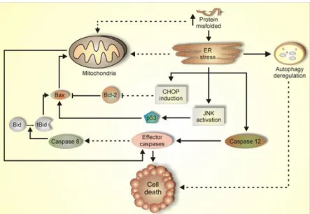
Figure 1.3. Molecular pathways of programmed cell death (PCD) operating in misfolding/aggregation diseases.
Apoptosis vs Necrosis
Apoptosis, also referred to as type I programmed cell death, is the most well defined type of cell death pathways, both morphologically and biochemically, occurring by a caspase cascade. It is characterized by membrane blebbing, cytoplasmic shrinkage and reduction of cellular volume (pyknosis), as well as condensation of the chromatin, and fragmentation of the nucleus (karyorrhexis), all of which ultimately lead to the formation of apoptotic bodies, a prominent morphological feature of apoptotic cell death [93]. As apoptosis typically does not induce inflammation or tissue scarring, it is well suited for a role in normal cell turnover during embryogenesis and in adult tissues [95]. Caspases are a family of proteins that are one of the main effectors of apoptosis. Their activation is a hallmark of apoptosis. Based on their function, the caspases can be classified into three groups: inflammatory (caspases 1, 5, 11, 12, 13, 14, involved in inflammation), initiator caspases (2 and 8 which contain a death effector receptor and 2 and 9 that possesses recruitment and activation domain) and finally effector caspases (3, 6, and 7, which are the executers) [96-97]. Recent estimates suggest that apoptosis deregulation contributes to about half of all the major medical illnesses for which adequate therapy or prevention is currently lacking [98].
The type of cell death in neurodegenerative disorders remains a matter of controversy, and it is possible that both apoptotic and nonapoptotic cell death coexist in the brains of affected patients [91]. Knock-out mice models have elucidated the researchers about the cell death pathways occurring in mammals and have revealed a prominent neuronal apoptosis defect in mice defective for caspase-3, caspase-9 and Apaf-1, suggesting that this pathway of caspase activation is important in regulating neuronal cell death in the developing brain [99].
Alzheimer’s, Huntington’s, Parkinson’s and Prion diseases, as well as ALS, all have features of misfolded proteins. These disturbances can cause accumulation of unfolded proteins in the ER, triggering an evolutionarily conserved response termed the unfolded protein response (UPR) [100-101]. It has been proposed that these protein aggregates exhaust cellular proteasome activity in a failed attempt to degrade them, resulting in a secondary accumulation of misfolded proteins in the ER, thereby triggering ER stress (see review [102]. The UPR controls the levels of molecular chaperones and enzymes involved in protein folding in the ER. ATF6 located at the ER membrane is one of the candidates for the UPR-specific transcription factor [14]. Grp78/Bip (Bip) protein is the chaperone that increases protein folding in the ER lumen [15]. Upon induction of ER stress, ATF6 is
14
processed and the processed cytoplasmic region translocates into the nucleus and activates transcription of the endogenous Bip gene [14]. When these stress signals are unable to rescue cells, the apoptotic pathway is activated. Caspase-12, which is located at the outer layer of the ER, is one of the apoptotic pathways of ER stress-mediated cell death [16-17].
Necrosis has traditionally been considered an unregulated, energy- independent form of cell death, and has been well-characterized in a wide range of pathologic states. Extensive failure of normal physiological pathways that are essential for maintaining cellular homeostasis, such as regulation of ion transport, energy production and pH balance can lead to necrosis. Necrosis is characterized morphologically by vacuolation of the cytoplasm, breakdown of the plasma membrane and induction of inflammation around the dying cell due to the release of cellular contents activating the resident phagocytes and attraction of leucocytes into the necrosis area [103][18]. A classic example of necrotic conditions is ischemia that leads to a drastic depletion of oxygen, glucose, and other trophic factors and evokes massive necrotic death of endothelial cells and nonproliferating cells of surrounding tissues. Necrosis can be induced by microbial infections, neuronal excitotoxins, or ROS [104].
Many human tumors carry mutations that inactivate apoptotic pathways. Inactivation of apoptosis allows tumor cells to proliferate beyond normal homeostatic control. Necrosis represents an alternative pathway for these cells to be eliminated. The inflammatory component of necrotic death has the potential advantage of stimulating an immune response that could increase the efficiency of tumor cell death [105]. The balance between apoptotic and necrotic cell death may be modulated to potentiate a patient's immune response to a tumor.
Adult neuronal death also occurs by necrosis, an unregulated cell death that is the direct result of external insults such as physical injury, energy depletion, toxic insults, hypoxia and/or ischemia. Necrosis of adult neurons is independent of caspases, and although the precise molecular mechanism underlying this type of cell death is unknown, it might be mediated by increases in intracellular calcium, which activates calpains and cathepsins (cytosolic calcium-activated cysteine proteases), leading to degradation of cytoplasmic proteins [106].
Another way to die: autophagic programmed cell death
Autophagy is an evolutionary conserved and genetically controlled turnover of cellular constituents that occurs in all eukaryotic cells [107]. Autophagy is defined morphologically by the appearance of numerous cytosolic autophagosomes, which are formed by the assembly and expansion of double layered, membrane bound structures of unknown origin around whole organelles and isolated proteins. The origins of autophagosomes are difficult to determine because they contain a mixture of markers from the ER, endosomes and lysosomes. The autophagosomes encapsulate cytosolic materials and subsequently dock and fuse with lysosomes or other vacuoles, resulting in the degradation of their contents [108].
Autophagic cell death is thought to represent an alternative pathway to cell death when apoptosis is impeded, but there are indications that autophagic cell death and apoptosis are not mutually exclusive death pathways, and can cross talk with each other [108]. Evidences for this purpose are (i) the fact that apoptosis regulating molecules such as Bcl 2, Bcl xL, Bax and Bak were implicated in the regulation of autophagy, both independently and through interaction with Beclin 1 [109-110] and (ii) that some of the endonucleases that take a part in apoptosis associated DNA fragmentation may originate in lysosomes [111].
Autophagy impairment has been reported in neurodegenerative diseases including Parkinson, Huntington, and Alzheimer diseases [112-114]. In these diseases, the pathological accumulation of autophagosomes/ autophagosome-like structures and abnormalities in the endosomal-lysosomal pathway were documented by electron microscopy (EM) in human postmortem brain tissue [115-119].
While the role of autophagy in neurodegenerative diseases is far from being understood, the available data indicate it plays an integral role in the cellular response to intracellular protein aggregation common to these diseases. The effect of autophagy in neurons during disease can be broadly divided into two classes: autophagosomal degradation is either impaired or excessively activated, leading to an apparent disruption of the intracellular organelle organization and accumulation of autophagosomes in neurons over long periods of time.
Autophagy plays a crucial role in maintaining neuronal homeostasis through clearance of defective organelles and unfolded/aggregating proteins.
16
1.6 Machado-Joseph Disease
Machado-Joseph disease (MJD), also known as Spinocerebellar Ataxia 3 (SCA3), is the most common autosomal dominant ataxia worldwide [24]. The disease was first described in families from the Azores Islands, being reported some years later in other countries and in families with no Portuguese ancestrality. This late-onset disorder was first described as an autosomal dominant ataxia in William Machado’s family, and thus named Machado disease [120]. In the same year, another case was reported (in the Thomas family) with similar clinical symptoms, and this disease was entitled as “Nigro-spino-dentatal degeneration with nuclar ophthalmoplegia” [121]. Later on, in 1976 was described a “particular type of autosomal dominant hereditary ataxia” in the family of Antone Joseph, which was designated as Joseph disease [122].
Two years later, after an intensive study in Azorean families and regarding the common features of the three families described above, a new “autosomal dominant system degeneration in Portuguese families of the Azores Islands” was introduced [123]. In the 80’s, this disease was named Machado-Joseph disease (MJD) and some clinical criteria for diagnosis were introduced [124]. The prevalence of this disease was from the beginning thought to be the highest among people of Portuguese/Azorean descent. For immigrants of Portuguese Azorean ancestry in New England, the prevalence was described to be around 1:4000. In Portugal the prevalence of Machado-Joseph disease is 3,1:100,000 (P.Coutinho, Personal Communication), with clusters in several mainland regions, reaching a value of 1:140, on the small Azorean island of Flores [125].
Later, however, researchers, based in DNA studies, have identified MJD cases in many ethnic backgrounds. Its relative frequency among the spinocerebellar ataxias is higher in Portugal (49%), China (49%), Brazil (44%), the Netherlands (44%), Japan (43%), and Germany (42%); its relative frequency is lower in France (33%), the United States (21%), and Australia (12%); and it is rare in the United Kingdom (5%), India (3%), and Italy (1%) [126-137].
1.6.1 Clinical definition
MJD patients suffer from a progressive neurodegenerative disorder appearing more frequently between the ages of 20 and 50 years, in which the intellect is preserved [123]. The
preservation of cognitive function is a key feature of MJD in its differential diagnosis among the vast group of spinocerebellar ataxias. MJD is characterized by motor uncoordination and weakness in the arms and legs, spasticity, gait ataxia, difficulty with speech and swallowing, altered eye movements, double vision, and frequent urination. Some patients have dystonia (sustained muscle contractions that cause twisting of the body and limbs, repetitive movements, abnormal postures, and/or rigidity) or symptoms similar to those of Parkinson's disease. Others have twitching of the face or tongue, or peculiar bulging eyes due to lid retraction. The severity of the disease is related to the age of onset, an earlier onset being associated with a more severe and rapidly progressive form of the disease. Symptoms can begin any time between early adolescence and old age up to 70 years of age [125]. MJD is also a progressive disease, meaning that symptoms get worse with time. Life expectancy ranges from the mid-thirties, for patients with severe forms of MJD, to a normal life expectancy for those with mild forms. For patients who die early from the disease, the cause of death is often aspiration pneumonia due to immobility and poor coordination of swallowing and breathing. The clinical spectrum of SCA3 is highly pleomorphic and led to the definition of four clinical sub-phenotypes: type I, characterised by the dominance of pyramidal and extrapyramidal anomalies, in addition to ataxia and other signs, with an early age-at-onset and fast progression; type II, with typical cerebellar ataxia, progressive external ophtalmoplegia and pyramidal signs appearing at an intermediate age; type III, with late onset and slow progression of peripheral signs, such as loss of proprioception and muscle atrophies; and type IV, the rarest, characterised by the presence of Parkinsonic signs, associated to the core clinical features [123-124, 127].
1.6.2 Pathology
Pathological examination of post-mortem MJD patients’ brains showed a depigmentation of the substantia nigra, a considerable atrophy of the cerebellum, pons, and medulla oblongata, as well as in motor cranial nuclei [3, 129]. More recently, it was shown that all the precerebellar nuclei and the thalamus were also affected in MJD patients [126, 130]. In addition, in the majority of these patients the post-mortem brain weight is lower than that of individuals without medical histories of neurological or psychiatric diseases [131].
Intracytoplasmic inclusions although, observed to a lesser extent than intranuclear ones, are found in MJD patients’ brains [128], constituting a pathological feature of MJD. In vivo and in vitro studies suggests that disease protein ataxin-3 accumulates in ubiquitinated intranuclear inclusions in
18
neurons of affected brain regions [6, 19-20]. Several MJD mouse models were generated in the last years and nuclear and /or cytoplasmic aggregates were observed in the mice brains [21-25].
1.6.3 Genetics
The fact that MJD is a hereditary autosomal dominant disease means that the presence of the mutation in one single allele is sufficient to cause the disease. This implies that an affected individual has a 50% chance of passing the disease on to their offspring. Like in other CAG repeat diseases the phenomenon of anticipation is quite common. The children of affected parents tend to develop symptoms of the disease earlier in life, have a faster progression of the disease and experience more severe symptoms. A longer expansion is associated with an earlier age-of-onset and a more severe form of the disease. However, it is impossible to predict precisely the course of the disease for an individual based solely on the repeat length. Many years after the first description of the disease, its causative gene was described and mapped in the chromosome 14.q32.1 [132]. The MJD1 gene (later named ATXN3) was cloned one year later, and the authors observed that the mutation (expansion) of the CAG tract was only present in patients [7]. This knowledge allowed the establishment of the molecular diagnosis of MJD, based on the determination of the CAG repeat length, and the consequent confirmation of the disease in families of different origins [24, 133]. The genomic structure of the ATXN3 gene and the subsequent knowledge of the number and size of exons and introns was only established in 2001. The gene has around 48 Kbp and was described to contain 11 exons, the CAG tract being in exon 10, and to encode at least four different transcripts with variable sizes: 1.4, 1.8, 4.5, and 7.5 Kb, probably due to differential splicing and polyadenylation signals. Very recently 56 additional splicing variants of ATXN3 gene, some of which observed only in MJD patient’s, were reported [134-135]. The biological relevance of these variants remains to be determined. In the brain, ATXN3 is preferentially transcribed in neurons although low levels are also present in glial cells. Most importantly, neurons susceptible to cell death in MJD/SCA3 express ATXN3, although not selectively. Also, ATXN3 mRNA levels do not differ between controls and patients and are not correlated with the clinical severity or repeat length [136].
1.6.4 ATXN3 gene product: ataxin-3 (ATXN3) protein
Ataxin-3 has an approximate molecular weight of 42 kDa in normal individuals, but is significantly larger in affected patients, confirming that the expanded CAG repeat is translated into a polyQ stretch. This polyQ tract is located in the C-terminus region that is variable in length, depending on the isoform, and whose expansion is intrinsically related with MJD. [7]
Different isoforms of ataxin-3 have been described: isoform 1 (NP_004984) has 361 aa, with a hydrophilic C-terminal and corresponds to the clones MJD1-1 and MJD5-1 [137]; isoform 2 (P54252) has a distinct C-terminal region when comparing with the first one and it has a hydrophobic nature. It is formed by 365 aa and matches the MJD2-1 clone [137]; the third variant, MJD1a (S50830) contains 349 aa (it lacks 16 aa within the C- terminus region comparatively to isoform 2, due to a premature stop codon [7]. Finally, the variant 4 (NP_1093376) contains less 55 aa than isoforms 1 due to the lack of exon 2 in the H2 clone [134]. This ATXN3 variant lacks the catalytic aminoacid (Cys14), essential for ATXN3 deubiquitylating in vitro activity against poly-ubiquitin chains [135, 138]. The human ataxin-3 protein has a conserved N-terminal josephin domain (1-198 aa) containing the putative catalytic triad aminoacids cysteine (C14), histidine (H119) and asparagine (N134). The josephin domain, which derives from the name of the disease, is followed by two or three (in isoforms 1 and 4) ubiquitin-interacting motifs (UIMs) and the polyQ stretch and also has a conserved nuclear-localization signal (NLS). Similarly, 6 putative nuclear export signal (NES) sequences were found within the ataxin-3 primary structure [135]. Considering the splicing variants recently reported [26], more ataxin-3 isoforms could exist, and given that fact, is still unclear which ones would be more relevant in the disease context.
Ataxin-3 is found in the genomes of several species, ranging from nematodes to human, and including plants [138]. It possesses a globular N-terminal domain with a sequence motif named Josephin (residues 1-198 in the human protein). The sequence of the C-terminus, which contains low complexity sequences, is less conserved among species.
The search for protein motifs suggested that ataxin-3 might be an ubiquitin-binding protein [139]. Functional assays in vitro supported this function and showed predominant binding to K48-linked tetra-ubiquitin through its ubiquitin interaction motifs (UIM) located near the polyglutamine domain [60, 140-141]. A crucial breakthrough in the understanding of ataxin-3 function was the discovery of its DUB activity in vitro [140, 142], however the biological consequence of this function hasn’t been characterized extensively yet and the cellular substrates of ataxin-3 remain unknown.
20
Histone ubiquitylation results in heterochromatin relaxation and assembly of transcription complexes on the promoter, and ubiquitylation of transcription factors enhances their transcriptional-activation function (see review [143]. Therefore, deubiquitylating enzymes as ataxin-3 can also modify transcriptional regulation through the removal of ubiquitin from histones. A role of ataxin-3 in proteasomal protein degradation has been supported by the identification of its interaction partner, valosin-containing protein (VCP) [142]. Recently, several reports have been described interactions between ataxin-3 and numerous other proteins, implicating it in various cellular functions and pathways [144-146].
1.7 Different mouse models for the same disease
Although there is no treatment available at present to cure or delay the onset of MJD, mouse models have been generated to facilitate the understanding of the disease and the development of a therapy. Mice are the preferred animal model because they are small and easy to genetically manipulate and can be generated in relatively large numbers, kept in a controlled environment, and used for invasive procedures. Being mammals, they have important genomic, anatomical, and physiological similarities to humans [147]. The technical problems encountered in the use of patient brain tissue, such as autolysis caused by long postmortem delays, can be avoided using mouse models.
The first transgenic mouse model of MJD was generated using truncated and full-length cDNAs of the ATXN3 gene, with the L7 promoter directing expression specifically in Purkinje cells (poorly affected in MJD). In these constructs, the cDNA encoded for Q79C, Q79, Q35C (truncated forms with or without the C-terminal) or MJD79 (full-length context). The transgenic mice expressing truncated forms (Q79C, Q79) of the protein with an expanded polyQ tract showed an ataxic phenotype, whereas the animals expressing the full-length protein (MJD79) or a polyQ length present in human control individuals (Q35C) did not reveal any characteristic symptom of the disease. The onset of the symptoms in the affected animals was around 4 weeks of age and the strong ataxic phenotype was more prominent in animals with a higher copy number of the transgene. Histological analysis of these ataxic mice brains showed an atrophic cerebellum and
massive loss of Purkinje cells. This work showed that the expanded polyglutamine tract is responsible for neuronal loss and degeneration [15].
Some years later, another mouse model was created in an attempt to properly mimic the temporal and spatial expression of the human disease gene, using yeast artificial chromosome (YAC) constructs carrying the full-length ATXN3 gene with expanded polyglutamine tracts, containing all the enhancers and long-range regulatory elements needed for cell-specific expression at physiological levels. Two additional genes were also part of the cloned genomic region.
Homologous recombination in the yeast host was used to generate three YAC constructs with 15, 76 and 84 repeats, corresponding to the human wild-type, intermediate and early-disease-onset MJD alleles, respectively. The mice carrying expanded alleles showed abnormal gait, tremor, hypoactivity, limb clasping, an inability to correct geotaxis, reduced grip strength, abnormal toe pinch responses and progressive loss of weight. These symptoms aggravated with repeat length and with the gene dosage. Regarding the onset of the disease, homozygous mice showed an earlier onset, with faster and worse disease progression. Analysis of brain sections showed degeneration, mild gliosis of the dentate and pontine nerve nuclei (affected in MJD patients) and increased number of reactive astrocytes in the cerebellum. Ubiquitinated nuclear inclusions were also shown in symptomatic mice [148]. These mice are representative of MJD and can be a valuable resource for the detailed analysis of the roles of repeat length, tissue specificity and level of expression in the neurodegenerative processes underlying MJD pathogenesis and could also be used to test therapeutic strategies. However, for this later purpose, their phenotype may be considered too mild.
In 2004, a third mouse model of MJD was published. This transgenic mouse expresses human mutant (Q71B and Q71C) or normal (Q20) ataxin-3 MJD1a under control of the mouse prion promoter, driving expression throughout the brain and spinal cord. Homozygous but not heterozygous (Q71B and C) animals displayed a phenotype that included progressive postural instability, gait and limb ataxia, weight loss, premature death, neuronal intranuclear inclusions, and decreased TH-positive neurons in the substantia nigra. The phenotype manifested only when the mutated protein was expressed above a critical concentration, i.e., only in their Q71C line. The heterozygous animals were indistinguishable in appearance and behavior from wild-type mice. This line also had problems in breeding, since homozygous animals were infertile. Brains from affected Q71-expressing transgenic mice contained an abundant mutant ataxin-3 putative-cleavage fragment
22
(Fragment ~35kDa), which was scarce in normal Q20 transgenic mice. Reactivity of the Fragment with several antibodies and analysis of co-migration with truncations of mutant ataxin-3 revealed that it contained C-terminal residues spanning from amino acid 221, including the polyglutamine expansion. The Fragment was also shown to be more abundant in the two affected brain regions in MJD patient’s post-mortem (dentate nuclei and substantia nigra). Thus these authors developed a new murine model for mutant ataxin-3 toxicity and identified a putative cleavage fragment of the disease protein that appeared to be cytotoxic above a critical concentration [149].
In the last few years, more mouse models were generated and other clues have arisen from these models. In 2007, a mouse model was generated using full-length ataxin-3 constructs (isoform 1, clone MJD1-1, with a third UIM at the C-terminus) containing 15, 70, or 148 CAG repeats under control of murine prion protein promoter. A nuclear signal (NLS) was also introduced as well as a nuclear export signal (NES) to transport ataxin-3 into the cytoplasm. Transgenic mice carrying 70 CAG repeats revealed a severe and rapidly progressive phenotype (tremor, wide-based hindlimbs to stabilize the body in a resting position as well as markedly reduced activity and grooming, resulting in a disheveled appearance) displaying a large number of NIIs and dying prematurely. This study demonstrated, not unexpectedly, that both the size of the expanded CAG repeat length and the level of transgene expression are of major importance for disease onset and disease progression in mice. Mice with a less strong expression of ataxin-3 with 70 CAG repeats developed a milder phenotype than lines with a stronger expression. Similar, transgenic mice but with 148 CAG repeats merely survived the first months and had major problems to produce offspring, whereas mice with stronger expression of this transgene died very early without producing any offspring. More important, the artificially induced nuclear localization of ataxin-3 with 148 polyglutamine repeats accelerated and intensified the phenotype of transgenic mice even further, whereas the addition of a nuclear export signal ameliorated the phenotype. This model thus demonstrated that nuclear localization of ataxin-3 plays an essential role in MJD [150].
In 2008, another transgenic mouse model was created using cDNA of human wild-type ataxin-3-Q22 or disease-causing ataxin-3-Q79 (ataxin-3 mjd1a isoform) under the control of mouse prion protein promoter [151]. Mice from two ataxin-3-Q79 transgenic lines displayed various symptoms of motor dysfunction with an onset age of about 5–6 months, and the severity of
![Table 1. Molecular and pathogenic features of polyglutamine diseases. Adapted from [1] [3] [4]](https://thumb-eu.123doks.com/thumbv2/123dok_br/17944396.853300/25.892.128.783.735.1124/table-molecular-pathogenic-features-polyglutamine-diseases-adapted.webp)