Universidade de Lisboa
Faculdade de Ciências
Departamento de Biologia Animal
Hox-code in Thymus Identity
Ana Sofia Salvador Santos
Dissertação
Mestrado em Biologia Evolutiva e do Desenvolvimento
Universidade de Lisboa
Faculdade de Ciências
Departamento de Biologia Animal
Hox-code in Thymus Identity
Ana Sofia Salvador Santos
Dissertação
Mestrado em Biologia Evolutiva e do Desenvolvimento
Orientadores
Professora Dra. Rita Maria Pulido Garcia Zilhão (DBV/FCUL)
Professora Dra. Hélia Cristina de Oliveira Neves (IHBD/FMUL)
II
Acknowledgments
Chegado o final desta etapa, é preciso relembrar e agradecer as todas as pessoas que participaram e tornaram possível a concretização deste projecto e desta fase da minha vida. Hélia, quero em primeiro lugar agradecer-lhe, pois sem si, nada teria sido possível. Aceitou-me como sua aluna para este projecto de Mestrado e acreditou no meu potencial para realizá-lo. Neste último ano fez-me crescer tanto a nível profissional como a nível pessoal. Obrigada pela sua dedicação, confiança e paciência, por tudo o que me ensinou e especialmente por me ter aberto as portas à investigação.
Prof. Rita, quero agradecer-lhe também por todo o apoio que me deu ao longo deste ano. Tal como a Hélia, acreditou no meu potencial e ajudou-me e acompanhou-me em muitas fases deste projecto. Obrigada por todo o conhecimento que partilhou comigo durante este ano e pela paciência, dedicação e motivação que me ofereceu.
Marta e Joana, tenho muito por que vos agradecer. Sem vocês, este ano e a realização deste projecto não teriam sido os mesmos. Foi um ano de muito trabalho mas também de muitas conversas, risos, partilhas e brincadeiras. Obrigada por todo o apoio! Espero um dia conseguir retribuir toda a ajuda e disponibilidade que ofereceram. Foram mais do que colegas, foram companheiras e amigas e marcaram de uma forma muito positiva este trabalho e este ano. Quero agradecer também aos restantes elementos da Unidade de Células Estaminais e Organogénese, especialmente à Isabel e ao Vítor. A vossa dedicação e disponibilidade foram essenciais para a finalização deste trabalho.
Deixo também um agradecimento especial à Raquel Andrade, por nos ter cedido as sondas que foram essenciais para a realização deste trabalho.
Família e amigos, agradeço-vos pela vossa força e o vosso carinho. Especialmente:
À Samanta, por tudo o que temos partilhado nos últimos anos. Estás sempre presente, nos bons e nos maus momentos, sempre com uma palavra amiga, um conselho, um sorriso. Obrigada por toda a força e preocupação, particularmente nas últimas semanas.
Ao Daniel, que nos últimos 5 anos, para além de Arquitectura tem-se “especializado” em Biologia. Obrigada por estares sempre do meu lado, por me acompanhares em todas as fases, dando-me sempre força para nunca desistir. Na fase final deste projecto, foste incansável e ajudaste-me em tudo o que te era possível. Obrigada pela dedicação, pelo carinho e pela paciência.
Aos meus pais. Tudo o que sou e consegui até hoje, devo-vos a vocês. Obrigada pelo vosso amor, carinho, compreensão, empenho, preocupação e dedicação, que me fizeram sentir segura e confiante em todas as fases da minha vida e em todas as pequenas conquistas.
III
Abstract
In jaw vertebrates, thymus is a primary hematopoietic organ responsible for T-cell differentiation. The thymus derives from the endoderm of the 3rd and 4th pharyngeal pouches (3/4 PP) in avian. However, in distinct species, the thymus can derive from other PP. Such anterior-posterior (AP) diversity of thymus positional origin has become of great interest to evolutionary developmental biology.
The transcription factors Homeobox (Hox) genes are responsible for positional identity during development and are ruled out by a spatially collinear of gene expression along the AP-axis of the embryo and by phenotypic suppression by posterior genes.
In this study we aimed to identify a potential Hox-code responsible for the positional identity of the thymus. For that, we first characterized the expression pattern of Hox-genes in the pharyngeal region of chick embryos, by whole-mount in situ hybridization, at stages prior to thymus formation. We observed that PP positional identity could be defined by an orderly expression of Hox genes with HoxA3 and HoxB1 defining the anterior frontiers of the 3 PP and 4PP, respectively. We hypothesised that HoxA3 and HoxB1 may be the potential Hox-code responsible for positional identity of thymic rudiment.
To test our hypothesis we intend to ectopically express HoxA3 and HoxB1, in the 2PP and 3PP respectively. To genetically modify the PP endoderm, we already produced a pT2K-HoxA3eGFP construct to be used in the combined system of vectors, “Tol2-mediated gene transfer” and “Tetracycline-dependent conditional expression”. To test the efficiency of this system of vectors we transfected the HEK 293T cell line with the control-vectors and we monitored its efficacy by fluorescence microscopic observation and flow cytometry analysis. Long-term culture of transfected cells showed modest genomic integration of these vectors and “leakage” of the system when modulated by doxycycline.
Keywords: thymus positional identity, pharyngeal pouch identity, endoderm, Hox-code, HoxA3, HoxB1
IV
Resumo
Em vertebrados que apresentam mandíbula, o timo é um órgão especializado do sistema imunitário adaptativo e o principal órgão hematopoiético responsável pela produção e diferenciação de células-T “auto-restritas” e “auto-tolerantes”. A produção destas células-T está dependente da interação entre as suas células percursoras, os timócitos, e as células epiteliais tímicas (CET), células especializadas do nicho tímico.
O desenvolvimento do timo é acompanhado pelo desenvolvimento das glândulas paratiróides, pois partilham a mesma origem embrionária, as bolsas faríngicas (BF). Em galinha, foi demonstrado que os domínios presuntivos do timo e das glândulas paratiróides são identificados na endoderme da 3ª e 4ª BFpela expressão dos factores de transcrição Foxn1 e Gcm2, respectivamente.
No entanto, entre as diferentes espécies de vertebrados, o timo pode derivar de diferentes bolsas faríngicas. Nos peixes cartilagíneos, considerados o grupo de espécies mais primitivo a desenvolver timo, como por exemplo o tubarão, o timo deriva conjuntamente das 2ª à 6ª BF. No caso dos anfíbios, a origem do timo é a 2ª BF e nos répteis desenvolve-se a partir da 2ª e da 3ªBF. Em aves, o timo deriva das 3ª e 4ª BF e nos mamíferos apenas da 3ªBF.
A extraordinária diversidade, ao longo do eixo anterior-posterior (A-P), no número de BF com potencial para o desenvolvimento do timo tornou-se uma questão de grande interesse para a biologia evolutiva e do desenvolvimento e remete para o estudo de mecanismos moleculares conservados evolutivamente, que ocorrem em estadios precoces do desenvolvimento das BF, nomeadamente da 3ª e da 4ª bolsas.
Os genes Homeobox (Hox) são factores de transcrição, evolutivamente conservados entre filos e são os genes responsáveis pela especificação do plano corporal, nomeadamente do eixo A-P. Estes genes por sua vez, são governados pelas regras da colinearidade espacial da sua expressão génica ao longo do eixo A-P do embrião e pela prevalência posterior ou supressão fenotípica dos genes posteriores. Diferentes estudos sobre o padrão de expressão dos genes Hox em ratinho e galinha sugerem a existência de um código formado por combinações específicas destes genes que determinam uma região específica do eixo A-P do embrião. De um ponto de vista evolutivo, a existência de um código-Hox a nível das BF, conservado entre vertebrados, poderia explicar a diversidade no número de BF com potencial para o desenvolvimento do timo.
Múltiplos estudos em ratinho e galinha demonstraram o papel fundamental de HoxA3 na formação do 3º arco e 3ª bolsa faríngicos e nos seus derivados. Surpreendentemente, o fenótipo da mutação HoxA3 em ratinho é muito semelhante ao fenótipo apresentado por doentes com síndrome de DiGeorge: não apresentam timo nem glândulas paratiróides, apresentam hipoplasia da tiroide e múltiplos defeitos na estrutura dos 3º e 4º arcos faríngicos. Estas evidências sugerem um importante papel deste gene na identidade da bolsa de origem do timo e possivelmente na própria identidade posicional do timo.
Neste projecto, o nosso príncipal objectivo foi a identificação de um possível código Hox responsável pela identidade posicional do timo na 3ª e 4ª BF no embrião de galinha. Para tal e
V com o objectivo de identificar quais os genes Hox expressos na endoderme da 3ª e 4ª BF, começamos por realizar hibridações in situ em embriões de galinha nos estadios E3 e E4, estadios anteriores à formação do timo. Foram caracterizados os padrões de expressão dos genes HoxA2, HoxA3, HoxB1, HoxB2, HoxB3 e HoxB4, alguns dos genes Hox expressos mais anteriormente no embrião. De forma a obtermos informação mais detalhada da expressão destes genes nos tecidos mais internos da região faríngica, os embriões foram processados pós-hibridação para cortes histológicos. Neste estudo foram também desenvolvidas novas sondas para os genes HoxA3, HoxB2 e HoxB4. A análise dos padrões de expressão génica na região faríngica revelou a expressão ordenada de genes HoxA ao longo do eixo A-P. Nomeadamente, observámos a expressão de HoxA2 a partir do 2º arco faríngico (AF) e especificamente na 2ª BF e expressão de HoxA3 a partir do 3º AF e especificamente na 3ª BF. Relativamente à expressão dos genes do grupo HoxB, não se observou uma expressão padronizada ao longo do eixo A-P. O padrão de expressão de HoxB1 foi observado unicamente na porção posterior da endoderme da 4ª BF. No caso dos genes HoxB2, HoxB3 e HoxB4, a sua expressão foi observada no mesênquima da região posterior à 4ª BF. Curiosamente, também foi observada expressão de HoxB4 ao nível da porção posterior da endoderme da 4ª BF. Estes resultados sugerem que a identidade posicional das diferentes BF pode ser definida por uma combinação específica de genes Hox. Em particular, sugerem que os genes HoxA3, HoxB1 e HoxB4 se encontram a definir as fronteiras anteriores da 3ª e 4ª BF. No entanto, estudos em ratinho demonstraram que HoxB4 está apenas envolvido na especificação da região ventral do embrião, sugerindo que este não está envolvido na identidade posicional das BF. Com estes resultados, levantamos assim a hipótese da combinação HoxA3+HoxB1- ser o código responsável pela identidade posicional do timo.
Para testar esta hipótese, desenhámos um ensaio funcional in vivo em que a endoderme de diferentes BF é modificada geneticamente para expressar de forma ectópica os genes Hox. Especificamente, o nosso objectivo era sobre-expressar HoxA3 e HoxB1, na 2ª BF e 3ª BF, respectivamente, por forma a modificar a identidade posicional das mesmas. Para modificar geneticamente a endoderme, utilizamos um sistema de vectores que combina a “Transferência génica mediada por Tol2” e a “expressão condicional dependente de tetraciclina” (desenvolvido por Y. Takahashi e colaboradores).
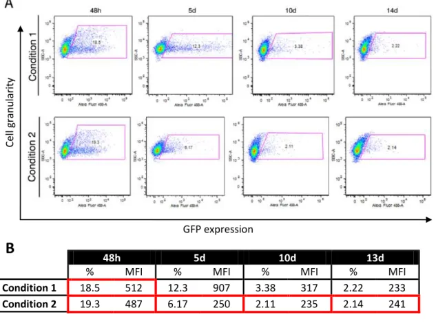
Para a utilização deste sistema no nosso trabalho, foram desenhados 2 novos vectores: pT2K-HoxA3eGFP, para a expressão de HoxA3 e pT2K-HoxB1eGFP para a expressão de HoxB1. Durante a realização desta tese, realizámos a construção do primeiro vector, encontrando-se o segundo em construção.Para testar a eficiência do sistema de vectores utilizamos uma linha celular humana, a linha HEK 293T. Esta foi transfectada com o sistema de vectores (controlo), em condições que permitissem avaliar a capacidade de integração genómica e a resposta deste sistema quando modulado por doxiciclina (doxy). A cultura de células transfectadas foi avaliada por observação ao microscópio de fluorescência e analisada por citometria de fluxo. Os resultados obtidos, ao longo de 14 dias, demonstraram uma rápida e drástica redução tanto na percentagem de células a expressar GFP como na intensidade média de fluorescência de GFP, sugerindo uma reduzida capacidade de integração dos vectores deste sistema no genoma. Para avaliar a resposta do sistema quando modulado pela presença/ausência de doxy, as células foram transfectadas na presença de doxy. Células transfectadas e cultivadas na presença de doxy apresentaram, aproximadamente, 20% de células GFP+ após 48h de cultura.
VI Este resultado demostrou que este sistema não bloqueia eficientemente a expressão de GFP, sugerindo a existência de um “leakage” na modulação pela doxy. No entanto, foi observado, que a presença continuada de doxy levou à redução da intensidade média de fluorescência das células GFP+, após 10-14 dias em cultura, sugerindo que este “leakage” é responsável por níveis muito baixos de expressão génica. Foi também observado, que a remoção de doxy às 48h de cultura levou a um aumento da intensidade média de fluorescência das células GFP+. Estes dados sugerem que este sistema de vectores é modulado negativamente pela doxy, pois na sua presença, o sistema bloqueia a expressão de GFP.
De futuro, com as construções de vectores finalizadas e as condições de utilização do sistema de vectores aferidas, iremos realizar os ensaios funcionais que permitam avaliar a possível mudança de identidade das bolsas por expressão ectópica dos genes HoxA3 e HoxB1 nas 2ª e 3ª BF, respectivamente. Resumidamente, as endodermes isoladas da 2ª e da 3ª BF serão geneticamente modificadas com os plasmídeos pT2K-HoxA3eGFP e pT2K-HoxB1eGFP (neste sistema de vectores), respectivamente. Serão depois cultivadas in vitro com mesênquima permissivo e posteriormente enxertadas na membrana corioalantóidepara testar a formação dum timo (desenvolvimento in ovo). Com este ensaio esperamos mostrar o “ganho-de-potencialidade” da 2ª BF e a “perda-de-“ganho-de-potencialidade” da 3ª BF, na formação do timo. A conjugação deste sistema de vectores com o ensaio funcional oferece uma abordagem experimental única para a identificação dum putativo código-Hox responsável pela identidade posicional do timo.
Palavras-chave: identidade posicional do timo, identidade das bolsas faríngicas, endoderme, código-Hox, HoxA3, HoxB1
VII
Index
Acknowledgments ... II Abstract ... III Resumo ... IV Abbreviations ... 1 I. Introduction... 2I.1. Development of the pharyngeal region in vertebrates ... 2
I.2. The thymus and parathyroid glands development ... 3
I.3. The Hox-code in the pharyngeal region ... 5
I.3.1. The hox-code in thymus organogenesis ... 6
II. Objective ... 9
III. Experimental design for the study of Hox-code in thymus identity ... 10
III.1. The “Tol2-mediated gene transfer” and “tetracycline-dependent conditional expression” system of vectors for the study of Hox genes in thymus development ... 10
III.2.Quail-chick chimeras for the study of Hox-code in the positional identity of the thymus - functional assay to thymus formation ... 11
III.3. Materials and Methods ... 11
III.3.1. Molecular biology procedures ... 11
III.3.2. Celular biology procedures ... 16
III.3.3. Developmental biology procedures ... 17
IV. Results ... 18
IV.1. Patterns of expression of Hox genes in the pharyngeal region of chicken embryos, at stages of development prior to formation of the thymus ... 18
IV.1.1. Production of new sense and antisense riboprobes ... 18
IV.1.2. HoxA2, HoxA3, HoxB1, HoxB2, HoxB3 and HoxB4 expression pattern at E3 and E4 chick embryos ... 18
IV.2. In vitro demonstration and validation of the “Tol2-mediated gene transfer” and “Tetracycline-dependent conditional expression” combined system of vectors ... 23
IV.3. In vivo modulation of the Hox-code in the 2nd and 3rd PP endoderm ... 28
IV.3.1. Production of the pT2K-HoxA3eGFP ... 29
V. Discussion ... 30
V.1. HoxA3 and HoxB1 as possible Hox-code for thymic rudiment positional identity ... 30
VIII V.3. In vitro modulation of HEK 293T by the “Tol2-mediated gene transfer” and
“Tetracycline-dependent conditional expression” combined system of vectors. ... 32
V.4. Technical considerations regarding the in situ hybridization procedures ... 32
VI. Final considerations and future perspectives ... 34
VII. References ... 35
APPENDIX I– BUFFERS, MEDIA AND OTHER SOLUTIONS ... 40
1
Abbreviations
bp – base pairs
d, h, m, sec – day, hour, minute, second doxy – Doxycycline
DMEM – Dulbecco's Modified Eagle Medium DNA – Deoxyribonucleic Acid
E – Embryonic day
EDTA – Ethylenediaminetetraacetic Acid FBS – Fetal Bovine Serum
GFP – Green Fluorescence Protein GFP+– GFP positive
MFI – median fluorescence intensity ng/mL – nanogram per milliliter o/n – overnight
PA – Pharyngeal Arch
PBS – Phosphate Buffered Saline Pen/Strep – Penicillin/Streptomycin PFA – Paraformaldehyde
PP – Pharyngeal Pouch RNA – Ribonucleic Acid rpm – Revolutions per minute PCR –Polymerase Chain Reaction U/μL – units per microliter μg/mL – microgram per milliliter v/v – volume/volume
2
I. Introduction
I.1. Development of the pharyngeal region in vertebrates
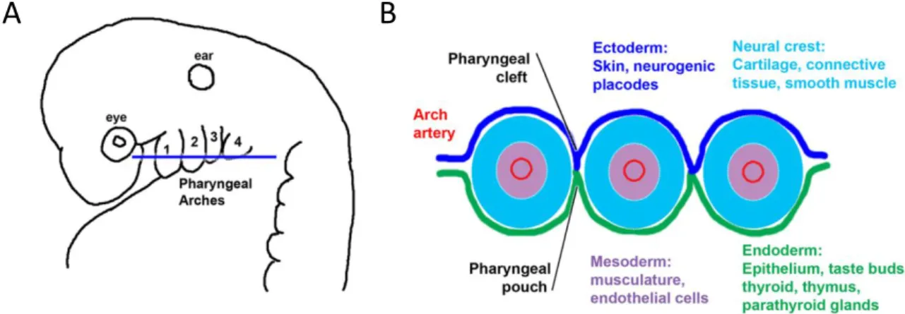
During vertebrate embryogenesis, the pharyngeal apparatus develops from a transient series of segmental structures, appearing as bulges on the cranial lateral side of the embryo, named Pharyngeal Arches (PA). Between adjacent arches, the pharynx endoderm evaginates laterally forming the pharyngeal pouches (PP). Externally, the ectoderm depresses forming the pharyngeal cleft. The PA comprise tissues derived from the 3 germ layers: externally the arch is composed by ectoderm, inner covered by endoderm and its core is thought to be composed of mesoderm surrounded by neural crest cells (NCCs). Each of these different embryonic populations will generate distinct derivatives (Figure 1): the NC will develop into cartilage, connective tissues and smooth muscle; the mesoderm, musculature and endothelial cells; the ectoderm will form the epidermis and neurogenic placodes; the endoderm will give rise to the epithelium, taste buds, thyroid, thymus and pharathyroid glands (Graham & Smith, 2001; Graham, 2001; Graham & Richardson, 2012; Grevellec & Tucker, 2010).
Figure 1| Schematic representation of the pharyngeal apparatus. (A) Lateral view of an amniote
embryo, with pharyngeal arches identified from 1-4 in an anterior to posterior manner. (B) Transverse section through the arch region represented in (A), showing the constituent tissues: ectoderm, dark blue; endoderm, green; neural crest, pale blue ; mesoderm, purple. [adapted from Graham & Richardson, 2012]
Although these structures are found in all vertebrates, it appears to be a trend towards the loss on the number of PA, reflecting the alteration in function of the different pharyngeal structures. For instances, shifting from filter feeding to a predatory behavior, which led to the appearance of the jaw. Also, in aquatic to terrestrial transition, during tetrapods’ evolution, the necessity for regulation of calcium and phosphorous led to the appearance of the parathyroid gland and the shift to lungs as the major respiratory organs. Therefore, caudal arches no longer had to support gills. This trend has been associated to the role of endoderm as the major organizer of the pharyngeal region development, and not the neural crest (Graham & Richardson, 2012; Grevellec & Tucker, 2010). Different developmental studies have shown that ablation of the neural crest does not prevent initial formation PP, suggesting it is
3 independent from the migrating crest (Veitch, Begbie, Schilling, Smith, & Graham, 1999). Also, Couly and colleagues demonstrated that removal of the foregut endoderm in the chick led to failure in development of the pharyngeal skeleton and ectopic grafts of foregut endoderm led to duplication of skeleton elements (Couly, Creuzet, Bennaceur, Vincent, & Le Douarin, 2002). The different PA and PP generate specific tissues and organs. For this project I’m going to focus in the formation of the thymus and the parathyroid glands, organs derived from the 3rd and 4th PP (3/4PP), in birds and mammals.
I.2. The thymus and parathyroid glands development
As a specialized organ of the adaptative immune system, the thymus is the primary site for T-lymphocytes (T-cell) development. The thymus is a capsulated epithelial organ, histological divided into two compartments, cortex and medulla containing distinct thymic epithelial cells (TECs) subtypes. This thymic architecture provides a unique environment for thymocytes (developing T-cells) differentiation. The parathyroids are endocrine glands responsible for the production of the parathyroid hormone (PTH), a peptidic hormone essential to regulate calcium and phosphate homeostasis (Julie Gordon & Manley, 2011; Grevellec & Tucker, 2010). Thymus and parathyroid glands have the same embryological origin: the endoderm of the PP (3/4PP chicken, 3PP in mammals), previously demonstrated (Blackburn & Manley, 2004; Cordier & Haumont, 1980; L. Douarin & Jotereau, 1975; Farley et al., 2013).
The expression domains of two transcription factors, Foxn1 (forkhead box N1) and Gcm2 (glial cells missing-2), were shown to identify the rudiments of the thymus and parathyroid glands in the pouches, respectively (Gordon, Bennett, Blackburn, & Manley, 2001). In mouse and human, thymic and parathyroid rudiments are located ventrally and dorsal-proximally in the 3PP, respectively (Farley et al., 2013; Gordon et al., 2001). Foxn1 is required cell-autonomously for thymic epithelium differentiation and LPC colonization. Loss-of function of Foxn1 in nude mice led to athymia, as LPCs fails to enter the primordium and remains in surrounding mesenchyme and the epithelial cells fail to expand within the primordium (Blackburn et al., 1996; Bleul et al., 2006; Gordon et al., 2001; Nehls et al., 1996). Deletion of Gcm2 marker leads to no formation of the parathyroid glands, with no interference in thymus development (Günther et al., 2000; Liu et al., 2007).
In chicken, transcription factors Foxn1 and Gcm2 were expressed in the prospective domains of the thymus and parathyroid glands, respectively. Foxn1 expression was identified in the thymus rudiment, in the most dorsal region of the 3/4 PP endoderm in chicken embryo at E4.5 (Figure 2A and C). Gcm2 expression, preceding the parathyroid glands rudiment, was identified ventral/anterior region of the 3/4 PP (Figure 2B and C) (Neves, Dupin, Parreira, & L. Douarin, 2012).
4
Figure 2|Expression of Foxn1 and Gcm2 during thymic and parathyroid glands development in chick embryos. In situ hybridization showing Foxn1 (A) and Gcm2 (B) expression in the isolated 3/4 PP
endoderm of E4.5 chicken embryos. Schematic representation of the expression domains of Foxn1 and
Gcm2 in the 3PP endoderm of E4.5 chicken embryos (C). A, anterior; D, dorsal; P, posterior; PP,
pharyngeal pouch; V, ventral. [adapted from Neves et al., 2012]
Thymus organogenesis is a dynamic process that occurs in two main phases: a thymocyte-independent phase followed by a thymocyte-dependent. In the first one, the endoderm and the surrounding mesenchyme (NC-derived) interact to direct TECs specification (Gordon & Manley, 2011; Rodewald, 2008). In the second phase, thymic anlage is dependent of lymphoid progenitor cells (LPCs) colonization which allows the maturation of the thymic epithelium into cortical (cTECs) and medullar (mTECs) compartments (Alves, Huntington, Rodewald, & Di Santo, 2009; Blackburn & Manley, 2004; Gordon & Manley, 2011; Rodewald, 2008).
Using the quail-chick chimera system, Le Douarin and colleagues showed that epithelial-mesenchymal interactions are essential in early – phase of thymic development. Furthermore, they showed that heterologous mesenchymal tissues (derived from the somatopleure or splanchnopleure) have the ability to mimic the role of neural crest-derived mesenchyme. Briefly, quail 3/4 PP endoderm grafted into heterologous mesenchyme from a chick at embryonic day 3 (E3) was able to develop into thymic epithelium. Moreover, the endoderm appeared to be capable of inducing the heterologous mesenchyme to participate in the formation of a fully developed thymus. Therefore, this mesenchyme was considered “permissive” to endoderm development (L. Douarin & Jotereau, 1975; L. Douarin, 1967). H. Neves and colleagues working on the thymocyte-independent stage of thymus organogenesis, unraveled some of the early molecular events occurring during this developmental window (Neves et al., 2012). Taking advantage of quail-chick chimeras, they demonstrated that cellular interactions between the endoderm and surrounding mesenchyme involved a sequential mesenchymal-derived expression of Bmp4 and Fgf10, essential for the development of the 3/4 PP endoderm into thymic and parathyroid glands epithelial. It was also observed a temporal regulation of Bmp4 expression in the mesenchymal compartment, suggesting Bmp4 levels to be tightly regulated in the developing pouches. It was proposed a model (see Figure 3) for the crosstalk between Bmp and Fgf signaling molecules during tissue interaction in thymic and parathyroid development, emphasizing the highly dynamic temporal and spatial dialogue between the PP endoderm and mesenchyme. Nevertheless, the signals from the early 3/4PP endoderm, which can induce the mesenchyme to participate in thymus and parathyroid glands development remain to be identified (Neves et al., 2012).
5
Figure 3 | Schematic model of Bmp and Fgf signaling crosstalk during epithelial-mesenchymal interactions in early thymic and parathyroid development. A highly dynamic temporal and spatial
dialogue occurs between the 3/4 PP endoderm and surrounding mesenchyme, in which signals from the endoderm induce the mesenchyme to participate in thymus and parathyroid glands formation. Arrows indicate putative signalling crosstalk involved in the epithelial-mesenchymal dialogue. [adapted from Neves et al., 2012]
Among vertebrate, the thymus is only present in jawed species and it can be derive from other PP than the 3rd and 4th PP, as it happens in bony fish, birds and mammals. In cartilaginous fish, the most primitive thymus-bearing species (for instances sharks), thymus anlagen are located in the 2nd to the 6th PP; in frogs, they are found in the 2 PP and in reptiles, the 2/3 PP (Rodewald, 2008). There are also differences in the number and in the final anatomical position of these organs among vertebrates. Why such anterior to posterior diversity in the number of PP with potential to form a thymus exists within nature has become of great interest for evolutionary developmental biology to study evolutionary conserved molecular mechanisms occurring in early stages of 3/4 PP development.
I.3. The Hox-code in the pharyngeal region
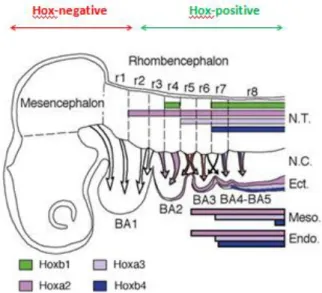
The specification of the body plan was unravelled with the discovery of a clustered family of transcriptional factors, the Homeobox (Hox) genes. They are homologous to the Drosophila homeotic selector genes (Hom-C genes) and highly conserved within phyla. In vertebrates, this complex is usually composed of 39 Hox genes organized into four separated chromosomal cluster, HoxA to HoxD, and the known mammalian genome has 4 copies of this Hox complex (Capecchi, 1997; De Robertis, 2008a,b; Holland & Takahashi, 2005; Holland, 2013; McGinnis & Krumlauf, 1992; Scott, 1992). The order of Hox genes on their respective chromosomes is remarkably similar to their expression pattern, suggesting these genes to be ruled out by a spatially collinear way of gene expression along the antero-posterior axis of the embryo and determined primarily by the most posterior Hox genes (Duboule & Dollé, 1989; Hunt & Krumlauf, 1992; Krumlauf, 1994; Wilkinson, 1989). Expression pattern of these transcription factors in mouse and chicken suggest the existence of a code whereby particular combinations of Hox genes specify a determined region of the anterior-posterior axis (Duboule & Dollé, 1989; Hunt & Krumlauf, 1991; McGinnis & Krumlauf, 1992; D. J. Roberts et al., 1995). For example, the anterior expression boundary of the paralogous group 2, 3 and 4 Hox genes coincide with the 2nd , 3rd and 4th PP regions, respectively (see Figure 4) (Creuzet, Couly, Vincent and L. Douarin, 2002; Hunt & Krumlauf, 1991; Manley & Capecchi, 1995). In figure 4, it is also possible to define two Hox-regions: a negative one throughout the midbrain and ending specifically in the neural tube 1st rhombomere (r) and the 1st PA; and a positive region starting
6 in the neural tube, specifically in rhombomere 2 and 2nd PA (Creuzet et al., 2002; N R Manley & Capecchi, 1995).
Figure 4 | Schematic representation of Hox genes expression in E3 chick and E2.5 quail embryos.
Pharyngeal arches (here described as branchial arches, BA) are being colonized by neural crest cells originated from the posterior half of the mesencephalon and the rhombencephalon, specifically from rhombomeres r1 to r8. Arrows indicate the origin of the neural crest cells migrating to each BA. Expression of Hox genes in the ectoderme, mesoderm and endoderm is also indicated. N.T., neural tube; r, rhomnomere; N.C., neural crest; Ect., ectoderm; Meso., mesoderm; Endo., endoderm. [adapted from Creuzet et al., 2002]
These evidences raised the hypothesis that PP identity, from the 2 PP onward, may be defined by an orderly Hox genes expression, and consequently specifying a Hox-code to thymus positional identity. In an evolutionary point of view, the diversity observed in different jawed vertebrate species, on the number of PP with thymus-potential, could be explained by the conservation of this code in the pouch, as a repetitive morphogenic unit.
I.3.1. The hox-code in thymus organogenesis
Many studies in mammal and in birds have unraveled to role of the Hox-code in NCCs and in the PA region. As described above, the expression pattern of the paralogous Hox group 3 is at the level of the 3PP, thymus and parathyroid glands’ embryonic origin.
In 1991, Chisaka and Capecchi demonstrated for the first time the phenotypic result of loss-of-function of HoxA3 in mice (previously identified as Hox-1.5). Similar to DiGeorge’s syndrome patients, HoxA3 mutant mice were athymic, with no paratyroids, with thyroid hypoplasia and multiple defects in the 3rd and 4th PA. Moreover, these mutants presented fusion of the 2nd and 3rd PA and impaired development of 4th PA (Chisaka & Capecchi, 1991). Later, Manley and Capecchi demonstrated that the thyroid defects included hemiagenesis (absence of one of the lobes) and reduction or absence of C cells in the thyroid lobes. C cells are calcitonin-producing cells that are produce in the ultimobranchial bodies. In HoxA3 mutants, these bodies fail to fuse with the thyroid, explaining the lacking of C cells. The expression pattern of the
7 paralogous Hox3 group in E10.5 mice embryos was also described: HoxA3 was strongly expressed in the 3/4 PP endoderm and throughout the mesenchymal cells of the 3 PA; HoxB3 is primarily expressed in more lateral regions of the 3rd PA mesenchyme; and HoxD3 is expressed in only a small subset of dorsally located cells in the 3rd PA mesenchyme. Double mutants HoxA3-/-HoxB3-/- and HoxA3-/-HoxD3-/- presented the same exacerbated defects in thyroid and ultimobranchial bodies as the HoxA3+/- mutant. Furthermore, HoxB3-/-HoxD3 -/-mice did not present obvious defects in the thymus and parathyroid glands. However, in HoxB3-/-HoxD3-/-HoxA3 +/- mutants both organ primordia failed to migrate correctly (N R Manley & Capecchi, 1995, 1998).
In chicken, a similar expression pattern was described for HoxA3. It was also demonstrated the similarity between chicken and mouse HoxA3 control regions and suggested a nested organization of early elements of segmental patterning (Manzanares et al., 2001). It was also demonstrated that depletion of HoxA3 resulted in defects in the branchial nerve of the 3 PA (Watari-Goshima & Chisaka, 2011).
The complex phenotype presented in mouse and chicken mutants for HoxA3 demonstrate how important this gene is in multiple processes in the pharyngeal region and how it can regulate interactions between different tissues. Even though some of the defects described are mostly related to alterations in the NCC, absent thymus and parathyroid glands in mutant HoxA3 could not be explained by NCC defects, since these organs epithelium is endoderm-derived. Moreover, HoxA3 was recently reviewed as one of the transcription factors controlling early steps of thymus development in mouse, before thymus-specific transcription factor Foxn1 is expressed (Manley & Condie, 2010). Therefore, we may hypothesize a boundary in the 3/4 PP in which HoxA3 is establishing the positional identity of the thymic and parathyroid epithelium. As already mentioned, Hox genes are ruled out by a posterior prevalence, meaning most posterior genes impose their expression, overlapping the anterior Hox gene, resulting in homeotic transformation. Considering our hypothesis that specific hox-code is determining different PP, we would expect duplication of the 2 PP identity in the 3rd, in Hox3 mutants, and duplication of the 3 PP in the 4th, in Hox4 mutants. Unfortunately, the complex phenotype these mutants present would make it impossible to reveal the effect of altering the Hox-code in that specific pouch, as the observable defects resulted from the alteration of multiple interacting-tissues in that region (endoderm, mesoderm, ectoderm, NCC derived mesenchyme) (Creuzet et al., 2002). Nevertheless, paralogous genes expressed in the specific pouch have to be taken in account, as they could have redundant roles.
However, homeotic transformation of other tissues of the arch region in chick embryos has already been described. Le Douarin and collaborators demonstrated that NCC of the 2 PA obey the “Hox-code posterior prevalence rule” as HoxA2 mutant mice exhibit homeotic transformation of the 1 PA (Hox-negative) in the 2 PA, giving rise to skeletal elements from cephalic NCC. Moreover, they demonstrated that overexpressing HoxA2 in the 1 PA led to the transformation of this arch in the 2nd one (Couly et al., 2002; Creuzet et al., 2002).
To sustain this hypothesis of a specific Hox-code established in endoderm is also important to consider the emerging importance of positive auto- and cross-regulatoryinteractions between Hox genes as a general mechanism for maintainingtheir correct spatial patterns, as previously
8 reported in the vertebrate nervous system (Manzanares et al., 2001). Also, trans-regulatory interaction between Hox genes, in order to establish pouch identity, should not be overlooked. Treatment with retinoic acid (RA) lead to an anterior shift of HoxB1 (normally restricted to the endoderm and mesoderm caudal to 4PP) to a level just below 1 PA, disrupting normal development of the most anterior pouches (Mark, Ghyselinck, & Chambon, 2004; Roberts, Ivins, Cook, Baldini, & Scambler, 2006)
How these “positional Hox-code” in endoderm relates with other early patterning transcription factors involved with thymus development is still unknown. Nevertheless, these evidences suggest the Hox-code responsible for thymus positional identity in the 3/4 PP should be HoxA3+HoxB1-.
9
II. Objective
The principal objective of this work was to unravel a possible Hox-code responsible for thymus positional identity.
To study the Hox-code, our first aim was to characterize the normal pattern of Hox genes expression in the pharyngeal region of chicken embryos. Specifically, we evaluated by in situ hybridization, Hox genes expression pattern in the prospective domain of the thymus, the 3/4PP endoderm of chicken embryos at E3 and E4 - stages prior to thymus formation.
Our second aim was to genetically modify Hox-code in distinct PP, using the Tet-off system from the “Tol2-mediated gene transfer” and “Tetracycline-dependent conditional expression” combined system of vectors. Specifically, our goal was to ectopically express HoxA3 (normally expressed in the 3 PP) in the 2 PP endoderm and to express HoxB1 (normally expressed in the 4 PP) in the 3 PP endoderm. With this approach, we intended to perform a “gain-of-potential” (HoxA3 overexpression) and “loss-of-potential” (HoxB1 overexpression) to produce a thymus in the 2nd and 3rd PP, respectively.
To achieve this second aim, we first produced a HoxA3 expressing vector, the pT2k-HoxA3eGFP, to be integrated in the combined Tet-off system of vectors, the“Tol2-mediated gene transfer” and “Tetracycline-dependent conditional expression”. Secondly, we evaluated the efficiency of this system of vectors by in vitro transfecting two different mammalian cell lines, the human embryonic kidney (HEK) 293T and the murine 3T3 lines. GFP expression was monitored by fluorescence microscopy observation and analysed by flow cytometry.
10
III. Experimental design for the study of Hox-code in thymus
identity
III.1. The “Tol2-mediated gene transfer” and “tetracycline-dependent
conditional expression” system of vectors for the study of Hox genes in
thymus development
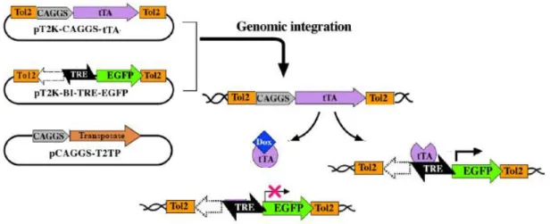
In 2007, Y. Takahashi and collaborators developed a transposon-mediated gene transfer technique, taking advantage of the Tol2 transposable element, and combined it with a tetracycline-induced conditional expression system (Sato et al., 2007; Watanabe et al., 2007) (Figure 5). This combined system, which will be used in this work, allows the stable integration and conditional expression of a transgene in chicken embryos, in a stage-specific fashion.
Figure 5 | The “Tol2-mediated gene transfer” and “tetracycline-dependent conditional expression”
Tet-off combined system. Transient activity of transposase (pCAGGS-T2TP) will induce the transposon
construct containing rtTA2S-M2 (pT2K-CAGGS-rtTA2SM2) and TRE-eGFP (pT2K-BI-TREeGFP) to be integrated into the host genome. Activation of transcription of the TRE-driven gene (“tet-on”) occurs only in the absence of doxycycline (Dox). [Adapted from Sato et al., 2007]
This system is composed of the following plasmids: 1) pT2K-CAGGS-tTA2, 2) pT2K-BI-TREeGFP and 3) pCAGGS-T2TP. Plasmids 1 and 2 were both constructed with a gene expression cassette delimited by Tol2 transposable elements. When these three plasmids are electroporated or transfected into a embryo or cell culture, the third plasmid pCAGGS-T2TP, which transiently expresses the transposase, will induce the transposon construct containing either CAGGS-tTA (plasmid 1) or TRE-eGFP (plasmid 2) to be integrated into the host genome. Then, the tet-controlled transcriptional activator (tTA – plasmid 1), under the control of CAGGS promoter, will act on the cis-element promoter, the tetracycline responsive element (TRE) of the second plasmid, therefore activating transcription of the TRE-driven gene. tTA can only bind to TRE in the absence of doxycycline (doxy, an analog of tetracycline). When doxy is present in this system, it will bind to tTA, blocking its ability to recognize and bind to TRE. The TRE promoter region of the pT2K-BI-TREeGFP is bidirectional with two minimal promoters of cytomegalovirus in both directions, having an eGFP sequence in one direction and a polylinker region on the
11 other one, where it is possible to clone the transgene of interest. Thus, cells expressing the transgene can be identified by GFP expression.
To investigate the possible Hox-code give thymus positional identity for it to develop we aim to modify the Hox-code in the 2nd and 3rd PP endoderm. These endoderm pouches will be genetically modified to express HoxA3 and HoxB1, respectively, using the “Tol2-mediated gene transfer” and “Tetracycline-dependent conditional expression” combined system of vectors, in a functional assay to thymus formation. Therefore, coding sequences for HoxA3
and HoxB1 are intended to be sub-cloned in the pT2K-BI-TREeGFP in order to produce the
pT2K-HoxA3eGFP and pT2K-HoxB1eGFP constructs. For this project, the pT2K-HoxA3eGFP construct was already developed (see section III.3.Materials and Methods for details).
III.2. Quail-chick chimeras for the study of Hox-code in the positional identity
of the thymus - functional assay to thymus formation
In order to genetically modify the Hox-code in the 2nd and 3rd PP endoderm, we designed a functional assay for evaluation of thymus formation. Isolation of the endoderm of the 2 PP and 3/4 PP would be performed in E2.5 and E3 quail embryos, respectively, as previously described (NM Le Douarin & Jotereau, 1975). These isolated endoderms would then be genetically modified by electroporation with pT2K-HoxA3eGFP and pT2K-HoxB1eGFP, respectively, integrated in the combined system of vectors described above. Afterwards, electroporated quail’s 2 PP and 3/4 PP endoderm would be cultured in association with somatopleural mesenchymal tissues isolated from E2.5 chick embryos, generating a chimeric tissue co-culture. After an incubation period, these chimeric co-cultures would be grafted onto a chorioallantoic membrane (CAM) and left for develop.
With this functional assay we expect the formation of a thymus from the genetically altered 2 PP (“gain-of-potential” by overexpression of HoxA3) and the genetically altered 3/4 PP not to produce a thymus (“loss-of-potential” by overexpression of HoxB1).
At the time of this thesis dissertation, the functional assay has not been performed.
III.3. Materials and Methods
III.3.1. Molecular biology procedures Bacteria preparation and transformation
All bacterial transformations in this study were performed using the DH5-α strain of E. coli. Before transformation, and to allow the intake of exogenous DNA, cells have to go through a process to become competent cells. Non-competent E. coli DH5-α from frozen glycerol stock were plated onto LB plates, supplemented with ampicilin (100 μg/mL) and grown o/n. One colony was selected for a starter culture. The next day a higher volume of LB was inoculated with 1/100 dilution of the starter culture and incubated until it reached 0.3 optical density at 600 nm (OD600). The cells were kept on ice and harvested by a series of centrifugations in the
12 presence of CaCl2 to generate chemically competent DH5-α cells. Transformation of DH5-α
cells was performed by heat shock treatment for 1 min. Detailed protocols for both preparation and transformation of DH5-α cells are shown in Appendix II.
Isolation of total RNA
Total RNA extraction from chicken embryos with 3 days of development was performedusing High Pure RNA Isolation Kit (Roche) according to the manufacturer specifications. Embryos were cut in pieces and resuspended in 200 μL PBS. After adding 400 μL ofLysis/Binding Buffer and mixing 15 sec with a vortex, the samples were maintained at -20°C until performing to the RNA extraction protocol. The RNA pellet was eluted in 50 μLof Elution Buffer). RNA samples were stored at -80°C.
Complementary deoxyribonucleic acid (cDNA) synthesis
The synthesis of the first-strand cDNA from total RNA (previous section) was carried out using the SuperScript® III First-Strand Synthesis System for RT-PCR (Invitrogen), according to the manufacturer instructions. 2 μg of total RNA were used in each reaction. cDNAs were stored at -20°C until needed.
PCR amplification
cDNA template synthesized from cE3 RNA (previous section) was amplified by PCR in a 25 μl reaction with 0.5 μM final concentration of primers, using the Phusion™ Master Mix with HF Buffer (Finnzymes), according to instructions from the manufacturer. The cycling conditions for HoxA3, HoxB2 and HoxB4 were: 1 cycle to initiate denaturation at 98°C for 15 sec; 30 cycles of denaturation at 98°C for 10 sec, annealing at optimal temperature (see below) for 30 sec, and extension at 72°C for 15 sec per 1Kb; and 1cycle of final extension at 72°C for 10 min. The optimal conditions of HoxA3 amplification was 65°C for annealing temperature and 15 sec for extension (1251 bp production). Optimal conditions for HoxB2 and HoxB4 were 58°C of annealing temperature and 15 sec for extension (579 bp production for HoxB2; 742 bp production for HoxB4). Samples were stored at -20°C. For the PCR reaction MyCycler™ Thermal Cycler (Bio-Rad) was used.
Primers selection for amplification of chicken HoxA3, HoxB2 and HoxB4 sequences Chicken HoxA3 sequence
HoxA3 mRNA sequence in chicken was identified by Watari-Goshima & Chisaka (2011). Partial nucleotide sequence of HoxA3 in Gallus gallus is shown below (the sequences chosen for primers construction are in bold, the coding sequence is underlined and the stop codon that corresponds to the end of the HOXA3 protein is in red):
HoxA3 Gallus gallus gene ID: NM_204548.1:
1681 agtttgcgaa ataaatattg ggaaacaacg aaatgcaaaa agcgacctac tacgacagct 1741 ctgcaatcta tggtgcctac ccctaccaag gagcaaatgg tttcacttat aatgcgagtc (…)
2881 catacacaga ccttacagct caccatcctt ctcagggaag aattcaggaa gcgcccaaac 2941 tcacccatct gtaggagcca ggagtcacta ggcggaacgc aaagccccaa ccttttaaag
13 To amplify the HoxA3 sequence, and allow direct cloning and its further expression the sequence of the 5’ primer was modified introducing a restriction site and a KOZAK sequence. The final primers were: forward 5’-GCTAGCCATGcaaaaagcgacctactacg-3’ (the inserted sequences are in capital letter; the restriction sequence of NheI is underlined; the KOZAK sequence is in bold) and reverse 5’-ctcctggctcctacagatgggtgag-3’. The expected amplified product is 1251 bp.
Chicken HoxB2 sequence
HoxB2 mRNA sequence in chicken was predicted by automated computational analysis. For human and mouse, this sequence was already identified and referenced so we performed a blast analysis between the predicted chicken HoxB2 sequence and the HoxB2 mRNA sequences for human and mouse. We found that only the 5´ region was homologous for the 3 species (approximately by 70%), so the reverse primer was designed from the most homologous stretch in 3’ termini.
Partial nucleotide sequence of HoxB2 in Gallus gallus is shown below (the sequences chosen for primers construction are in bold and the coding sequence is underlined):
HoxB2 Gallus gallus gene ID: XM_003642792.1:
61 cctccttcct cccccctccc gctttttaaa ccctgggccc tggaaaagcc atgaattttg 121 aatttgagag ggagatcggg tttataaata gccagccttc gctcgcagag tgcctgacgt
(...)
601 acctctgcag gccccgtcgg gtggaaatcg cggctttgct cgacctgacc gagcgacaag 661 tcaaagtgtg gttccagaac cgcaggatga agcacaagag gcaaacacag tacaaagaac
To amplify the HoxB2 sequence, and allow direct cloning and its further expression the sequence of the 5’ primer was modified introducing a restriction site and a KOZAK sequence. The final primers were: forward 5’-GCTAGCCATGaattttgaatttgagagggagatcggg-3’ (the inserted sequences are in capital letter; the restriction sequence of NheI is underlined; the KOZAK sequence is in bold) and reverse 5’-tcatcctgcggttctggaaccacactttgac-3’. The expected amplified product is 579 bp.
Chicken HoxB4 sequence
HoxB4 mRNA sequence in chicken was identified by Attia et al. (2009). Partial nucleotide sequence of HoxB4 in Gallus gallus is shown below (the sequences chosen for primers construction are in bold, the coding sequence is underlined and the stop codon that corresponds to the end of the HOXB4 protein is in red):
HoxB4 Gallus gallus gene ID: NM_205293.1:
241 atatatatat atatattttt cgtgtgtgca attctaagaa attaatggcc atgagctcgt 301 ttttgatcaa ctccaactat gtggacccca agttcccacc ctgtgaagag tattcccaca (…)
961 gcctgcagat cccaccggca gcttctcaaa gccgatccag cggaccagcc agcagcctat 1021 aactattccc tggaggattt cagggcccgt tgtcgtatgg cagtgccgga ggtgggggtg
14 To amplify the HoxB4 sequence, and allow direct cloning and its further expression the sequence of the 5’ primer was modified introducing a restriction site and a KOZAK sequence. The final primers were: forward 5’-GCTAGCCATGgccatgagctcgtttttgatcaactcc -3’ (the inserted sequences are in capital letter; the restriction sequence of NheI is underlined; the KOZAK sequence is in bold) and reverse 5’-aatagttataggctgctggctggtccgctggatc-3’. The expected amplified product is 742 bp.
DNA Restrictions
Enzymatic restriction of DNA was performed using commercially available restriction enzymes and respective buffers (Promega, New England Biolabs), at 37°C, for 2h to 3h, the volume of reaction depended on the quantity of DNA (as a rule final volume should be 10x higher (in μL) than the quantity of DNA (in μg)). For each reaction the volume of enzyme used never surpassed 10% of the total reaction volume.
TOPO II PCR cloning
PCR products were cloned into TOPO II PCR vector using Zero Blunt® TOPO® PCR Cloning Kit (Invitrogen) according to manufacturer instructions. Afterwards, the mix was used to transform commercial competent DH5α bacteria. It was the added 250 μL of S.O.C. medium (TOPO® PCR Cloning Kit) and transformed cells were incubated in a 37°C shaker for 45 min (200 rpm). Bacteria were plated (20 μL and 200 μL) on solid LB agar medium supplemented with ampicillin (100μg/mL) (Sigma) and 30 μL of X-Gal (50mg/mL) (Promega) and were incubated at 37°C o/n. Plasmid DNA was extracted (see section Plasmid DNA Mini Preparation and Midi Preparation). Restriction analysis was performed to confirm the correct construction and correct PCR amplification product.
Cloning of HoxA3 into the pT2K-BI-TREeGFP to generate pT2K-HoxA3eGFP
A second cloning reaction was performed to create the final recombinant plasmids. 4 μg of TOPO-HoxA3 and 5 μg of pT2K-BI-TREeGFP were digested with EcoRV and NheI restriction enzymes, in a total volume reaction of 80 μL and 70 μL, respectively. Afterwards, reaction products were loaded on an agarose gel (see section Agarose gel electrophoresis) and DNA fragments of interest were recovered and purified (see section QIAquick gel extraction kit). The ligation reaction of the linearized vector pT2K-BI-TREeGFP and insert HoxA3 (with the correct termini) was performed according to the ratio: μg of vector/vector’s dimension = 10x (μg of insert/insert’s dimension). For the ligation reactions it was used 150 ng for both pT2K-BI-TREeGFP (8.7kb) and HoxA3 insert (1,25kb) in a total volume of 20 μL. The reaction was performed in 1x Buffer for T4 DNA Ligase (Biolabs), with 1μL T4 DNA ligase (Biolabs 400U/μL) at room temperature (22°C) o/n. The ligation product was directly used to transform DH5-α competent cells. Plasmid DNA was extracted and isolated (see section Plasmid DNA mini- and midi-preparation) from transformant colonies. The insertion of donor DNA into the vector (pT2K-BI-TREeGFP) was confirmed by double digestion with EcoRV and NheI.
15 Plasmid DNA mini- and midi-preparation
For mini-preparation of plasmid DNA, single colonies of transformed bacteria were collected and inoculated into 5 mL of liquid LB medium supplemented with ampicillin (100μg/mL) and incubated in a 37°C shaker (225 rpm) o/n. The purification of plasmid DNA was carried out using the QIAprep® Spin Miniprep Kit (QIAGEN) according to the protocol recommended by the manufacturer.
For midi-preparation of plasmid DNA, single colonies of transformed bacteria were growno/n in 50 or 100 mL (high or low copy, respectively) of liquid LB medium supplemented with ampicillin (100 μg/mL) at 37°C (225 rpm) o/n. The purification of plasmid DNA wascarried out using the QIAfilter Plasmid Midi kit (QIAGEN) according to the protocolrecommended by the manufacturer. DNA samples were stored at -20°C.
QIAquick Gel extraction kit
DNA extraction from agarose gel was carried out using the QIAquick Gel Extraction Kit (QIAGEN), according to manufacturer instructions. DNA was eluted with 30μL of DNase and RNase free water.
DNA and RNA quantification
The concentration of nucleic acids was determined by spectrophotometry using the NanoDrop® ND-1000 Spectrophotometer (Thermo Scientific). One A260 unit corresponds to 50
μg/ml of double-stranded DNA and to 40 μg/ml of single-stranded RNA. DNA and RNA samples purity was evaluated based on A260/A280 and A260/A230 ratios. For the A260/A280 ratio, ~1.8 is
generally accepted as pure DNA and ~2.0 as pure RNA. The A260/A230 is a secondary measure
for nucleic acid purity. In this ratio, pure DNA and RNA values must higher than the ones in A260/A280, commonly in the range of 1.8-2.2, respectively. Lower ratio values are due to
proteins, phenol or co-purified contaminants. Preparation of riboprobes for in situ hybridization
To generate the antisense and sense transcripts, TOPO II PCR, pGEM-T and pGEM-T easy vectors containing the sequences of interest were linearized with the appropriate restriction enzyme (see Table 1 in Appendix II). The digestion reaction was performed in a total volume of 150 μL containing 20 μg of DNA, 15 μL of 10x enzyme buffer, 5 μL of restriction enzyme (6 U-20 U/μL) and RNase-free water. After digestion, linearized plasmid DNA was purified using phenol-chloroform extraction and ethanol precipitation (see Appendix II). The synthesis of digoxigenin (DIG) -labelled antisense and sense RNA probes was carried out by in vitro transcription at 37°C for 2h using Riboprobe® Combination System SP6/T7 or T3/T7 (Promega). The reaction contained 8 μL of Transcription Optimized 5x Buffer, 4 μL of 0.1M DTT, 2 μL of each rGTP, rATP, rCTP (10 mM), 1.3 μL of rUTP (10 mM), 2 μL of RNasin® Ribonuclease Inhibitor, 0.7 μL of Digoxigenin-11-UTP (10 mM) (Roche) and 8 μL of RNAse free water. To this mixture solution and 2 μL of the appropriate RNA polymerase (see Table 1 in Appendix II), 2 μL (2μg) of the linearized templates were added. After incubation for riboprobe synthesis, the sample was treated with 6 μL of DNase I recombinant RNase-free (10 U/μL) (Roche) at 37°C for 15 min. Purification of the probe was performed using illustra MicroSpin G-50 Columns (GE
16 Healthcare) according to manufacturer instructions. To check for probe quality and success of transcription reaction, 2 μL of reaction product were analyzed by agarose gel electrophoresis (see below).The samples were stored at -20°C.
Agarose gel electrophoresis
Electrophoresis in agarose gel was performed to confirm PCR amplification products, complete digestions with restriction enzymes, to recover and purify specific DNA fragments using extraction kit (see section QIAquick Gel extraction kit) and to check riboprobes and DNA samples integrity. UltraPureTM Agarose (Invitrogen) was dissolved by heating in 1x TAE buffer (composition provided in Appendix I) to a final concentration of 0.8-1.5% (depending on the required resolution for DNA fragment). To check for the presence of nuclear acids, GelRedTM Nucleic Acid Gel Stain (Biotium) was added to dissolved agarose in a 1:10 proportion. Samples were mixed with 6x Loading Dye or 6x MassRuler™ Loading Dye (both from Fermentas) in 6:1 proportion and were loaded into the gel. Electrophoresis was performed in 1x TAE at 5-10 V/cm of gel length. Samples were observed under UV light and images acquired with AlphaImager HP (Alpha Innotech) or ChemiDoc XRS+ system (Bio-rad). Fragments size was estimated by comparison with the DNA ladders (FastRuler™ Low Range DNA Ladder, FastRuler™ Middle Range DNA Ladder or O'GeneRuler™ 1 kb DNA Ladder, Fermentas) ran along with DNA samples.
III.3.2. Celular biology procedures Maintenance of HEK 293T and 3T3 cell lines
The Human Embryonic Kidney (HEK) 293T cell line (provided by João Barata’s group, Unidade de Biologia do Cancro, Instituto de Medicina Molecular, Lisbon, Portugal; (Zenatti et al., 2011)) and the murine 3T3 cell line (Santos et al., 2007) were maintained in culture medium containing Dulbecco’s modified Eagle medium (DMEM) (Gibco) supplemented with 10% FBS (Gibco), 1x Pen/Strep (Gibco), 1x Glutamine (Gibco) and 1x Non Essential Amino Acids (NEAA) (Gibco). The cells were cultured on T25, T75 or T175 flasks (Nunc) using a media volume to surface area ratio of 0.1-0.2 mL/cm2. All cultures were grown in a humidified incubator (Heraeus® HERAcell®) at 37°C with 5% of CO2. 293T cells were provided cultured in T25 or T75
flasks. 3T3 cell line aliquotes were taken from the liquid nitrogen and put in a 37°C bath, without immerging, long enough to start to thaw. Immediately after, the cells were resuspended in maintenance medium and plated onto a T75 flask. The medium was changed regularly until cells reached 40-50% confluence. For this, the media was removed and the cells were washed with PBS. In order to detach the cells, pre-warmed 1-2 mL of 0.25% Trypsin/EDTA (Gibco) was added and the cells were incubated at 37°C for about 5 min. When cells detached, trypsin was inactivated by addition of medium. Dilutions were made with the appropriate volume and plated.
Transfection of HEK 293T and 3T3 cell lines
Both HEK 293T and 3T3 cell lines were transfected with a “Tol2-mediated gene transfer” and “Tetracycline-dependent conditional expression” system of vectors, composed by:
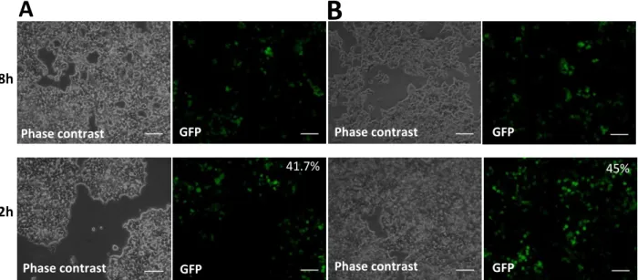
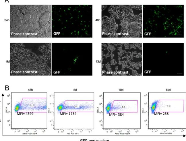
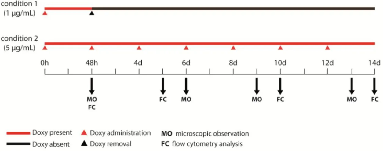
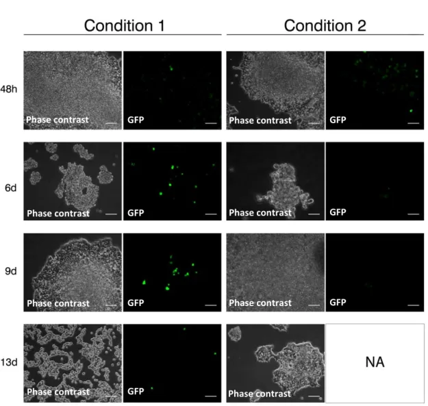
pCAGGS-17 T2TP, pT2k-CAGGS-EGFP, pT2k-BI-TRE-EGFP and pT2k-CAGGS-tTA-M2 (Sato et al., 2007), in 2 different concentrations and in the presence and absence of 2 different concentrations of Doxycycline, in order to study and validate it for our work. A standard calcium phosphate-mediated transfection protocol for mammalian cell lines was used (detailed protocol in Appendix II). For both cell lines, analysis of transfected cells expressing GFP and fluorescence intensity of GFP was performed a different time-points, preliminary by eye observation using a Leica DMIL inverted microscope and quantified by flow cytometry analysis using a BD LSRFortessa cell analyzer. For each condition, ≥1x10⁴ cells were analysed with a minimum of 10000 events acquired. Data was analysed with FlowJo (Tree Star, Inc. Ashland, OR). A negative control was employed, where cells went through this transfection protocol but without the addition of plasmid DNA.
III.3.3. Developmental biology procedures Chicken embryo manipulation
Fertilized chicken (Gallus gallus) eggs, obtained from Sociedade Agrícola Quinta da Freiria, S.A., Portugal, were stored at 16°C and incubated at 38°C to initiate development. Embryos age was estimated by incubation time and staged according to Hamburger and Hamilton (Hamburger & Hamilton, 1951). At specific stages of development, embryos were removed and dissected from the egg and the extra-embryonic membranes. Isolated embryos were further processed to whole-mount in situ hybridization
Whole-mount in situ hybridization
Whole-mount In situ hybridization of E3 and E4 chick embryos were performed as previously described (Henrique, 1995 and Etchevers, 2001) (detailed protocol in Appendix II). Whole-mount preparations were hybridized with the follow riboprobes: antisense and sense for HoxA3, HoxB2 and HoxB4; antisense for HoxA2, HoxB1 and HoxB3. Pictures were taken under a Leica Z6 APO equipped with a Leica DFC490 camera.
Whole-mount post-hybridized E3 and E4 chick embryos were fixated and included in paraffin for sectioning throughout embryo’s coronal axis. Afterwards, these sections went through Hematoxyline-Eosine (HE) staining. HE staining in paraffin sections was done using Harris Hematoxylin (Merck Millipore) and Eosin Y alcoholic (Thermo scientific) according to manufacturer’s instructions. Final preparations were observed and photographed using an Upright Brightfield microscope Leica DM2500, equipped with a color camera for brightfield and differential interference contrast imaging.
18
IV. Results
IV.1. Patterns of expression of Hox genes in the pharyngeal region of chicken
embryos, at stages of development prior to formation of the thymus
To identify the Hox-genes that may be involved in the Hox-code responsible for thymus (and parathyroid glands) positional identity, we first study the expression of hox genes by whole-mount in situ hybridization in chicken embryos at E3 and E4.
IV.1.1. Production of new sense and antisense riboprobes
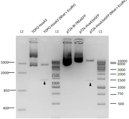
New HoxB2 and HoxB4 riboprobes were produced (sense and antisense). A 579 bp and a 742 bp PCR amplification products of HoxB2 and HoxB4 were cloned into TOPO II PCR (Figure 6A), respectively (detailed in III.3 Materials and Methods, section Molecular biology procedures). New HoxA3 riboprobe was produced by cloning a 1251 bp product derived from PCR amplification of HoxA3 into TOPO II PCR (Figure 6B). This plasmid was further used for the production of pT2K-HoxA3eGFP vector (for functional assay to test “gain-of-potential” of the 2 PP to produce a thymus, detailed in section V.2.1).
IV.1.2. HoxA2, HoxA3, HoxB1, HoxB2, HoxB3 and HoxB4 expression pattern at E3 and E4 chick embryos
HoxA2
In figure 7A, we observed the expression of HoxA2 in chicken embryos with 3 days of development. HoxA2 is expressed in the neural tube, at the level of the rhombomeres (r) 2 onwards. As shown in fig.7A’ (amplification of the pharyngeal region observed in 7A), HoxA2 is strongly expressed in the ectoderm and/or mesenchyme of the 2 PA. A more faint expression is observed from the 3 PA onwards. Notice the absence of HoxA2 expression in the otic vesicle (ov), dorsally positioned to 2 PA. Similar patterns of expression are observed for HoxA2 at E4 (Fig. 7I and 7I’). These results are in agreement with the expression pattern previously reported (Prince & Lumsden, 1994).
In Fig. 7B and 7J, is depicted the coronal sections of E3 and E4 whole-mount hybridization. In the 2 PA, is confirmed the HoxA2 expression in mesenchymal cells. Interestingly, the endoderm of the 2 PP showed no HoxA2 expression in anterior half of the pouch while the posterior half is positive (see green arrows in Fig. 7B). At the level of 3/4 PA, endoderm of the 3/4 PP and mesenchymal cells are faintly positive for HoxA2.
19 Figure 6 | Agarose gels showing the several steps involved on the generation of TOPO constructs for development of riboprobes. 1.5% (wt/vol) agarose gels showing the steps involved in the construction
of TOPO-HoxB2, TOPO-HoxB4 (A) and TOPO-HoxA3 (B). (A) PCR amplification of HoxB2 generated a 579 bp product and PCR amplification of HoxB4 generated a 742 bp product; Cloning of these products in the TOPO vector was confirmed by DNA restriction with NheI and EcoRV of both TOPO-HoxB2 and TOPO-HoxB4, showing a 4000 bp band corresponding to the vector and the band corresponding to the insert. (B) PCR amplification of HoxA3 generated a 1251 bp product. Cloning of this product in the TOPO vector was confirmed by DNA restriction with NheI and EcoRV, showing a 4000 bp band corresponding to the vector and the band corresponding to the 1251 bp insert. Fragment sizes were determined by comparison with O'GeneRuler™ 1 kb DNA Ladder (L1) and FastRuler™ Middle Range DNA Ladder (L2). DNA molecular weight markers are indicated (bp). Black arrows indicate the insert correspondent bands.
B
A
20 HoxA3
Expression pattern of HoxA3 at E3 is shown in Fig. 7C and 7E (new probe, (t)HoxA3). HoxA3 is expressed in the neural tube, at r3 level onwards. HoxA3 is strongly expressed in the ectoderm and/or mesenchyme of the 3/4 PA and in the region posterior to 4 PA, as observed in fig. 7C´. Posterior to this region, HoxA3 is faintly expressed. At E4, HoxA3 expression pattern is more anteriorly positioned in the pharyngeal region and seems to be highly expressed in the 2/3 PP boundary. HoxA3 is also expressed in the 3/4 PA (Fig. 7K and 7K’). These results are in agreement with the expression pattern previously reported (Watari-Goshima & Chisaka, 2011). The coronal sections of E3 and E4 whole-mount hybridization for HoxA3 are depicted in Fig. 7D and 7L, and 7F and 7N for the new probe. At E3, HoxA3 expression is confirmed in the mesenchyme cells of the 3PA and, interestingly, in both halves the endoderm of the 3 PP (see orange arrows in fig. 7D and 7F). With the new probe, we also observed and unexpected faint expression of HoxA3 in the posterior half of the 2 PP (fig. 7F, green arrow). In the most posterior region of the arches, HoxA3 is also weakly expressed in the mesenchyme of the 4 PA and endoderm of the 4 PP (fig. 7D,L,F,N). In figure 7F (new probe), gene expression is restricted to the most external region of the embryo indicating a permeabilization problem (insufficient proteinase K treatment). At E4, HoxA3 expression is maintained in the mesenchymal cells of the 3 PA but appears to be absent or faintly expressed in the 3 PP endoderm. The same pattern is observable in the 4 PA and PP.
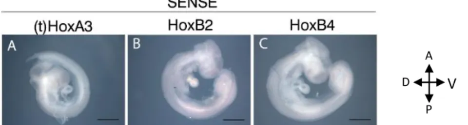
In figure 9 A is depicted the in situ hybridization of the sense riboprobe developed from the new HoxA3 probe ((t)HoxA3).
HoxB1
HoxB1 expression pattern in chick embryos at E3 and E4 is described for the first time in this dissertation (Figures 7G and 7O). As observed in fig. 7G, HoxB1 is strongly expressed at r4, is absent in r5, and faintly expressed from r6 onwards. This pattern in the neural tube was previously reported in earlier stages of development (Bothe et al., 2011). In the pharyngeal region, HoxB1 is only expressed at the level of the 4 PA and onward (Fig. 7G’ and O’). In figures 7H and 7P, coronal sections of E3 and E4 whole-mount hybridization are depicted. HoxB1, in both stages, is strongly expressed and limited to the most posterior half of the endoderm of the 4 PP (see blue arrows).
HoxB2
Expression pattern of HoxB2 in chick embryos at E3 and E4 is also described for the first time in this project (Figures 8A and 8G). At the neural tube, HoxB2 is expressed in r2 onwards which is in agreement with previously reported in earlier stages of development (Barak et al., 2012). In the pharyngeal region, both stages have HoxB2 expressed in the posterior region to the 4 PA (Fig. 8A’ and G’). Coronal sections depicted in figure 8B and 8H confirm that at E3 and E4, HoxB2 is expressed in the mesenchyme posterior to the 4 PP. HoxB2 also appears to be weakly expressed in the endoderm of the 4 PP at E4 (fig. 8H, blue arrow).
In figure 9 B is depicted the in situ hybridization of the sense riboprobe developed from the new HoxB2 probe.
21
Figure 7 | Expression pattern of HoxA2, HoxA3 and HoxB1 at E3 and E4 chick embryos. Whole-mount in
situ hybridization and corresponding post-hybridized coronal section showing the expression pattern of HoxA2, HoxA3 and HoxB1. New probe for HoxA3 is identified as (t)HoxA3. Coronal post-hybridization
sections have 5-7 μm and went through HE staining. Different colored arrows indicate a specific PP: green for 2 PP, orange for 3 PP and blue for 4 PP; asterisks indicate the ventral aorta. Scale bars 500 μm (rows A-G’ and I-O’) and 100 μm (rows B-H and J-P). (A, anterior; D, dorsal; ov, otic vesicle; P, posterior; PP, pharyngeal pouch; PA, pharyngeal arch; r, rhombomere; V, ventral)
r2 ov