UNIVERSIDADE NOVA DE LISBOA
INSTITUTO DE HIGIENE E MEDICINA TROPICAL
Master in Ciências Biomédicas
Biological activity of well defined hydantoin derivatives on
efflux pump systems of bacteria and cancer cells
Miguel José Simas do Rosário Evaristo
Biological activity of well defined hydantoin derivatives on
efflux pump systems of bacteria and cancer cells
Miguel José Simas do Rosário Evaristo
Presentation of the thesis for the master degree in Biomedical Sciences, speciality in Molecular
Biology
Supervisor
Gabriella Spengler
Publications related to the master thesis
- Gabriella Spengler, Miguel Evaristo, Jadwiga Handzlik, Joseph Molnár, Miguel Viveiros,
Katarzyna Kieć-Kononowicz, Leonard Amaral: Biological activity of hydantoin derivatives on efflux pump systems of bacteria and cancer cells. 462nd WE Heraeus Seminar Transport across membranes:Multiple drug resistance, mechanisms and new tools. Jacobs University, 4-10 July 2010, Bremen Germany (poster presentation)
- Gabriella Spengler, Miguel Evaristo, Jadwiga Handzlik, Julianna Serly, Joseph Molnár, Miguel Viveiros, Katarzyna Kieć-Kononowicz, Leonard Amaral: Biological activity of hydantoin derivatives on P-glycoprotein (ABCB1) of mouse lymphoma cells. (2010) Anticancer Research 30: xxx-xxx (accepted manuscript)
Acknowledgements
I would like to acknowledge Dr. Gabriella Spengler PhD for the opportunity to be supervised by such an expert in the area. She was a supervisor and sometimes an English teacher as well. Dr. Gabriella Spengler’s advices helped me a lot to construct my scientific work and thesis. I would like to thank her for all the revisions, discussions during my master work. And finally I am grateful for her ever-availability during all this time.
I would like to acknowledge Professor Dr. Leonard Amaral to have given me the opportunity to be part of his working group in the Unidade de Micobactérias of the Instituto de Higiene e Medicina Tropical. I would like to thank Professor Dr. Leonard Amaral for all the suggestions and critical remarks.
I would like to acknowledge the absolute availability and support of the director of the Unidade de Micobactérias, Prof. Dr. Miguel Viveiros PhD. His interest from the first meeting until the delivery of the thesis was very important. His enthusiasm influenced me to choose his unit to do my master thesis there. He made me understand how exciting is to make science.
I would like to acknowledge Professor Dr. Isabel Couto PhD for her support throughout the thesis. She was present many times until very late, helping me to solve my problems that occurred in my work. She is also an absolute influence for me.
I would like to acknowledge the friendship shown by all the students of the Unidade de Micobactérias, in particular Sofia Costa for her several advices and Diana Machado for her help and company on the long weekdays and weekends at the institute.
I would like to acknowledge Dr. Jadwiga Handzlik and Professor Dr. Katarzyna Kiec-Kononowicz for providing the thirty hydantoin compounds. I greatly appreciate their full support.
I would like to acknowledge Fernanda Dias who works tirelessly to provide the proper tools necessary for the laboratory work of the students.
Table of contents Acknowledgements………..i General index………..iii Index of figures...viii Index of tables...xi Resumo...xvii I - INTRODUCTION………..…1
1. Historical background of the control of bacterial infections………1
2. Antibiotics and their importance to human medicine………...3
2.1 Importance of the discovery of antibiotics………...3
2.2 Mode of action of antibiotics………..4
2.3 Antibiotic resistance: emerging problems………..5
2.3.1 Types of antibiotic resistance………...6
3. Absorption, distribution, metabolization and excretion (ADME) system and mechanisms of transport………9
4. Permeability barriers as bacterial defense systems………..9
4.1 Difference between gram-positive and gram-negative bacteria………..10
5. Families of transporters………..14
5.1 Primary multidrug transporters……….15
5.1.1 ABC transporters………15
5.2 Secondary multidrug transporters………16
5.2.1 Major Facilitator Superfamily (MFS)…..……….16
5.2.2 Small Drug Resistance Superfamily (SMR)……….17
5.2.3 Resistance-Nodulation-Cell Division Superfamily (RND)………..18
5.2.4 Multidrug and Toxic Compound Extrusion Superfamily (MATE)…………..19
6. Multidrug resistance in cancer………21
6.1 Cancer burden………...21
6.2 Causes of cancer………...22
7. Relationship between bacterial infection and cancer……….22
8. Carcinogenesis – the development of cancer……….23
9. Cancer therapy and the problems of resistance mechanisms to anticancer agents……….24
9.1 Main groups of anticancer agents……….24
9.2 Resistance to anticancer agents………25
10. The importance of ABC transporters in cancer………26
10.1 Physiological localization of ABCB1………27
10.2.1 Structure of ABCB1………30
11. Efflux pump inhibitors……….30
11.1 Efflux pump inhibitors in bacteria………..30
11.2 Efflux pump inhibitors in eukaryotes……….32
12. Hydantoin derivatives and their pharmacological use………..35
12.1 Physiological role of hydantoin derivatives……….………..35
12.2 Structure……….36
II - OBJECTIVES OF THE PRESENT PROJECT………...37
III – MATERIAL AND METHODS……….39
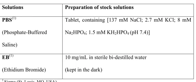
1 – Materials………...39
1.1 Compounds tested………39
1.2 Solutions………...39
1.3 Bacterial strains………40
1.4 Cell lines………...40
1.5 Broths and culture media….……….41
1.6 Cell culture media……….41
2 – Methods………43
2.1 Determination of minimum inhibitory concentrations (MIC) of bacteria...43
2.3 Real-time semi-automated fluorometric method: accumulation assay to monitor efflux
pump activity in bacteria………44
2.4 Real-time semi-automated fluorometric method: accumulation assay to monitor ABCB1 (P-glycoprotein) pump activity in mouse lymphoma cells……….46
IV – RESULTS………...48
1 - Multidrug resistance in bacteria...48
2 - Accumulation of EB by bacterial efflux systems...49
2.1 Effect of hydantoins on Gram-negative bacteria………..52
2.1.1 Accumulation of EB by Salmonella Enteritidis NCTC 13349...52
2.1.2 Accumulation of EB by Escherichia coli AG 100……….55
2.2 Effect of hydantoins on Gram-positive bacteria………...58
2.2.1 Accumulation of EB by Staphylococcus aureus ATCC 25923...58
2.2.2 Accumulation of EB by Enterococcus faecalis ATCC 29212...60
3 - Efflux pump modulating activity of hydantoin derivatives on cancer cells………..62
3.1 Antiproliferative and cytotoxic effects of hydantoin derivatives on L5178 (parental, PAR) mouse T-cell lymphoma cells and its human ABCB1 (MDR1)-transfected subline (MDR)...63
3.2 Accumulation assay of EB in efflux systems of eukaryotic cells...66
3.2.1 Effective compounds on PAR cells………...68
3.2.2 Effective compounds on MDR cells……….71
V – DISCUSSION AND CONCLUSIONS………..76
1.1 Evaluation of hydantoin derivatives on Gram-negative and Gram-positive bacteria...76
1.2 Future perspectives...77 2.1 Evaluation of hydantoin derivatives on the activity of P-glycoprotein in mouse T-lymphoma cells...78 2.2Future perspectives...81
Index of figures
Figure 1. Schematic representation of the Gram-negative cell envelope………..11
Figure 2. Schematic representation of Gram-positive cell wall……….12
Figure 3. Schematic representation of uniport, symport and antiport systems………..14
Figure 4. Schematic representation of the RND superfamily: the structure of AcrAB-TolC………19
Figure 5. Schematic representation of the five superfamilies of multidrug transporters…………...20
Figure 6. 2008 statistics of the cancer incidence and mortality rates in EU for both sexes………...21
Figure 7. Cellular division in normal and cancer cells...24
Figure 8. Schematic representation of the metastization process………...24
Figure 9: Physiological distribution of ABCB1………27
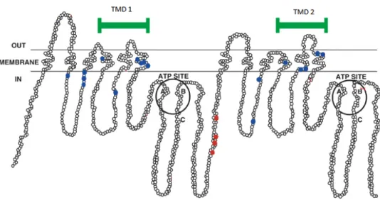
Figure 10. Schematic representation of the organization of ABC proteins……….………...29
Figure 11. Structure of the Hydantoin ring and his double activity………...36
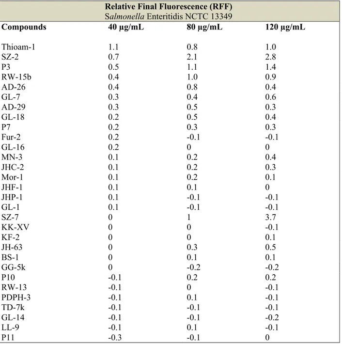
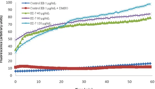
Figure 12. Accumulation of EB (1µg/mL) in the presence of SZ-7 (40, 80 and 120 µg/mL) on Salmonella Enteritidis NCTC 13349………..53
Figure 13. Accumulation of EB (1µg/mL) in the presence of P3 (40, 80 and 120 µg/mL) on Salmonella Enteritidis NCTC 13349………..54
Figure 14. Accumulation of EB (1µg/mL) in the presence of SZ-2 (40, 80 and 120 µg/mL) on Salmonella Enteritidis NCTC 13349………..54
Figure 15. Accumulation of EB (1µg/mL) by E. coli AG 100 in the presence of AD-29 (40, 80 and 120 µg/mL)……….56
Figure 16. Accumulation of EB (1 µg/mL) by E. coli AG 100 in the presence of RW-15b (40, 80 and 120 µg/mL)………..57
Figure 17. Accumulation of EB (1µg/mL) in the presence of AD-26 (40, 80 and 120 µg/mL) on Escherichia coli AG100……….57
Figure 18. Accumulation of EB (0.25 µg/mL) in the presence of SZ-7 (40, 80 and 120 µg/mL) on Staphylococcus aureus ATCC 25923...59
Figure 19. Accumulation of EB (0.5 µg/mL) in the presence of GL-14 (40, 80 and 120 µg/mL) on Enterococcus faecalis ATCC 29212………..61
Figure 20. Precipitation shown by PAR cells in McCoy’s 5A medium in the presence of AD-26 (200 µg/mL) ………...………….65
Figure 21. Accumulation of EB (1 µg/mL) in the presence of Thioam-1 (4 and 40 µg/mL) on PAR cells……….68
Figure 22. Accumulation of EB (1 µg/mL) in the presence of Fur-2 (4 and 40 µg/mL) on PAR cells……….69
Figure 23. Accumulation of EB (1 µg/mL) in the presence of RW-15b (4 and 40 µg/mL) on MDR cells……….71
Figure 24. Accumulation of EB (1 µg/mL) in the presence of RW-13 (4 and 40 µg/mL) on MDR cells……….71
Figure 25. Accumulation of EB (1µg/mL) in the presence of AD-29 (4 and 40 µg/mL) on PAR (A) and MDR (B) cells………..73
Figure 26. Accumulation of EB (1 µg/mL) in the presence of MN-3 (4 and 40 µg/mL) on PAR (A) and MDR (B) cells………..74
Figure 27. Accumulation of EB (1 µg/mL) in the presence of JH-63 (4 and 40 µg/mL) on PAR (A) and MDR (B) cells………..75
Index of tables
Table 1. Mode of action of antibiotics………5
Table 2. Examples of bacterial EPIs, their targets and antibacterials with enhanced activity……..31
Table 3. ABCB1 substrates and inhibitors……….32
Table 4. Used solutions………..39
Table 5. Media………41
Table 6. Relative final fluorescence (RFF) based on the accumulation of EB (1 µg/mL) by Salmonella Enteritidis NCTC 13349 in the presence of hydantoin derivatives...52
Table 7. Relative final fluorescence (RFF) based on the accumulation of EB (1 µg/mL) by E. coli AG100 in the presence of hydantoin derivatives...55
Table 8. Relative final fluorescence (RFF) based on the accumulation of EB (0.25 µg/mL) by Staphylococcus aureus ATCC 25923 in the presence of hydantoin derivatives...58
Table 9. Relative final fluorescence (RFF) based on the accumulation of EB (0.5 µg/mL) by Enteroccocus faecalis ATCC 29212 in the presence of hydantoin derivatives...60
Table 10. Antiproliferative and cytotoxic effects of hydantoin derivatives on L5178 (parental, PAR) mouse T-cell lymphoma cells and its human ABCB1 (MDR1)-transfected subline (MDR)...64
Table 11. Relative final fluorescence (RFF) based on the accumulation of EB (1 µg/mL) by PAR cells in the presence of hydantoin derivatives...67 Table 12. Relative final fluorescence (RFF) based on the accumulation of EB (1 µg/mL) by MDR cells in the presence of hydantoin derivatives...70 Table 13. Most effective hydantoin derivatives on the selected bacterial strains...77
Table 14. The most active hydantoin derivatives on parental (PAR) and multidrug resistant (MDR) human MDR1 (ABCB1) gene-transfected mouse lymphoma cells………...79
Abbreviation list
ABC-transporter - ABC-binding cassette transporter superfamily
A. baumannii - Acinetobacter baumannii
ADME – Absorption, distribution, metabolization and excretion kinetics of the organism
B. subtilis - Bacillus subtilis
CCCP - Carbonyl cyanide m-chlorophenylhydrazone
CDC – Center for disease prevention and control
CFTR - Cystic fibrosis transmembrane regulator protein
CGS - Cellular glutathione system
DMSO –Dimethyl sulfoxide
DNA – Desoxyribonucleic acid
EB – Ethidium bromide
E. coli – Escherichia coli
ECDC – European Centre for Disease Prevention and Control
E. faecalis – Enterococcus faecalis
E. aerogenes – Enterobacter aerogenes
EPI – Efflux pump inhibitor
FACS - Fluorescence Activated Cell Sorting
H. pylori – Helicobacter pylori
ICU – Intensive care unit
IM - Inner membrane
K. pneumoniae – Klebsiella pneumoniae
L. lactis – Lactococcus lactis
LPS - Lipopolysaccharide
MALT - mucosa-associated lymphoid tissue lymphoma
MATE - Multidrug and toxic compound extrusion superfamily
MDR – Multidrug resistance
MFP – Membrane fusion protein
MFS – Major facilitator superfamily
MIC – Minimum inhibitory concentration
MRSA - Methicillin-resistant Staphylococcus aureus
MTT - Thiazolyl blue tetrazolium bromide
M. tuberculosis – Mycobacterium tuberculosis
NBD – Nucleotide binding domain
OM – Outer membrane
OMF – Outer membrane factor
PAR – Parental cells
P. aeruginosa – Pseudomonas aeruginosa
PDR – Pandrug-resistance
P-gp – P-glycoprotein
PMH – phenyl-methylene hydantoin
PPI - Proton pump inhibitor
PMF – Proton motive force
QSAR - Quantitative structure-activity relationship
RF – Relative fluorescence
RFF – Relative final fluorescence
RNA – Ribonucleic acid
RND – Resistance-nodulation division superfamily
S. aureus – Staphyloccocus aureus
SDS - Sodium dodecyl sulphate
S. Enteritidis – Salmonella Enteritidis
SMR – Small multidrug resistance superfamily
S. pneumoniae – Streptococcus pneumoniae
S. typhi – Salmonella typhi
TM - Transmembrane
TMD – Transmembrane domain
TMH - Transmembrane helix
TMS – Transmembrane segments
VEGF - Vascular endothelial growth factor
V. cholera – Vibrio cholerae
XDR – Extensive drug-resistance
Resumo
A multi-resistência a antibióticos e medicamentos usados em quimioterapia é um dos grandes problemas com os quais as instituições de saúde se debatem hoje em dia. A acção provocada por bombas de efluxo é uma das suas causas. Estas bombas têm uma importância fundamental, uma vez que, ao expelirem todo o tipo de tóxicos para o exterior das células, também expelem medicamentos, fazendo com que estes não tenham o efeito desejado dentro delas.
As bombas de efluxo são transportadores que se encontram nas membranas de todo o tipo de células. Existem dois grandes tipos de bombas de efluxo: as primárias e as secundárias. As primeiras conferem multi-resistência principalmente em células eucariotas, como as células do cancro em humanos, tendo como função a mediação da repulsa de substâncias tóxicas por intermédio da hidrólise de ATP. A primeira a ser descoberta e mais estudada destas bombas foi a ABCB1 que é o gene que codifica a glicoproteína-P (P de permeabilidade). Enquanto as secundárias, que são a maior fonte de multi-resistência em bactérias, promovem a extrusão de substâncias tóxicas através da força motriz de protões. Neste tipo de bombas são conhecidas quatro famílias principais, das quais uma das mais importantes é a superfamília RND, uma vez que inclui a bomba AcrAB-TolC, que é muito importante no metabolismo xenobiótico de bactérias Gram-negativas, nomeadamente a E.coli.
Com o objectivo de reverter a multi-resistência, tanto em células eucariotas como procariotas, têm-se detêm-senvolvido estratégias de combate que envolvem a descoberta de substâncias que inibam as bombas de efluxo. Assim sendo, ao longo dos tempos têm sido descobertas variadas substâncias que cumprem este objectivo. É o caso, por exemplo, dos derivados de fluoroquinolonas usados como inibidores de bombas de efluxo em bactérias ou do Tamoxifen, utilizado na terapia de pacientes com cancro da mama.
Um dos grupos de substâncias estudados para o desenvolvimento de possíveis compostos que actuem como reversores de multi-resistência são os compostos derivados de hidantoínas. Estes, são conhecidos por possuírem uma grande variedade de propriedades bioquímicas e farmacológicas, sendo portanto usados para tratarem algumas doenças em humanos, como a epilepsia. Nestes, estão englobados compostos com actividade anti-convulsão que constitui a sua grande mais-valia e, dependente da substituição no anel que os constitui, uma grande variedade de outras propriedades farmacológicas como a anti-fungica, a anti-arritmica, a anti-viral, a anti-diabética ou por exemplo a antagonização de determinados receptores, como os da serotonina. Apesar de pouco usados em estudos experimentais para desenvolver substâncias anti-carcinogénicas, existem alguns estudos com este efeito.
Objectivos: O presente projecto envolve o estudo de bombas de efluxo primárias e secundárias, em células eucariotas e procariotas, respectivamente. Em bactérias, foram usados quatro modelos experimentais: Staphylococcus aureus ATCC 25923, Enterococcus faecalis ATCC 29212, E. coli AG 100 e Salmonella Enteritidis NCTC 13349. Em células de cancro foram usadas, células T de linfoma de rato parentais e células T de linfoma de rato transfectadas com o gene humano MDR-1.
O principal objectivo deste estudo foi a pesquisa de novos moduladores de bombas de efluxo presentes em bactérias e células do cancro, tentando assim contribuir para o desenvolvimento de novos agentes farmacológicos que consigam reverter a multi-resistência a medicamentos. Assim sendo foram testados trinta compostos derivados de hidantoínas: SZ-2, SZ-7, LL-9, BS-1, JH-63,
MN-3, TD-7k, GG-5k, P3, P7, P10, P11, RW-15b, AD-26, RW-13, AD-29, KF-2, PDPH-3, Mor-1, KK-XV, Thioam-1, JHF-1, JHC-2, JHP-1, Fur-2, GL-1, GL-7, GL-14, GL-16, GL-18.
Como forma de atingir estes objectivos, a actividade biológica dos trinta compostos derivados de hidantoínas foi avaliada nas quatro estirpes de bactérias da seguinte forma: foram determinadas as concentrações mínimas inibitórias dos trinta compostos como forma de definir as concentrações em
que os compostos seriam utilizados. Os compostos foram posteriormente testadas com um método fluorométrico de acumulação de brometo de etídeo, que é um substrato comum em bombas de efluxo bacterianas, desenvolvido por Viveiros et al.
A actividade biológica dos compostos derivados de hidantoínas nas células de cancro foi demonstrada por diferentes métodos. O efeito anti-proliferativo e citotóxico dos trinta compostos foi avaliado nas células T de linfoma de rato transfectadas com o gene humano MDR-1 pelo método de thiazolyl de tetrazólio (MTT). Como o brometo de etídeo também é expelido pelos transportadores ABC, estes compostos foram posteriormente testados com um método fluorométrico de acumulação de brometo de etídeo desenvolvido por Spengler et al nos dois diferentes tipos de células eucariotas.
Resultados: A maioria dos compostos derivados de hidantoínas foi eficaz na modulação de bombas de efluxo, nas duas estirpes de bactérias Gram-negativas e nos dois diferentes tipos de células T de linfoma. Em contraste com estes resultados, nas duas estirpes de células Gram-positivas, a maioria dos compostos tiveram pouco efeito na inibição de bombas de efluxo ou até nenhum, em muitos dos casos. De uma maneira geral os melhores compostos nas diferentes estirpes de bactérias foram:
Thioam-1, SZ-2, P3, Rw-15b, AD-26, AD-29, 18, 7, KF-2, SZ-7, MN-3, 16 e GL-14. Foram portanto estes os compostos que provocaram maior acumulação de brometo de etídeo,
inibindo assim com maior eficácia as bombas de efluxo.
No presente estudo, a maioria dos compostos conseguiu inibir a resistência provocada pela bomba de efluxo ABCB1, tanto nas células parentais bem como nas células que sobre-expressam esta bomba, causando a acumulação de brometo de etídeo dentro das células. As células que sobre-expressam a bomba ABCB1 foram posteriormente testadas com citometria de fluxo que é a técnica padrão para pesquisa de inibidores de bombas de efluxo.
Os compostos que foram mais efectivos na inibição da bomba ABCB1, causando assim maior acumulação de brometo de etídeo nas células que sobre-expressam esta bomba foram: PDPH-3,
GL-7, KK-XV, AD-29, Thioam-1, SZ-7, KF-2, MN-3, 13, LL-9, P3, AD-26, JH-63 e RW-15b. Este facto não corroborou totalmente os resultados da citometria de fluxo uma vez que os
moduladores que provocaram maior inibição da bomba ABCB1 foram o MN-3, JH-63 e o BS-1, sendo que o último não foi seleccionado como um bom composto usando o método fluorométrico de acumulação de brometo de etídeo.
Conclusão: Os compostos derivados de hidantoínas testados tiveram maior efeito nas estirpes de bactérias Gram-negativas do que nas Gram-positivas. Relativamente às células eucariotas, as estruturas mais activas apresentam substituintes aromáticos bem como alguns fragmentos aminicos terciários.
I – INTRODUCTION
1. Historical background of the control of bacterial infections
In spite of the great efforts on health development, to combat infectious diseases is one of the great concerns of health care institutions. Taking in consideration the United Nations Millenium objectives, one of the most important needs for human populations is the fight against these microorganisms, reducing thus the deaths of thousands of lives (WHO). From their discovery, more or less sixty years ago, antibiotics played a huge role in the development of medicine, contributing astonishingly to the wellbeing and evolution of the society, becoming thus one of the pillars of modern medicine (MILLER et al, 2008).
In the year of 1546, Hieronymus Fracastorius published a book called “De cotagione”, where he described three modes of disease spread: direct contact with infected persons, indirect contact with fomites and airborne transmission. In the XIX century, Ignaz Philipp Semmelweis, endeavoured the practise of hands washing before surgeries in order to reduce the spread of infection, which was the start of antiseptic prophylaxis. These measures were not well received at that time; however many deaths by hospital gangrene would have been avoided. It was in the end of the XIX century when infection control measures, such as hands washing or boiling of surgical instruments before use started to be common (nobelprize.org).
In 1864, Pasteur, with his work on fermentation, unveiled the role of microorganisms as agents of infectious disease. Shortly after, Joseph Lister was the first to denote the connection between Pasteur’s work and the suppuration of wounds in surgical settings. In 1867 he published his work on antisepsis and then started to apply carbolic acid to open fractures. With this new measure, the healed suppurations plummeted, which dropped the deaths from amputation from 45 to 15 % (nobelprize.org).
Paul Ehrlich is one of the greatest contributors to the research on infectious diseases. In the beginning of the XX century he presented an idea in his doctoral thesis: “The chemical constitution of drugs used must be studied in relation to their mode of action and their affinity for the cells of the organisms against which they were directed”. In collaboration with Hata, he tested a drug on syphilis-infected rabbits that was very effective and known under the name Salvarsan (SYKES, 2010).
In 1929, Sir Alexander Fleming was searching for potential antibacterial compounds. He noted that a patch of the mould Penicillium notatum could grow on a Staphylococcus plate. He also denoted that around the mould, the Staphylococcus strain could not grow. However, just Ernst Chain and Howard Florey could make the mass production of penicillin possible. The substance, highly effective against some of the most prevalent infectious agents, was then used in World War II, saving thousands of lives. This discovery remains one of the most remarkable advents in medicine. In parallel, Gerhard Domagk discovered the first synthetic molecule, a sulphonamide derivative with antibacterial properties. He used this drug to cure his daughter’s streptococcal infection, revealing thus the powerful properties of the sulphonamide derivative, which came into clinical usage in the 30’s, under the name Prontosil. Both penicillin and prontosil discoveries paved the way to the advent of more natural and synthetic antibiotics (SYKES, 2010; TODAR, 2004).
Previously, in the XX century, an antibiotic was defined as a chemical substance that was produced by a microorganism and, in dilute solutions, could inhibit the growth of other microorganisms. That definition was then broaden in order to include similar inhibitory substances produced not solely by microorganisms. Today, the antibiotics combined with improvements in sanitation, housing and nutrition alongside the advent of widespread vaccination programs, have led to a dramatic drop in the once common infectious diseases that formerly killed lots of people. Since 1977, the WHO has been publishing the so called Model Lists of Essential Drugs, regularly updated, which have been of great help, decreasing the number of malpractices regarding antimicrobial therapy (WHO).
However, the world has been assisting to the re-emergence of infectious diseases, mainly because of the increasing antibiotic resistance. The key factors driving this threat are the following: migration of people, animals and goods, increased industrialization and increased antibiotic usage (HAWKEY & JONES, 2009). While in the most developed countries the problem is related to the overuse and abuse of antibiotics (agriculture, tetracyclines in animal husbandry), in poorer countries it is related to the lack of proper medication (WHO).
In order to overcome the emerging antibiotic resistance, the World Health Organization launched the WHO Global Strategy for Containment of Antimicrobial Resistance. This strategy is directed to reduce the spread of resistance in various ways. The interventions should be centered in groups of people whose behavior contribute to resistance, which have significant impact at both national and international levels. These interventions are thus directed to people and institutions as well, e.g. from the pharmaceutical industry to consumers and managers of hospitals. On the other hand, the introduction of legislation governing the development, licensing, distribution and sale of antimicrobials is also of great importance (WHO).
2. Antibiotics and their importance to human medicine
2.1 Importance of the discovery of antibiotics
Physicians around the globe have realized soon that bacteria might become resistant to antibiotics. Concomitant with the first description of the clinical use of penicillin there was a report of an enzyme (named penicillinase) that conferred resistance to penicillin (HAWKEY, 2008a). But at that time, a period of ever more number of successfully introduced drugs into clinical practice, it was not too much alarming. The 1940’s and 1950’s were the most prolific to the antibiotics discovery, with the introduction of several classes of drugs: aminoglycosides, β-lactams,
chloramphenicol, tetracycline, macrolides, glycopeptides, streptogramins and lincosamides. Rifamycin and nalidixic acid have been used since the 1960’s. The arrival of new classes of therapeutically useful antibiotics lasted two more decades: oxazolidinones (linezolid) and lipopetides (daptomycin) (ECKER & CHIBA, 2010; REDDY et al, 2009).
The accelerated resistance of important human pathogens has been the main cause for the lack of antibiotics discovery. The increasing morbidity and mortality rates associated with bacterial infections are some of the consequences (HAWKEY & JONES, 2009; SAIER et al, 1998; NIKAIDO, 2001). Two of the strategies that have been used to overcome this situation is the improvement of old antibiotics and the design of new generations of antibiotics, with the help of new tools, e.g., bacterial genomics (NIKAIDO, 2001).
2.2 Mode of action of antibiotics
Most of the antimicrobials used for the treatment of bacterial infections can be categorized according to their principal mode of action: 1) interference with cell wall synthesis, 2) inhibition of protein synthesis, 3) interference with nucleic acid synthesis, 4) inhibition of a metabolic pathway and 5) disruption of the bacterial membrane structure (TENOVER, 2006). Table 1 presents some examples according to the antibiotics mode of action.
Table 1: Mode of action of antibiotics (TENOVER, 2006)
Mode of action
Examples
Interference with cell wall synthesis
β-Lactams: penicillins, cephalosporins, carbapenems, monobactams
Glycopeptides: vancomycin, teicoplanin Protein synthesis inhibition macrolides1, cloramphenicol1, clindamycin1,
linezolid1, aminoglycosides2, tetracyclines2, mupirocin3
Interference with nucleic acid synthesis fluoroquinolones4, rifampin5
Inhibition of a metabolic pathway sulfonamides, folic acid analogues
Disruption of bacterial membrane structure
Polymyxins, daptomycin
1bind to 50S ribosomal subunit, 2bind to 30S ribosomal subunit, 3bind to bacterial isoleucyl-transfer RNA synthetase, 4inhibit DNA synthesis, 5inhibit RNA synthesis
2.3 Antibiotic resistance: emerging problems
WHO defines antimicrobial resistance as the use of an antimicrobial, in any dose and over any time period that causes a selective pressure on microbial populations. Antimicrobials become thus less effective against resistant microorganisms that can spread (WHO).
Selective pressure, which is the driving force of evolution and natural selection caused by the prolonged use of antibiotics, had big consequences on the increase of resistance (HAWKEY, 2008a; FALAGAS & BLIZIOTIS, 2007). This contributed to unveil strains of bacteria that are no longer susceptible to the conventional antimicrobial therapy. In particular, staphylococci, enterococci, Klebsiella pneumoniae and Pseudomonas ssp, that are becoming commonplace in healthcare institutions.
Concerning the so called “Dawn of the post-antibiotic era” (FALAGAS & BLIZIOTIS, 2007) there are an ongoing international debate regarding the terms multidrug resistance (MDR), extensive drug-resistance (XDR) and pandrug-resistance (PDR). Experts from the European Centre for
Disease Prevention and Control (ECDC) and the Center for Disease Control and Prevention (CDC) met to discuss these terms. It was then proposed that MDR is defined as non-susceptible to at least one agent in three or more antimicrobial categories. XDR is defined as non-susceptible to at least one agent in all but two or fewer antimicrobial categories, i.e. bacterial isolates remain susceptible to only one or two categories. PDR is defined as non-susceptible to all agents in all antimicrobial categories (MGIORAKOS et al, 2008).
Regarding the worldwide migration of the populations, control of these infections is also ever more difficult, not just in health care institutions, but also, for example, in children attending day care centers (HAWKEY & JONES, 2009).
2.3.1 Types of antibiotic resistance
Bacteria develop resistance to antimicrobials through a variety of resistance mechanisms that fall in three major categories: 1) antimicrobial target and receptor alteration, 2) antimicrobial modification or destruction and 3) prevention of the antimicrobial from reaching its intended target by either a decrease of permeability due to the LPS or the decrease of porins present in the OM (restricted to Gram-negatives) or overexpression of EPs that extrude the antimicrobial (VIVEIROS et al, 2010). These mechanisms can be intrinsic or acquired.
Innately resistant bacteria: Some species of bacteria, as an intrinsic property, have inherent resistance to two or more different classes of antibiotics. They are, thus likewise resistant to all members of those antibacterial classes. Intrinsic resistance is conferred mostly by drug impermeability through the cell wall. E.g. P. aeruginosa has intrinsic resistance to structurally unrelated antimicrobial agents and enterococci are resistant to cephalosporins (TENOVER, 2006; STRATEVA & YORDANOV, 2009).
Acquired resistance bacteria: Initially, susceptible bacteria become resistant to an antibacterial agent, under the selective pressure of the prolonged use of that agent (MACPHERSON et al, 2009).
This can be the result of spontaneous mutations – vertical evolution - in the existing DNA or acquisition of foreign DNA that encodes resistance – horizontal evolution. Respectively, it may occur in the chromosomal DNA or through transformation, conjugation or transduction (THOMAS & NIELSEN, 2005). The above-mentioned two types of acquired resistance give bacteria an extremely high genetic flexibility (SYKES, 2010).
Horizontal evolution is the main mechanism through which bacteria evolve into resistant organisms (HAWKEY, 2008a; SYKES, 2010; HAWKEY & JONES, 2009; HAWKEY, 2008b). Each of the mechanisms of horizontal evolution, which are conjugation, transduction and transformation, can introduce DNA with little or no homology with those of the recipient cell. These external DNA is obtained with the help of three major entities: plasmids, transposons and integrons.
Plasmids, which are linear or circular DNA molecules, found in both prokaryotes and eukaryotes are capable of autonomous replication. They can self-transfer between strains and species forming a mosaic-like structure with different resistant genes (HAWKEY, 2008b). Transposons are mobile DNA sequences that can migrate to different regions of the genome of bacteria or even to plasmids. There are two types of transposons. The Class I mobile elements or retrotransposons that are first transcribed to RNA and then again to DNA by reverse transcriptase enzyme; The Class II mobile elements, encode an enzyme called transposase, which regulates transposition (STRACHAN & READ, 2004). Integrons that are found in chromosomal DNA, plasmids and transposons have pieces of DNA called gene cassettes, which can be incorporated and expressed (SYKES, 2010; TENOVER, 2006).
Mechanisms of resistance
The most typical mechanisms of resistance are:
• Mutational alteration of the target protein: bacteria can become resistant through mutations that turn the target protein less susceptible to an agent. E.g. fluoroquinolone resistance is often due to alteration in the targets, i.e. DNA topoisomerases (NIKAIDO, 2009);
• Enzymatic inactivation of the drug: secretion of enzymes that degrade the drug are common against antibiotics of natural origin, e.g. through enzymatic phosphorylation, and against β-lactams, which are inactivated by β-lactamases (NIKAIDO, 2009);
• Acquisition of genes for less susceptible target proteins from other species: the production of mosaic proteins give bacteria more possibilities to avoid the noxious effect of antibiotics, e.g. penicillin resistance in Streptococcus pneumoniae is related to the production of mosaic proteins that in part came from other species (NIKAIDO, 2009);
• Prevention of drug access to targets: Drug access to a target can be reduced by an active efflux pump activity, e.g. Tet (K) encoded protein in S. aureus, or can be reduced locally. Local inhibition can be due, for example, to the action of Tet (M) and Tet (S) that by changing the ribosomal conformation can prevent the association of tetracyclines to ribosomes (ROBERTS, 2005). Another mean of preventing drug access to its target is the non-specific inhibition of drug access. Through decreased outer membrane permeability (only on Gram-negative bacteria), antibiotics cannot enter bacterial cells, e.g. Enterobacter aerogenes (NIKAIDO, 2001; NIKAIDO, 2009).
3. Absorption, Distribution, Metabolization and Excretion (ADME) system and transport mechanisms
Membrane transporters perform important functions for the cell such as providing nutrients, protecting the cell against noxious agents and establishing electrochemical gradients across membranes (ECKER & CHIBA, 2010).
The ADME kinetics of the organism, describes the disposition of a pharmaceutical compound within the organism, influencing thus the drug level and kinetics of drug exposure to the tissues. It is applicable to the pharmacokinetics and metabolism investigations in humans and animals, being thus critical in all phases of a fully integrated drug development program (www.netsci.org).
Efflux pumps are determinants for the absorption, distribution and excretion of drugs, toxic compounds and their metabolites (VAN BAMBEKE et al, 2000; HULS et al, 2009). During evolution, transport pathways emerged, allowing molecules, such as nutrients and metabolites, to cross cell membranes. As a result, the transport of small molecules is mediated by transmembrane proteins (ECKER & CHIBA, 2010). Numerous disorders caused by mutations in transporter genes underscore the physiological role of efflux pumps, such as Dubin-Johnson syndrome, sitosterolemia, or Tangier disease (ECKER & CHIBA, 2010).
4. Permeability barriers as bacterial defense systems
In order to survive, bacteria evolved a complex cell wall. This structure protects them against the exterior and permits the entrance of nutrients and extrusion of waste products (SILHAVY et al, 2010).
The first barrier to antibiotics in bacterial cells is the cell wall, which provides strength, rigidity and shape (VIVEIROS et al, 2007). The balance of membrane permeability is of great importance to bacteria because it limits the intracellular concentration of antibiotics (NIKAIDO, 2001).
4.1 Difference between Gram-positive and Gram-negative bacteria
Gram-negative cell wall:
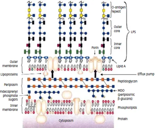
In general, Gram-negative bacteria are more resistant to antibiotics because of the more complex architecture of their cell envelope, which includes an outer and inner membrane with the periplasmic space in between (SILHAVY et al, 2010) (Figure 1).
The outer membrane (OM) is a distinguishing feature of negative bacteria, because Gram-positives lack this structure. OM gives an additional protection from the environment by excluding toxic molecules and providing an additional stabilizing layer. It is a lipid bilayer that contains glycoplipids, mainly lipopolysaccharide (LPS). LPS is a virulence factor that typically consists of a hydrophobic domain known as lipid A or endotoxin, a non-repeating core oligosaccharide and a distal polysaccharide or O-antigen. The lipid A of LPS is responsible for the septic shock related to septicaemia, caused by these microorganisms (SILHAVY et al, 2010).
The outer membrane proteins (OMPs) of Gram-negatives are divided into two classes: lipoproteins and β-barrel proteins. The majority of the OMPs are transmembrane proteins. For example, OmpF is a transmembrane protein called porin that allows the passive diffusion of small molecules (SILHAVY et al, 2010).
As demonstrated in Figure 1, the periplasmic space is delimited by the OM and the inner membrane (IM). This structure gives Gram-negative bacteria a great evolutionary advantage because it entraps, for example, the potentially harmful molecules, such as degradative enzymes. In contrast to the Gram-positives, which lack this narrow space, Gram-negatives can pick toxic compounds here and expel them directly to the external medium through efflux pumps (represented by the vertical bold arrows in Figure 1), strongly reducing the number of molecules reaching their cytoplasmic targets. Within the periplasmic space there is the peptidoglycan whose main function is to give rigidity and shape to the cell wall. Peptidoglycan is made up of repeating units of the disaccharide N-acetyl
glucosamine –N-acetyl muramic acid which is cross-linked by pentapetide side chains. The amount of this molecule in Gram-negatives is much lower compared to Gram-positive bacteria (NIKAIDO, 2001; SILHAVY et al, 2010).
As widely known, bacteria do not have various organelles, that eukaryotic cells contain. These organelles perform a myriad of very important tasks to the cell wellbeing. In bacteria, all these “household tasks”, like energy production, protein or lipid synthesis are performed in IM that is a phospholypid bilayer. Protein channels involved in the efflux mechanisms are also part of the bacterial cell envelope. Within the five superfamilies of transporters (chapter 5), MFS and RND are the most abundant. RND transporters (Figure 4), which are found so far exclusively in Gram-negative bacteria, are the major reason for antibiotic resistance (NIKAIDO, 2001).
Figure 1- Schematic representation of the Gram-negative cell envelope (5).
Gram-positive cell wall:
Gram-positives lack the OM, which is present in Gram-negatives; the peptidoglycan layer is thicker in Gram-positive bacteria, as shown by Figure 2. Interspersed in this layer, there are numerous polymers, called teichoic acids (TAs). This polymers account for 60 % of the mass of Gram-positives envelope, being thus major contributors to cell envelope structure and function (SILHAVY et al, 2010).
In addition to the TAs, there are proteins attached to the surfaces of Gram-positives. Some of these proteins have similar properties to those found in Gram-negative periplasmic space, furthermore the adhesion to extracellular noxious molecules is a task performed by these proteins (SILHAVY et al, 2010).
Because of their less complex cell envelope, Gram-positives, are considered much more susceptible to certain antimicrobials than Gram-negatives, which have been showing greater resistance to drugs (CORNAGLIA, 2009).
4.2 Transport systems (TENOVER, 2006):
For most polar molecules, such as nutrients or metabolites, the lipid bilayer (Figure 1) of a cell membrane represents an impermeable membrane. The transport of small molecules is mediated by transmembrane proteins, whereas macromolecules and small particles cross membranes by various cytotic mechanisms (ECKER & CHIBA, 2010).
Diffusion is the translocation of molecules from a region of higher concentration to a region of lower concentration. This is mediated by pore forming channels. There are also membrane proteins that mediate the translocation of molecules that are too polar or too large to move across a membrane by diffusion. In contrast to pore forming channel proteins, carrier proteins bind their substrates at specific binding sites, resulting in the so called “facilitated diffusion” (ECKER & CHIBA, 2010). There are three types of membrane transporter proteins (ECKER & CHIBA, 2010) (Figure 3):
• Uniport, when the carriers mediate transport of a single substrate;
• Symport, when the carriers bind to different substrates and transport them together across the membrane;
• Antiport refers to those transporters that exchange one substrate for another across the membrane.
The so-called cotransport systems – symporters and antiporters - permit the simultaneous or sequential passive transfer of molecules or ions across biological membranes (Figure 3). Cotransporters are classified in symporters and antiporters depending on the direction of the second substrate. Symport refers to migration of molecules in the same direction, whereas antiport in opposite directions. Bmr and Blt from Bacillus subtilis are examples of antiporters (LI & NIKAIDO, 2009) whereas E.coli LacY functions as a symporter (GUAN & KABACK, 2006).
In contrast with the facilitated diffusion, active transport systems couple the transport with energy. There are two types of active transport. In primary active transport the energy comes from the ATP hydrolysis, whereas in the secondary active transport the energy is provided by the flow of ions, such as Na+ (Sodium), H+ (Hidrogen) or K+ (Potassium).
5. Families of transporters
Efflux pump systems are present in all living systems, providing a frontline nonspecific defense, with standard responses to the external stimuli (RICE, 2007).
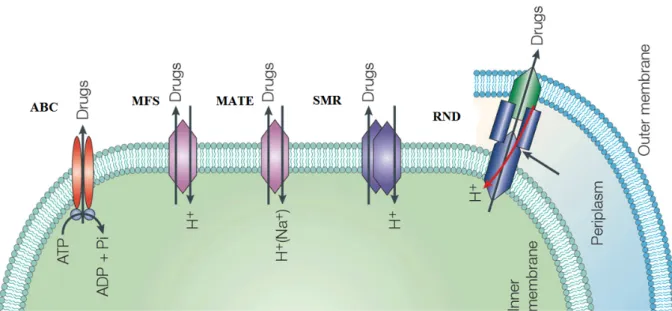
Substrate specific transporters constitute systems that are responsible for the extrusion of a given drug or class of drugs. On the contrary, the multidrug transporters are capable to handle a wide variety of structurally unrelated compounds. MDR transporters can be divided in two groups based on their structure and bioenergetics: 1) secondary multidrug transporters, that are predominant in bacteria, use the transmembrane electrochemical gradient of protons or sodium ions to extrude the drugs from the cell. Instead, the 2) primary multidrug transporters extrude their substrates with the energy of ATP hydrolysis. (ECKER & CHIBA, 2010; BORGES-WALMSLEY et al, 2003).
Although both primary and secondary transporters (Figure 5) are ubiquitous in bacteria, their relative presence seems to correlate with energy generation: fermentative bacteria tend to rely more
Figure 3 – Schematic representation of uniport, symport and antiport systems
The arrows represent the direction of transported ions or molecules through the transport system.
Source: vydavatelstvi.vscht.cz
on the primary transporters while aerobic bacteria contain somewhat more secondary transporters in their genomes (PAULSEN et al, 2000).
Bacteria appear to be particularly endowed with multidrug resistance transporters. Considering predictions from the bioinformatic analysis of more than 200 available bacterial genomes, putative MDRs comprise 2 to 7 % of the total bacterial protein complement (SAIER & PAULSEN, 2001). Among these bacterial genomes, not all have been confirmed to be polyspecific, but quite a few of them are, underscoring the impressive resistance capacity of bacteria.
Functional studies and subsequent phylogenetic analysis demonstrated that bacterial MDR transporters could be organized into five evolutionary distinct protein superfamilies that significantly differ in bioenergetics, structure and transport mechanism (SAIER & PAULSEN, 2001). This structural and functional diversity gives bacteria a big potential to combat noxious agents (SAIER et al, 1998 ).
5.1 Primary multidrug transporters in bacteria:
5.1.1 ABC transporters in bacteria:
In bacteria, these transporters are predominantly involved in the import of essential compounds that cannot be obtained by diffusion (sugars, vitamins, metal ions and others). In spite of the importance of these transporters in eukaryotic cells, in bacteria their role is more limited. However, both antibiotic-specific and polyspecific ABC transporters have been identified (ECKER & CHIBA, 2010; NIKAIDO, 2009).
The most well-studied bacterial ABC multidrug transporters are LmrA and LmrCD, both from Lactococcus lactis (ECKER & CHIBA, 2010). LmrA is homologous to one half of the mammalian MDR1 (NIKAIDO, 2009; PUTMAN et al, 2000). Instead of the minor role played by LmrA in L.
lactis, its overproduction in E. coli confers resistance to cationic dyes, daunomycin and triphenylphosphonium (NIKAIDO, 2009).
The data available suggests that ABC transporters are more present in Gram-positive bacteria (ECKER & CHIBA, 2010). Another example of this superfamily of transporters is S. aureus Sav1866 that is homologue to LmrA (NIKAIDO, 2009; LI & NIKAIDO, 2009).
5.2 Secondary multidrug transporters
The majority of the multidrug efflux systems known to date are sensitive to agents that dissipate the proton motive force (PMF). Thus, these transporters couple the extrusion of drugs and other toxic agents with the exchange of protons or sodium ions. Related to their size and structures, secondary transporters can be subdivided into distinct superfamilies: the major facilitator superfamily (MFS), the small multidrug resistance (SMR) superfamily, the resistance-nodulation-cell division (RND) superfamily and the multidrug and toxic compound extrusion (MATE) superfamily (ECKER & CHIBA, 2010).
5.2.1 Major facilitator superfamily (MFS)
The MFS is the largest characterized superfamily of transporters (LI & NIKAIDO, 2009; FLUMAN & BIBI, 2009). These transporters are found in all living organisms and are implicated in the symport, antiport or uniport of various substrates, such as sugars, drugs, neurotransmitters or amino acids (FLUMAN & BIBI, 2009). This superfamily contains transporters that extrude one specific substrate and transporters that extrude a variety of them. Based on phylogenetic analysis, it appears that specific and MDR transporters in the MFS appeared randomly on the evolutionary tree, indicating that the broadening and narrowing of specificity toward particular drugs occurred repeatedly during evolution. For example, a single amino acid change in the S. aureus QacB
transporter enables it to recognize not only monovalent but also divalent cationic compounds (PAULSEN et al, 1996). In addition, mutations that alter the substrate specificity of the transporters are thus of great importance as though, usually influence the transport of a subset of substrates. Therefore, these mutations usually occur in residues that participate in substrate binding. For example, Bmr, found in Bacillus subtilis, is one of the MFS transporters that harbor some of these residues, typically found inside of transmembrane domains (TMDs). Some of the substrate binding sites in this family are very large, thus with the ability to bind more than one substrates simultaneously (FLUMAN & BIBI, 2009).
These proteins can be clustered into two different groups, with either twelve or fourteen transmembrane segments (TMS). It has been proposed to function by an alternating access, rocker switch- type- structural mechanism, in which the substrate binding site is alternatively accessible from both sides of the membrane. For example, the EmrB of E. coli, where the MFS systems can function as components of tripartite systems (LI & NIKAIDO, 2009).
Other examples: NorA was first discovered in S. aureus and is from the 12-TMS cluster. It confers resistance to hydrophilic compounds and none to hydrophobic ones (FLUMAN & BIBI, 2009). Bmr, from B. subtilis, is structural and functionally homolog to NorA (AHMED et al 1995).
5.2.2 Small multidrug resistance superfamily (SMR)
The SMR superfamily, most found in Eubacteria (BAY et al, 2008), is the smallest secondary MDR transporter known, composed of around 100 amino acids. SMR transporters are believed to span the cytoplasmic membrane as four transmembrane (TM) α-helices with short hydrophilic loops, thus making them highly hydrophobic.
Members of this superfamily have been identified, encoded by a variety of plasmids and transposable members giving high resistance to a wide range of antibiotics. E.g. QacH in Staphylococcus saprophyticus. (PUTMAN et al, 2000).
Among Gram-negative bacteria, EmrE from E. coli, is considered the structural archetype of all SMR proteins (SCHULDINER et al, 2001). The scientific data available reveals that these transporters function as an oligomer, most likely a dimer. There are other examples, like the Mmr of Mycobacteria tuberculosis that confers resistance to ethidium bromide, erythromycin, or the antiseptic resistance multidrug transporter QacE present in Klebsiella aerogenes (PUTMAN et al, 2000).
5.2.3 Resistance-Nodulation-Cell Division Superfamily
The RND transporters (Figure 4 and Figure 5) are responsible for the high intrinsic antibiotic resistance found in negative bacteria (MURAKAMI, 2008). They are also found in Gram-positive bacteria as well as in M. tuberculosis where one of these RND type pumps is responsible for the efflux of the first-line antimycobacterial drug, isonazide (PASCA et al, 2005). These transporters are composed of approximately 1000 amino acid residues, adopting a 12-helical structure, comprising large periplasmic or extracytoplasmic domains between helices 1 and 2 and between helices 7 and 8.
RND transporters contain an astonishing wide range of substrate specificity surpassing even the ABC transporters (ZGURSKAYA & NIKAIDO, 2002). These pumps require three units: a transporter protein in the IM, called AcrB, in association with two more classes of proteins, such as the OMF (outer membrane factor) Tol C and the MFP (membrane fusion protein) AcrA from E. coli (Figure 4) (MURAKAMI, 2008). These transporters are very complex because without one of the three parts available, the complex turns non-functional.
It was shown that by inactivating the AcrB, bacteria become susceptible to some drugs (LI & NIKAIDO, 2009). Based on former results it can be supposed that the periplasmic loops of RND transporters are critical in substrate determination, possibly containing multiple binding sites for structurally unrelated compounds (ECKER & CHIBA, 2010; MURAKAMI, 2008).
5.2.4 Multidrug and Toxic Compound Extrusion Superfamily (MATE)
There are twenty MATE transporters characterized to date. Most members of this family consist of 400-500-residue polypeptides with 12 putative transmembrane helices (TMHs) (LI & NIKAIDO, 2009). Phylogenetic analyses classify these transporters into three subfamilies. Little is known about the molecular mechanism underlying their organic cation transport. However, there are some similarities with the MFS superfamily (LI & NIKAIDO, 2009; MCALEESE et al, 2005).
Examples: NorM from Vibrio parahaemolyticus and MepA from S. aureus are both MATE transporters implicated in the resistance to antibiotics, conferring resistance to fluoriquinolones and tigecycline, respectively (LI & NIKAIDO, 2009; MCALEESE et al, 2005).
Figure 4 – Schematic representation of the RND superfamily: the structure of AcrAB-Tol C (POS, 2009).
The tripartite complex is represented by the different colours: in blue, the inner membrane protein AcrB, in red the membrane fusion protein AcrA and in yellow the outer membrane factor TolC.
Figure 5 – Schematic representation of the five superfamilies of multidrug transporters.
The ATP-binding cassette (ABC) superfamily is the only one belonging to the primary multidrug transporters. The other four superfamilies belong to the secondary multidrug transporters.
6 – Multidrug resistance in cancer
Cancer is one of the most fatal diseases worldwide (WHO). In poor and developed nations, cancer is a leading cause of death. 70 % of all cancer deaths occurred in middle and low-income countries. The WHO projections for 2030 are around 11.5 million deaths. Limited improvement in diagnosis, surgical techniques, patient care and adjuvant therapies are among the top causes of this projection (FIEDLER, 2002)
6.1 Cancer burden
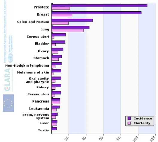
In Europe, 3.2 million people are diagnosed with cancer every year (FERLAY et al, 2010). In both sexes, in EU, cancers with the higher rates of incidence are: prostate cancer, breast cancer, colorectal cancer and lung cancer. Lung and breast cancers have the highest mortality rates (Figure 6).
There are also differences in the incidence between men and women. In men the types of cancer with higher mortality rate are: prostate cancer, lung cancer, and colorectal cancer. In women, the most common types of cancer are breast cancer, colorectal cancer and lung cancer (FERLAY et al, 2010).
6.2 Causes of cancer
The transformation from a normal cell into a cancer one is a multistage process that begins from one single cell (STEVENS & LOWE, 2000; LUO & ELLEDGE, 2008). These changes can be due to genetic factors, ageing and three categories of external agents (www.cancer.org) such as:
• Physical carcinogens (e.g UV light);
• Chemical carcinogens (e.g tobacco smoke); • Biological carcinogens (e.g HPV virus).
7. Relationship between bacterial infection and cancer
Bacterial infections can also be risk factors for cancer development, for instance Helicobacter pylori and Salmonella typhi can be involved in cancer development. H. pylori infects, at least 50 % of worldwide population. Within other malignancies, such as chronic inflammation or peptic ulcer disease, it is a risk factor for gastric cancer and mucosa-associated lymphoid tissue (MALT) lymphoma. In order to combat H. pylori an initial treatment has been developed consisting of a triple therapy that contains a proton pump-inhibitor (PPI). The treatment consists of clarithromycin, metronidazole or amoxicillin with a PPI, such as esomeprazole (BERGMAN & D’ELIOS, 2010; FISCHBACH et al, 2009). Resistance to clarithromycin has yet been reported in various countries (FISCHBACH et al, 2009).
Chronic infection with S. typhi can be related to gallbladder cancer. Carriers of this pathogen have an eight-fold higher risk of developing this type of cancer than non-carriers. The exact pathogenesis is yet to be understood but there is increasing evidence that the products of the degradation of bile-salts may contribute to tumorogenesis (SAMARAS et al, 2010).
8. Carcinogenesis - the development of cancer
In the human body, cells grow and divide in a controlled fashion to produce more cells, in order to keep the body healthy. Certain stimuli cause changes in genetic material that result in permanent alteration of the normal cellular growth pattern (STEVENS & LOWE, 2000). These distorted cells fail to respond normally to signals controlling cell growth; they are termed neoplastic (STEVENS & LOWE, 2000), because they proliferate uncontrolled, forming a tissue called neoplasm, which means “new growth”.
As shown by Figure 7, there is a difference between the normal and cancer cell division. Apoptosis or programmed cell death is a normal component of the development and health of multicellular organisms. Through apoptosis cells die in a controlled and regulated manner.
The main reason for the difficulties in cancer therapy is that cancer cells can spread to other organs. This process is called metastasis and refers to the growing of a secondary tumor that was originated from a primitive neoplasm (GUPTA & MASSAGUE, 2006).
As seen in Figure 8, the invasive cancer cells migrate from the primary tumour site into the surrounding tissue through blood vessels. Cells are thus carried to distant organs where they lodge in their small capillaries. They form secondary tumours by extravasating to the surrounding tissue (STEVENS & LOWE, 2000). As neoplastic cells are continually growing, they need an appropriate set of support tissues, particularly an adequate set of vascular supply that is why angiogenesis is essential for the growth of neoplasms (SCHEIDER et al, 2009).
9. Cancer therapy and the problem of resistance mechanisms to anticancer agents
Chemotherapy is the treatment of choice for ~50 % of all types of cancer(AMBUDKAR et al, 2005), the effectiveness of chemotherapy – a cancer treatment that uses drugs to destroy cancer cells - is of big concern for medicine. Combination chemotherapy is the use of different chemical agents simultaneously. It was first introduced in 1963 for the treatment of resistant cancers (SHEPS & LING, 2007).
9.1. Main groups of anticancer drugs
Among the great amount of cells that forms a neoplasm, not all are in the same development stage or respond in the same way to the same stimulus. In order to prevent selection of resistant cancer
Figure 7. Cellular division in normal and cancer cells.
A – Normal cell division; B – Cancer cell division; Source: http://www.jyi.org
A
B
Figure 8. Schematic representation of the metastasizing process.
1 – Attachment of the tumour cells; 2 – Influx of tumour cells through a vessel breakdown; 3 – Locomotion of the tumour cells through the circulatory system; 4 – Adhesion of the tumour cell mass in a different body location.
cells, contemporary chemotherapy uses combinations of various drugs of different targets. There are five main families of anticancer drugs:
• Alkylating agents: drugs that bind to the DNA, e.g. cisplatin, ifosfamide (STAVROVSKAYA, 2000; TASCILAR et al, 2007);
• Anticancer antibiotics: These agents act by topoisomerase inhibition, e.g. daunorubicin, doxorubicin (STAVROVSKAYA, 2000; KURUVILLA, 2009 );
• Antimitotic agents: Depolymerization of microtubules and damage to mitotic spindle, e.g. vinca alkaloids and taxanes (STAVROVSKAYA, 2000; PEREZ, 2009);
• Antimetabolites: The main mechanism of action of these agents is the inhibition of enzymes participating in DNA or RNA synthesis, e.g. methotrexate, gemcitabine (STAVROVSKAYA, 2000; MERL et al, 2010).
• Hormones: Hormones can be a natural product of an organ or represent abnormal synthesis reflecting unregulated cancer cell metabolism. Drugs, such as tamoxifen are antagonists of the estrogen receptor, obstructing the effect of the hormones (JORDAN, 2006).
9.2. Resistance to anticancer agents:
In order to resist to the effects of anticancer drugs, tumour cells have various ways to resist their noxious effects. There are thus four main mechanisms of drug resistance:
• Decrease of drug accumulation by the cells: drugs inside the cells can be extruded due to the activation of transporter proteins. Otherwise, these drugs remain inside the cells, able to exert their toxic effect. E.g. P-gp. (STAVROVSKAYA, 2000; LAPENSEE & BEN-JONATHAN, 2010 ).
• Detoxification of the drug in the cell: Cells have mechanisms to turn drugs ineffective. One of these mechanisms is the cellular glutathione system (CGS). Glutathione forms a conjugate with the reactive site of the drug that is more water-soluble and less active. This conjugate is then extruded from the cell through transporter proteins, such as GS-X (including MRP) (STAVROVSKAYA, 2000; MOSKAUG et al, 2005 ).
• Alteration of drug targets: Gene mutations are common in cancer cells that often render drug targets unrecognizable. Sometimes, during the progression of cancer, some targets are lost, for example the estrogen receptor in the progression of breast cancer. Another way of drug target alteration is the increase of target proteins, which is caused by the over-expression of the gene that controls the target molecule (STAVROVSKAYA, 2000; LO & SUKUMAR, 2008).
• Key genes that control apoptosis: One of these genes is p53, also known as “guardian of the genome”. p53 is often altered in tumour cells, resulting in impaired function and inducing apoptosis (STRACHAN & READ, 2004; STAVROVSKAYA, 2000; BELL & RYAN, 2007 ).
10. The importance of ABC transporters in cancer
Multidrug resistance is a major obstacle to the success of cancer chemotherapy. It is mainly related to the expression of ABC transporters (HALL et al, 2009). Forty-nine members of this superfamily have already been identified and classified in seven subfamilies based on their phylogenetic similarity (KLEIN et al, 1999).
P-glycoprotein (P-gp), the product of MDR-1 (ABCB1) gene, was the first to be discovered and is the most known efflux pump within ABC transporters (HALL et al, 2009). ABCB1 is found in various resistant tumour cell lines and is also naturally expressed in many tissues (MARCHETTI et al, 2007).
10.1 Physiological localization of ABCB1 (P-glycoprotein)
As seen in Figure 9, ABCB1 is expressed in the lumen of the endothelial cells, which comprises the blood-brain barrier. It is found in the canalicular membrane of hepatocytes, brush-border membrane of proximal tubules in the kidney, mediating thus the expression into the urine. ABCB1 also prevents the absorption of substrates into the human body because it is present in the intestine epithelial cells. ABCB1 is also present in the syncytiotrophoblasts and bone marrow (HULS et al, 2009).
In humans, defective ABC transporters are associated with several diseases, including Dubin-Johnson syndrome, sisterolemia and Tangier disease (ECKER & CHIBA, 2010). ABC transporters are often responsible for clinical MDR; consequently these transporters are also often a sign of poor
Figure 9. Physiological distribution of ABCB1 (P-glycoprotein) (HULS et al, 2009).
Representation of the various organs where ABCB1 has a normal physiological role.
prognosis (STAVROVSKAYA & STROMSKAYA, 2008). Mutations in genes encoding ABC transporters can induce a multitude of defects, presenting as autosomal recessive conditions. A good example is the cystic fibrosis transmembrane regulator protein (CFTR), a member of the ABCC subfamily. A mutation in the encoding gene on chromosome 7 impairs the synthesis of CFTR resulting in cystic fibrosis (LEONARD et al, 2003).
Like MRP1 – the second drug transporter in humans (COLE et al, 1993) – ABCB1 is thought to provide protection to normal tissues, preventing the accumulation of or exposure to toxic substances (HOLLAND et al, 2003; LEONARD et al, 2003). Mice lacking ABCB1 homologue (mdr1a and mdr1b) have a subtle phenotype indicating a role for ABCB1 in the physiological defense against xenotoxins (SCHINKEL, 1997).
10.2 Structure of ABC transporters
In contrast to prokaryotes, in eukaryotes the major mechanism of efflux is dependent on proteins that derive their transport energy from the hydrolysis of ATP. The majority of these transporters belong to the ABC superfamily (BORGES-WALMSLEY et al, 2003), such as ABCB1 (also known as P-gp), ABCC1 [also known as multi-drug resistance-associated protein (MRP1)] or ABCG2 [also known as breast cancer-resistance protein (BCRP)] (LIU, 2009).
The members of this superfamily translocate a wide range of substances (including sugars, amino acids, sterols, peptides, antibiotics, xenobiotics between others) across cytoplasmic and organellar membranes (LEONARD et al, 2003).
In eukaryotes, molecules are mostly transported from the cytoplasm to the outside of the cell or into an intracellular compartment (endoplasmic reticulum, mitochondria, peroxisome) (DEAN et al, 2001).
The topology of these transporters indicates the existence of highly hydrophobic transmembrane domains (TMDs) with between 4 and 10 TM α-helices – typically six per domain - and hydrophilic