online | memorias.ioc.fiocruz.br
Biofilm production by multiresistant
Corynebacterium striatum
associated with nosocomial outbreak
Cassius de Souza1, Yuri Vieira Faria1, Lincoln de Oliveira Sant’Anna1, Vanilda Gonçalves Viana1,
Sérgio Henrique Seabra2, Mônica Cristina de Souza1, Verônica Viana Vieira3,
Raphael Hirata Júnior1, Lílian de Oliveira Moreira4/+, Ana Luíza de Mattos-Guaraldi1
1Laboratório de Difteria e Corinebactérias de Importância Clínica, Centro Colaborador de Referência e Pesquisa em Difteria/Fundação
Nacional de Saúde/Ministério da Saúde, Universidade do Estado do Rio de Janeiro, Rio de Janeiro, RJ, Brasil
2Laboratório de Tecnologia em Bioquímica e Microscopia, Centro Universitário Estadual da Zona Oeste, Rio de Janeiro, RJ, Brasil 3Laboratório de Genética Molecular de Microrganismos, Instituto Oswaldo Cruz-Fiocruz, Rio de Janeiro, RJ, Brasil 4Laboratório de Bacteriologia e Imunologia Clínica, Universidade Federal do Rio de Janeiro, Rio de Janeiro, RJ, Brasil
Corynebacterium striatum is a potentially pathogenic microorganism that causes nosocomial outbreaks. How-ever, little is known about its virulence factors that may contribute to healthcare-associated infections (HAIs). We investigated the biofilm production on abiotic surfaces of multidrug-resistant (MDR) and multidrug-susceptible (MDS) strains of C. striatum of pulsed-field gel electrophoresis types I-MDR, II-MDR, III-MDS and IV-MDS iso-lated during a nosocomial outbreak in Rio de Janeiro, Brazil. The results showed that C. striatum was able to adhere to hydrophilic and hydrophobic abiotic surfaces. The C. striatum1987/I-MDR strain, predominantly isolated from patients undergoing endotracheal intubation procedures, showed the greatest ability to adhere to all surfaces. C. striatum bound fibrinogen to its surface, which contributed to biofilm formation. Scanning electron microscopy showed the production of mature biofilms on polyurethane catheters by all pulsotypes. In conclusion, biofilm pro-duction may contribute to the establishment of HAIs caused by C. striatum.
Key words: biofilm - Corynebacterium striatum - epidemic clone - fibrinogen - multi-resistance - nosocomial outbreak
doi: 10.1590/0074-02760140373
Financial support: CAPES (E26/101.937/2009), FAPERJ (E26/110.735/2012), CNPq (300236/2010-7),
SR-2/UERJ, PNPD-CAPES/MEC (0370088) + Corresponding author: lilian@pharma.ufrj.br Received 10 October 2014
Accepted 29 January 2015
Corynebacterium striatum is an emerging multidrug-resistant (MDR) potentially pathogenic microorganism that causes nosocomial infection in patients who have experienced long hospital admissions, those who have received several courses of antibiotics (Camello et al. 2003, Otsuka et al. 2006, Renom et al. 2007, Baio et al. 2013), those with acquired immune deficiency syndrome (AIDS) or cancer and those who have received a trans-plant (Tarr et al. 2003, Martins et al. 2009). Cases of se-vere infections in both immunocompromised and immu-nocompetent individuals and nosocomial outbreaks due to MDR C. striatum are increasing in both industrialised and developing countries. C. striatum has been associ-ated with cases of pulmonary infections, sepsis, endo-carditis, meningitis, osteomyelitis, arthritis, sinusitis, skin wounds and intrauterine infections (Rufael & Cohn 1994, Weiss et al. 1996, Fernández-Ayala et al. 2001, Ca-mello et al. 2003, Renom et al. 2007, Scholle 2007, Boltin et al. 2009, Campanile et al. 2009, Martins et al. 2009, Moore et al. 2010, Oliva et al. 2010, Baio et al. 2013).
Genotyping analysis by pulsed-field gel electropho-resis (PFGE) has identified PFGE types associated with nosocomial outbreaks of respiratory origin and with resistance to a broad range of antibiotics (MDR pheno-type). In Italy, MDR C. striatum isolates have been re-covered from hospitalised patients who have undergone surgery or have been admitted to intensive care units (ICUs). These isolates have been responsible for cases of ventilator-associated pneumonia and tracheobronchitis, catheter-related sepsis and wound infections. Infections caused by this species have been strongly associated with devices, including not only tubes and catheters, but also sternal surgical wound wires (Campanile et al. 2009).
C. striatum has also been isolated from other materials for hospital use, such as endotracheal tubes (Martinez-Martinez et al. 1995). Earlier genotype studies have con-firmed that C. striatum may be transmitted between pa-tients, from person to person and via caretakers (Leonard et al. 1994). Recently, a nosocomial outbreak caused by
C. striatum was documented in Rio de Janeiro (RJ), Bra-zil. PFGE analysis indicated the presence of four PFGE profiles, including two related clones of MDR strains (PFGE I and II). The results of these studies demonstrate the predominance of PFGE-type I MDR isolates that are mainly isolated from ICUs and surgical wards. C. stri-atum strains have largely been isolated in pure culture from tracheal aspirates of patients undergoing endotra-cheal intubation procedures (Baio et al. 2013).
several corynebacterial species (Martins et al. 2009). Op-portunistic pathogens may be endowed with an array of vir-ulence factors that facilitate their ability to survive within host tissues and confer resistance to clearance by host im-mune mechanisms and antimicrobial killing. The ability to form biofilms may be a prerequisite for the pathogeneses of nosocomial diseases associated (or not) with the use of medical devices (Bonifait et al. 2008). Biofilms have been previously described in Corynebacterium diphtheriae,
Corynebacterium pseudotuberculosis, Corynebacterium renale, Corynebacterium urealyticum and Corynebacteri-um jeikeiCorynebacteri-um (Mattos-Guaraldi & Formiga 1991, Soriano et al. 1993, Mattos-Guaraldi et al. 1999a, b, Olson et al. 2002, Kwaszewska et al. 2006, Gomes et al. 2009, 2013, Soriano et al. 2009). Thus, the better recognition and understand-ing of the biology and virulence potential of C. striatum
strains may help to effectively prevent infections caused by them. Therefore, we investigated the in vitro capacities for biofilm formation of C. striatum strains representative of four different PFGE types isolated during a nosocomial outbreak in RJ (Baio et al. 2013).
SUBJECTS, MATERIALS AND METHODS
Bacterial strains - Table shows the epidemiological and microbiological features of the partially studied C. striatum strains used in this investigation (Baio et al. 2013). C. striatum identification was established by 16S rRNA and rpoB gene sequencing. C. striatum pulsotypes I and II exhibited MDR profiles showing susceptibility only to vancomycin, linezolid and tetracycline, while C. striatum pulsotypes III and IV showed susceptibility to most of the 21 antimicrobial agents tested and resistance only to fosfomycin and ticarcillin/clavulanate. The C. diphtheriae CAT5003748 strain was used as a positive control in all experiments (Gomes et al. 2009).
Biofilm formation on hydrophilic surfaces of glass tubes -Microorganisms were inoculated in glass tubes (13 x 100 mm) containing 4 mL of trypticase soy broth (TSB) and incubated at 37°C for 24 h without shaking. The tubes were gently shaken and supernatants with non-adherent bacterial cells were discarded. TSB (4 mL) was then added and the tubes were reincubated at 37°C for 24 h. This procedure was repeated twice. Glass-adherent
bacteria created a confluent coat of cells on the sides of the tube. Quantitative analysis of viable sessile cells was based on previously described methods (Mattos-Guaral-di & Formiga 1991, Dooley et al. 1996).
Quantitative and semiquantitative analyses of biofilm formation on catheter - Polyurethane 16-gauge percuta-neous nephrostomy catheters (Intracath; Deseret Pharma-ceutical Co, USA) were used for an evaluation of bacterial adherence and biofilm formation on catheter surfaces. Sterile 4-cm segments of polyurethane catheters were im-mersed in TSB containing 106 colony-forming unit (CFU)
mL-1 and incubated at 37°C for 24 h (Gomes et al. 2009).
Then, quantitative catheter culturing (Dooley et al. 1996) and a semiquantitative roll-plate technique (Maki et al. 1977) were performed using Columbia agar medium sup-plemented with 5% sheep blood at 37°C for 24 h.
Scanning electron microscopy (SEM) - Sections of glass coverslips and polyurethane catheters were fixed in 2.5% glutaraldehyde, post-fixed in 1% osmium tetroxide and dehydrated in a graded series of ethanol. Subsequently, catheter segments were subjected to critical point drying with carbon dioxide, covered with a 10 nm layer of gold palladium and examined with a JEOL JSM 5310 scanning electron microscope. Sterile unused polyurethane cath-eters were also processed by SEM directly upon removal from commercial packaging (Gomes et al. 2009).
Biofilm formation on hydrophobic polystyrene sur-faces -Biofilm formation on negatively charged polysty-rene surfaces was determined quantitatively in 96-well flat-bottomed microtitre plates according to previously described methods (Stepanovic et al. 2000, Gomes et al. 2009). Aliquots of 200 μL of bacterial suspensions [0.2 optical density (OD) at λ=570 nm] were added to the mi -croplate wells. After incubation at 37°C for 24 h, the con-tents of each well were aspirated and washed three times with 200 μL phosphate-buffered saline (0.01 M, pH 7.2). The remaining attached bacteria were fixed with 200 μL of 99% methanol and stained with 2% crystal violet. The negative controls contained TSB only. The bound dye was then solubilised with 160 μL of 33% glacial acetic acid and the OD of the solution was measured at λ = 570 nm using an enzyme immunosorbent assay reader (BioRad, TABLE
Origin and pulsed-field gel electrophoresis (PFGE)-types of partially studied Corynebacterium striatum strains isolated from patients during a nosocomial outbreak in the metropolitan area, Rio de Janeiro, Brazil used in this study
Strains/year
Gender/ age
Hospital wards
Isolation
site Outcome
Antimicrobial susceptibility patterns
PFGE-types
1987 BR-RJ/09 F/50 Nursery 18 BAL Death MDR I
2369 BR-RJ/09 M/NI General ICU TA Cure MDR II
1961 BR-RJ/09 F/37 Infectious diseases Urine NI MDS III
1954 BR-RJ/09 M/NI Thoracic MSU Surgical wound NI MDS IV
model 550). The cut-off OD (ODc) was defined as the mean OD of the negative control. All strains were classi-fied into the following categories based on the ODs of the bacterial films: nonadherent (0: OD ≤ ODc) or weakly (+: ODc < OD ≤ 2 x ODc), moderately (++: 2 x ODc < OD ≤ 4 x ODc) or strongly (+++: 4 x ODc ≤ OD) adherent.
Influence of fibrinogen (Fbg) on biofilm formation
- Biofilm formation (24 h) was determined in 96-well flat-bottom polystyrene microtitre plates as described above, with some modifications. In these experiments, the wells of the microplates were pre-treated (or not) with human plasmatic Fbg (Sigma Chemical Co, USA) at a concentration of 50 µg mL-1 overnight at 4°C.
Fbg-coated wells containing 200 μL of TSB medium without bacteria were used as negative controls (Lembke et al. 2006, Gomes et al. 2009).
Statistical analysis - Each experiment was carried out in triplicate and repeated three times. The biofilm forma-tion by each representative pulsotype strain was compared by ANOVA with Tukey’s post test. Student’s t test was used to compare the means of biofilm formation (OD) in the presence of Fbg for each pulsotype and a p < 0.05 was considered to be statistically significant. Statistical analy-ses were performed using GraphPad Prism v.6 (USA).
RESULTS
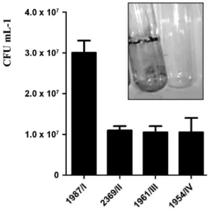
Bacterial adherence to hydrophilic surface of glass tube - Successful bacteria may survive in the hospital environment due to their ability to adhere to different substrates. To determine whether C. striatum is able to adhere to glass, we quantified the amount of viable sile forms of bacteria associated with glass. Viable ses-sile bacterial cells were observed on glass surfaces at 48 h post-incubation with C. striatum strains of PFGE types
I, II, III and IV at different levels (Fig. 1). In Fig. 1, a rep-resentative side figure illustrates sessile bacteria stained with crystal violet, indicating the formation of a positive slime/biofilm on the glass surface. All strains were able to strongly adhere to the glass surface, however the C. striatum 1987/I-MDR strain showed the highest ability to adhere to this hydrophilic abiotic surface (p < 0.05).
Biofilm formation on polyurethane catheter surface
- The ability to adhere to catheter materials for intra-venous use and medical devices inserted into the body are important characteristics of bacteria associated with healthcare-associated infections. To determine whether
C. striatum is able to adhere and form biofilms on cath-eter surfaces, segments of polyurethane cathcath-eters were colonised in vitro by C. striatum 1987/I, 2369/II, 1961/III and 1954/IV strains. The evaluation of the adherence and viability of microorganisms on the polyurethane catheter segments by the semiquantitative roll plate method (> 15 CFU) and by quantitative catheter culture assays (> 1.5 x 10⁶ CFU) showed that viable C. striatum cells were
ex-tensively adherent to and multiplied on the polyurethane catheter surface (Fig. 2). In Fig. 2A, the representative side figure illustrates bacterial growth on an agar plate after catheter colonisation, as assessed by the roll-plate technique. Although all strains were able to adhere to the catheter surface, the C. striatum 1987/I-MDR strain again exhibited significantly greater adherence (p < 0.05) to this abiotic hydrophobic surface compared with the representative strains of pulsotypes II, III and IV.
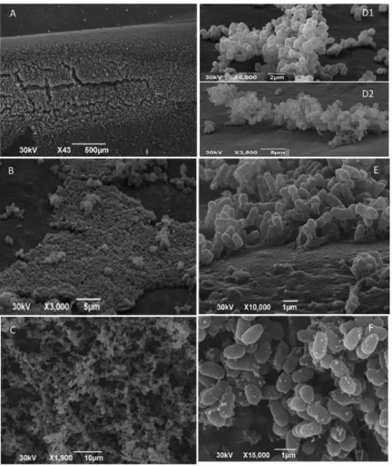
Morphological aspects of biofilm formation on the surface of polyurethane medical device as evaluated by SEM - After determining that C. striatum was able to ad-here to glass and catheter surfaces, forming a visible bio-film, the morphological aspects of the biofilm were inves-Fig. 1: Corynebacterium striatum adherence to glass surfaces (24 h
in-cubation) evaluated by quantitative and semiquantitative tests. In detail, glass tubes with 1987/I strain exhibiting confluent coat of sessile forms on the surface of glass tube wall and with negative control (trypticase soy broth medium without bacteria). CFU: colony-forming unit.
tigated by SEM. SEM assay confirmed that all C. striatum
strains were able to colonise the polyurethane catheter and form mature biofilms (Fig. 3). Fig. 3A-C shows the large amount of biofilm material that was observed. Bacterial microcolonies were present (Fig. 3B, D) and amorphous material on the catheter surface was evident (Fig. 3E). In addition, hollow voids were present (Fig. 3C, F). The presence of autoaggregative microcolonies, extracellular slime and hollow voids on the polyurethane surfacewere all indicative of mature biofilm structure.
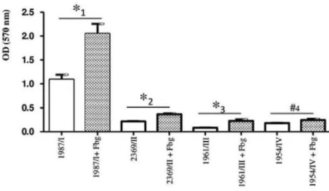
Bacterial adherence to hydrophobic polystyrene surface andinfluence of Fbg on biofilm formation - In addition to glass and catheter surfaces, we also investi-gated the ability of C. striatum to adhere to plastic us-ing negatively charged polystyrene 96-well flat-bottom microtitre plates. The quantification of biofilm revealed thatall C. striatum strains of PFGE types I, II, III and IV were able to adhere to the negatively charged plas-tic (polystyrene) surface at different intensities (Fig. 4). Again, the C. striatum 1987/I-MDR strain exhibited a significantly (p < 0.05) greater level of adherence to
this abiotic hydrophobic surface compared with the C. striatum 2369/II-MDR, 1961/III-multidrug-susceptible (MDS) and 1954/IV-MDS strains.
Several microorganisms use extracellular matrix proteins, such as Fbg, to increase their ability to inter-act with different cells and also with abiotic surfaces. To investigate the influence of Fbg on the ability of C. stri-atum to adhere to plastic microtitre plates, plates were pre-treated with human plasmatic Fbg prior to bacterial colonisation.As shown in Fig. 4, human Fbg enhanced biofilm formation on the polystyrene surfaces by the C. striatum 1987/I-MDR (p = 0.00362), 2369/II-MDR (p =
0.0022) and 1961/III-MDS (p = 0.0105) strains, suggest-ing that this matrix protein may contribute to the inter-action of different clones of this human pathogen with abiotic hydrophobic surfaces.
DISCUSSION
Bacterial biofilms form in association with many hu-man activities, including food processing, transportation, public infrastructure and, most importantly, healthcare (Rzhepishevska et al. 2013). Biofilms have been found on Fig. 3: scanning electron micrographs illustrating biofilm formation on polyurethane catheter surfaces after 24 h incubation with different
numerous medical devices (e.g., urinary catheters, central venous catheters and endoscopes). Their presence can have serious implications for immunocompromised pa-tients and those with indwelling medical devices (Brown & Williams 1985, Russell & Russell 1995, Rutala et al. 2008, Al Akhrass et al. 2012). The acquisition of the abil-ity to form biofilms may represent a good strategy for a microorganism to acquire enhanced survival under con-ditions of stress, e.g., during host invasion or following antibiotic treatment, because cells growing in biofilms are highly resistant to components of the human immune system and to numerous types of antimicrobial agents. In addition, the ability of bacterial cells to transfer genes horizontally is enhanced within biofilm communities, thereby facilitating the spread of antibiotic resistance (Stewart & Costerton 2001, Lee et al. 2008).
Our results reveal the capacities of diverse C. stria-tum isolates to adhere to various abiotic surfaces and to form biofilms in an in vitro catheter model. The data re-vealed variations among the capacities of diverse clones of MDR and MDS C. striatum strains identified during a nosocomial outbreak in RJ to adhere to and survive on positively and negatively charged abiotic surfaces. No-tably, we identified an association of increased biofilm formation, antimicrobial multiresistance and clonality of the C. striatum strains. In the present study, C. striatum
MDR PFGE types I and II were predominantly isolated during the nosocomial outbreak from in-patients under-going endotracheal intubation procedures in the ICU or in surgical wards. The clinical isolates of these PFGE types expressed a high capacity to form biofilms on hydrophilic (glass; positively charged) and hydrophobic (polystyrene; negatively charged) abiotic surfaces, including polyure-thane (positively charged) catheter surfaces.
The C. striatum strains showed properties similar to other pathogenic biofilm producers. The results of the semiquantitative roll-plate method and quantitative catheter culture assays showed that viable bacterial cells
extensively adhered to and multiplied on the surfaces of polyurethane catheters. SEM revealed a large amount of biofilm on polyurethane catheter surfaces produced by all C. striatum strains tested.
The developmental biology of biofilm formation can be characterised into three stages: initial attachment, development of microcolony formation and detachment (O’Toole et al. 2000). Similar to C. diphtheriae (Gomes et al. 2013), Acinetobacter baumannii (Rao et al. 2008) and other nondiphtherial Corynebacterium species (So-riano et al. 1993, Gomes et al. 2009), autoaggregative
C. striatum strains were able to attach to and form mi-crocolonies (a hallmark of biofilm formation) on abiotic surfaces. C. striatum also formed matrix-enclosed mi-crocolonies on in vitro colonised polyurethane surfaces. Hollow voids indicative of mature biofilm formation on the surfaces of polyurethane catheters were also ob-served. The formation of hollow voids seems to be in-volved in the dispersion of sessile bacterial cells during the final stage of biofilm formation, which can increase bacterial virulence (Rice et al. 2009).
Some bacterial properties are associated with biofilm production, including the increased synthesis of exopoly-saccharides, hydrophobic properties and the development of antibiotic resistance (Olson et al. 2002, Costerton et al. 2003, Rao et al. 2008). A previous study has addressed the prevention of biofilms and has shown that the surface charge of an abiotic substrate may influence the morphol-ogy and physiolmorphol-ogy of a biofilm (Rzhepishevska et al. 2013). In the present study, the MDR and MDS C. stria-tum strains were able to adhere at different levels to nega-tively charged plastic (polystyrene) and posinega-tively charged (glass) surfaces, as previously observed with C. diphthe-riae and/or C. urealyticum (Mattos-Guaraldi & Formiga 1991, Mattos-Guaraldi et al. 1999a, b, Soriano et al. 2009, Gomes et al. 2013). Polyurethane implanted subcutane-ously into mice led to an infiltration of erythrocytes and subsequent haemolysis, possibly due to the attraction of this positively charged plastic to negatively charged cells (Rigdon 1970). In accordance with previous observations of C. diphtheriae (Mattos-Guaraldi & Formiga 1991, Mattos-Guaraldi et al. 1999a, b, Gomes et al. 2013), the negatively charged cell surfaces of C. striatum strains and their adherence to polyurethane may be partially ex-plained by the positive electric charge associated with this polymer. Moreover, the amorphous deposited substances or glycocalyx noted surrounding C. striatum microcolo-nies on the surfaces of the polyurethane catheters suggest that this bacteria may produce or attract substances that strengthen their attachment to inert surfaces in vitro.
Hydrophobicity has been significantly associated with biofilm formation of lipophilic skin corynebacteria on solid surfaces (Kwaszewska et al. 2006). For C. diph-theriae strains, bacterial autoaggregation and hydropho-bicity are mainly related to biofilm formation on poly-styrene surfaces (Mattos-Guaraldi et al. 1999a). The cell surface hydrophobicity of C. striatum strains was dem-onstrated by their ability to adhere to polystyrene sur-faces. Therefore, C. striatum strains should be included among bacterial species that have a natural tendency to adhere to available biotic and/or abiotic surfaces and to Fig. 4: biofilm formation (24 h incubation) on polystyrene surface
form biofilm (Olson et al. 2002, Kwaszewska et al. 2006, Soriano et al. 2009) and that are also capable of rapid physiological responses following exposure to surfaces with varying physicochemical characteristics, enabling some bacterial colonisation on negatively charged sur-faces (Kwaszewska et al. 2006).
In natural environments, bacteria typically adhere to the layer of adsorbed molecules that coats inert sur-faces, the so-called “conditioning film” and not directly to the substratum. In vivo, any material surface is rap-idly covered by plasma and matrix proteins, to which bacteria may display specific adhesins. The stimulation of bacterial biofilm formation by exogenous mammalian proteins has been reported for many human pathogens (Bonifait et al. 2008, Gomes et al. 2009, 2013). Fbg is a major protein in human plas ma and is primarily in-volved in the coagulation cascade system through its conversion to insoluble fibrin. Fbg synthesis is dramati-cally upregulated during inflammation or under stress conditions, such as systemic infections. Fbg and fibrin play overlapping roles in blood clotting, fibrinolysis, the inflammatory response, cellular and matrix interactions and wound healing (Mosesson 2005). The Fbg bind-ing properties of Staphylococcus aureus (O’Neill et al. 2008), Streptococcus suis (Bonifait et al. 2008)and C. diphtheriae (Gomes et al. 2009, Sabbadini et al. 2010) allow them to attach to each other through Fbg-mediated cross-bridging, contributing to biofilm production. The ability of C. striatum strains to bind to Fbg was also dem-onstrated in the present study. In addition to the ability to form biofilms directly on hydrophilic and hydrophobic abiotic surfaces, C. striatum also produced biofilms on Fbg-associated “conditioning films”. Compared with the formation of biofilms on the uncoated polystyrene sur-faces, the Fbg-coated surfaces showed enhanced biofilm formation by the C. striatum 1987/I-MDR strain, which was responsible for a previous nosocomial outbreak. The enhancement occurred at a typical in vivo concentration of Fbg in blood plasma of approximately 2.5 mg/mL. Therefore, the expression of Fbg-binding adhesins at dif-ferent levels may be implicated in biofilm formation on “conditioning films” by C. striatum strains, as has been previously reported for S. suis (Bonifait et al. 2008).
In conclusion, C. striatum may form biofilms in vivo by an adherent biofilm mode of growth in vitro, as was demonstrated on hydrophilic and hydrophobic abiotic surfaces, including polyurethane catheters. The affinity of C. striatum for human Fbg was determined to be an ad-ditional potential virulence trait of this organism. In ad-dition to its multi-resistance to antimicrobial agents used in therapy, the ability to produce a “conditioning film” may contribute to the establishment and dissemination of nosocomial infections caused by this organism, includ-ing those in patients with indwellinclud-ing medical devices.
Thus, C. striatum strains capable of forming biofilms may be selected under antibiotic pressure, or conversely,
C. striatum may acquire resistance to multiple drugs with-in biofilm communities. In either event, the high colonis-ing capacity of C. striatum combined with its resistance to multiple drugs will contribute to the survival and further dissemination of this organism in the hospital setting.
REFERENCES
Al Akhrass F, Al Wohoush I, Chaftari AM, Reitzel R, Jiang Y,
Ghan-noum M, Tarrand J, Hachem R, Raad I 2012. Rhodococcus bacte-remia in cancer patients is mostly catheter related and associated with biofilm formation. PLoS ONE 7: e32945.
Baio PVP, Mota HF, Freitas AD, Gomes DLR, Ramos JN, Sant’Anna LO, Souza MC, Camello TCF, Hirata Jr R, Vieira VV,
Mattos-Guaraldi AL 2013. Clonal multidrug-resistant Corynebacterium striatum within a nosocomial environment, Rio de Janeiro, Bra-zil. Mem Inst Oswaldo Cruz 108: 23-29.
Boltin D, Katzir M, Bugoslavsky V, Yalashvili I, Brosh-Nissimov T, Fried M, Elkayam O 2009. Corynebacterium striatum - a classic pathogen eluding diagnosis. Eur J Intern Med20: 49-52.
Bonifait L, Grignon L, Grenier D 2008. Fibrinogen induces biofilm formation by Streptococcus suis and enhances its antibiotic re-sistance. Appl Environm Microbiol 74: 4969-4972.
Brown MRW, Williams P 1985. The influence of environment on envelope properties affecting survival of bacteria in infections.
Annu Rev Microbiol39: 527-556.
Camello TCF, Mattos-Guaraldi AL, Formiga LCD, Marques EA 2003. Nondiphtherial Corynebacterium species isolated from clinical specimens of patients in a university hospital, Rio de Ja-neiro, Brazil. Braz J Microbiol34: 39-44.
Campanile F, Carretto E, Barbarini D, Grigis A, Falcone M, Goglio A, Venditti M, Stefani S 2009. Clonal multidrug-resistant Coryne-bacterium striatum strains, Italy. Emerg Infect Dis 15: 75-78.
Costerton JW, Veeh R, Shirtliff M 2003. The application of biofilm science to the study of and control of chronic bacterial infections.
J Clin Inv 112: 1446-1477.
Dooley DP, Garcia A, Kelly JW, Longfield RN, Harrison L 1996.
Validation of catheter semiquantitative culture technique for non-staphylococcal organisms. J Clin Microbiol 34: 409-412.
Fernández-Ayala M, Nan DN, Fariñas MC 2001. Vertebral osteomy-Vertebral osteomy-elitis due to Corynebacterium striatum. Am J Med 111: 167.
Gomes DL, Martins CA, Faria LM, Santos LS, Santos CS, Sabba-dini PS, Souza MC, Alves GB, Rosa AC, Nagao PE, Pereira GA,
Hirata Jr R, Mattos-Guaraldi AL 2009. Corynebacterium diph-Corynebacterium diph-theriae as an emerging pathogen in nephrostomy catheter-related infection: evaluation of traits associated with bacterial virulence.
J Med Microbiol 58: 1419-1427.
Gomes DL, Peixoto RS, Barbosa EA, Napoleão F, Sabbadini PS, dos
Santos KR, Mattos-Guaraldi AL, Hirata Jr R 2013. SubMICs of penicillin and erythromycin enhance biofilm formation and hydrophobicity of Corynebacterium diphtheriae strains. J Med Microbiol 62: 754-760.
Kwaszewska AK, Brewczyńska A, Szewczyk EM 2006. Hydropho -bicity and biofilm formation of lipophilic skin corynebacteria.
Pol J Microbiol 55: 189-193.
Lee HW, Koh YM, Kim J, Lee JC, Lee YC, Seol SY, Cho DT, Kim J
2008. Capacity of multidrug-resistant clinical isolates of Acineto-bacter baumannii to form biofilm and adhere to epithelial cell surfaces. Clin Microbiol Infect 14: 49-54.
Lembke C, Podbielski A, Hidalgo-Grass C, Jonas L, Hanski E,
Kreikemeyer B 2006. Characterization of biofilm formation by clinically relevant serotypes of group A streptococci. Appl Envi-ronm Microbiol 72: 2864-2875.
Maki DG, Weise CE, Sarafin HW 1977. A semi-quantitative culture
method for identifying intravenous-catheter-related infection. N Engl J Med 296: 1305-1309.
Martinez-Martinez L, Suarez AI, Winstanley J, Ortega MC, Bernard K 1995. Phenotypic characteristics of 31 strains of Corynebac-terium striatum isolated from clinical samples. J Clin Microbiol 33: 2458-2461.
Martins CAS, Faria LMD, Souza MC, Camello TCF, Velasco E, Hira -ta Jr R, Thuler LCS, Mattos-Guaraldi AL 2009. Microbiological and host features associated with corynebacteriosis in cancer pa-tients: a five-year study. Mem Inst Oswaldo Cruz 104: 905-913.
Mattos-Guaraldi AL, Cappelli EA, Previato JO, Formiga LC, Andrade AF 1999a. Characterization of surface saccharides in two Coryne-bacterium diphtheriae strains. FEMS Microbiol Lett 170: 159-166.
Mattos-Guaraldi AL, Formiga LC 1991. Relationship of biotype and source to the hemagglutination and adhesive properties of Co-rynebacterium diphtheriae. Braz J Med Biol Res24: 399-406.
Mattos-Guaraldi AL, Formiga LC, Andrade AF 1999b. Cell surface hydrophobicity of sucrose fermenting and nonfermenting Co-rynebacterium diphtheriae strains evaluated by different meth-ods. Curr Microbiol 38: 37-42.
Moore K, Hall V, Paull A, Morris T, Brown S, McCulloch D, Rich
-ardson MC, Harding KG 2010. Surface bacteriology of venous leg
ulcers and healing outcome. J Clin Pathol 63: 830-834.
Mosesson MW 2005. Fibrinogen and fibrin structure and functions. J Thromb Haemost 3: 1894-1904.
O’Toole G, Kaplan HB, Kolter R 2000. Biofilm formation as micro -bial development. Annu Rev Microbiol 54: 49-79.
Oliva A, Belvisi V, Iannetta M, Andreoni C, Mascellino MT, Lichtner M, Vullo V, Mastroianni CM 2010. Pacemaker lead endocarditis due to multidrug-resistant Corynebacterium striatum detected with sonication of the device. J Clin Microbiol 48: 4669-4671.
Olson ME, Ceri H, Morck DW, Buret AG, Read RR 2002. Biofilm
bacteria: formation and comparative susceptibility to antibiotics. Can J Vet Res 66: 86-92.
O’Neill E, Pozzi C, Houston P, Humphreys H, Robinson DA, Lough -man A, Foster TJ, O’Gara JP 2008. A novel Staphylococcus au-reus biofilm phenotype mediated by the fibronectin-binding pro-teins, FnBPA and FnBPB. J Bacteriol 190: 3835-3850.
Otsuka Y, Ohkusu K, Kawamura Y, Baba S, Ezaki T, Kimura S 2006. Emergence of multidrug-resistant Corynebacterium striatum as a nosocomial pathogen in long-term hospitalized patients with underlying diseases. Diagn Microbiol Infect Dis 54: 109-114.
Rao RS, Karthika RU, Singh SP, Shashikala P, Kanungo R, Jayachan-dran S, Prashanth K 2008. Correlation between biofilm production and multiple drug resistance in imipenem resistant clinical isolates of Acinetobacter baumannii. Indian J Med Microbiol 26: 333-337.
Renom F, Garau M, Rubí M, Ramis F, Galmés A, Soriano JB 2007. Nosocomial outbreak of Corynebacterium striatum infection in patients with chronic obstructive pulmonary disease. J Clin Microbiol 45: 2064-2067.
Rice SA, Tan CH, Mikkelsen PJ, Kung V, Woo J, Tay M, Hauser A,
McDougald D, Webb JS, Kjelleberg S 2009. The biofilm life cycle and virulence of Pseudomonas aeruginosa are dependent on a filamentous prophage. ISME J 3: 271-282.
Rigdon RH 1970. Hemolysis associated with plastics - A histopatho -logic study with polyurethane. J Biomed Mater Res 4: 57-71.
Rufael DW, Cohn SE 1994. Native valve endocarditis due to Coryne-bacterium striatum: case report and review. Clin Infect Dis 19: 1054-1061.
Russell AD, Russell NJ 1995. Biocides: activity, action and resistance.
Symp Soc Gen Microbiol 53: 327-365.
Rutala WA, Weber DJ, Healthcare Infection Control Practices Advi-Healthcare Infection Control Practices Advi
-sory Committee (HICPAC) 2008. Guidelines for disinfection and sterilization in healthcare facilities, 2008, Centers for Disease control and Prevention, Atlanta, 158 pp.
Rzhepishevska O, Hakobyan S, Ruhal R, Gautrot J, Barberoc D, Ram -stedt M 2013. The surface charge of anti-bacterial coatings alters motility and biofilm architecture. Biomater Sci 1: 589-602.
Sabbadini PS, Genovez MRN, da Silva CF, Adelino TLN, dos Santos CS,
Pereira GA, Nagao PE, Dias AASO, Mattos-Guaraldi AL, Hirata Jr
R 2010. Fibrinogen binds to nontoxigenic and toxigenic Corynebac-terium diphtheriae strains. Mem Inst Oswaldo Cruz 105: 706-711.
Scholle DA 2007. Spontaneous joint infection with Corynebacterium striatum. J Clin Microbiol 45: 656-658.
Soriano F, Huelves L, Naves P, Rodríguez-Cerrato V, del Prado G,
Ruiz V, Ponte C 2009. In vitro activity of ciprofloxacin, moxi-floxacin, vancomycin and erythromycin against planktonic and biofilm forms of Corynebacterium urealyticum. J Antimicrob Chemother 63: 353-356.
Soriano F, Ponte C, Galiano MJ 1993. Adherence of Corynebacterium urealyticum (CDC group D2) and Corynebacterium jeikeium to intravascular and urinary catheters. Eur J Clin Microbiol Infect Dis12: 453-456.
Stepanovic S, Vukovic D, Davic I, Savic B, Svabic-Vlahovic M 2000. A modified microtiter-plate test for quantification of staphyloco-ccal biofilm formation. J Microb Meth 40: 175-179.
Stewart PS, Costerton JW 2001. Antibiotic resistance of bacteria in biofilms. Lancet 358: 135-138.
Tarr PE, Stock F, Cooke RH, Fedorko DP, Lucey DR 2003. Multidrug
resistant Corynebacterium striatum pneumonia in a heart trans-plant recipient. Transplant Infect Dis5: 53-58.
Weiss K, Labbé AC, Laverdière M 1996. Corynebacterium striatum
meningitis: case report and review of an increasingly important