Open Access
Research article
Characterization of a β-glucanase produced by Rhizopus microsporus
var. microsporus, and its potential for application in the brewing
industry
Klecius R Silveira Celestino
1, Ricardo B Cunha
2and Carlos R Felix*
1Address: 1Laboratório de Enzimologia, Departamento de Biologia Celular, Universidade de Brasília, Brasília, DF, CEP 70910-900, Brazil and 2Centro Brasileiro de Serviços e Pesquisas em Proteínas (LB QP)-Divisão de Química Analítica, Instituto de Química, Universidade de Brasília, Brasília, DF, CEP 70910-900, Brazil
Email: Klecius R Silveira Celestino - [email protected]; Ricardo B Cunha - [email protected]; Carlos R Felix* - [email protected] * Corresponding author
Abstract
Background: In the barley malting process, partial hydrolysis of β-glucans begins with seed germination.
However, the endogenous 1,3-1,4-β-glucanases are heat inactivated, and the remaining high molecular weight β-glucans may cause severe problems such as increased brewer mash viscosity and turbidity. Increased viscosity impairs pumping and filtration, resulting in lower efficiency, reduced yields of extracts, and lower filtration rates, as well as the appearance of gelatinous precipitates in the finished beer. Therefore, the use of exogenous β-glucanases to reduce the β-glucans already present in the malt barley is highly desirable.
Results: The zygomycete microfungus Rhizopus microsporus var. microsporus secreted substantial amounts
of β-glucanase in liquid culture medium containing 0.5% chitin. An active protein was isolated by gel filtration and ion exchange chromatographies of the β-glucanase activity-containing culture supernatant. This isolated protein hydrolyzed 1,3-1,4-β-glucan (barley β-glucan), but showed only residual activity against 1,3-β-glucan (laminarin), or no activity at all against 1,4-β-glucan (cellulose), indicating that the R.
microsporus var. microsporus enzyme is a member of the EC 3.2.1.73 category. The purified protein had a
molecular mass of 33.7 kDa, as determined by mass spectrometry. The optimal pH and temperature for hydrolysis of 1,3-1,4-β-glucan were in the ranges of 4–5, and 50–60°C, respectively. The Km and Vmax values for hydrolysis of β-glucan at pH 5.0 and 50°C were 22.39 mg.mL-1 and 16.46 mg.min-1, respectively.
The purified enzyme was highly sensitive to Cu+2, but showed less or no sensitivity to other divalent ions,
and was able to reduce both the viscosity and the filtration time of a sample of brewer mash. In comparison to the values determined for the mash treated with two commercial glucanases, the relative viscosity value for the mash treated with the 1,3-1,4-β-glucanase produced by R. microsporus var. microsporus. was determined to be consistently lower.
Conclusion: The zygomycete microfungus R. microsporus var. microsporus produced a 1,3-1,4-β-D-glucan
4-glucanhydrolase (EC 3.2.1.73) which is able to hydrolyze β-D-glucan that contains both the 1,3- and 1,4-bonds (barley β-glucans). Its molecular mass was 33.7 kDa. Maximum activity was detected at pH values in the range of 4–5, and temperatures in the range of 50–60°C. The enzyme was able to reduce both the viscosity of the brewer mash and the filtration time, indicating its potential value for the brewing industry.
Published: 05 December 2006
BMC Biochemistry 2006, 7:23 doi:10.1186/1471-2091-7-23
Received: 03 July 2006 Accepted: 05 December 2006
This article is available from: http://www.biomedcentral.com/1471-2091/7/23 © 2006 Celestino et al; licensee BioMed Central Ltd.
This is an Open Access article distributed under the terms of the Creative Commons Attribution License (http://creativecommons.org/licenses/by/2.0), which permits unrestricted use, distribution, and reproduction in any medium, provided the original work is properly cited.
Background
1,3-1,4-β-Glucans are polysaccharides, components of the cell walls of higher members of the Poaceae family. They areparticularly abundant in the endosperm cell walls of commercially valuable cereals such as barley, rye, sor-ghum, oats and wheat [1]. Structurally, these polysaccha-rides are linear glucans of up to 1,200 β-D-glucosyl residues linked through β-1,3 and β-1,4 glycosyl bonds. Variations in the proportions of 1,3-(25–30%) and β-1,4-linkages, and in the length of the mixed-linked seg-ments are currrently reported [2]. During malt produc-tion, partial hydrolysis of barley β-glucans begins with seed germination [13]. However, the endogenous 1,3-1,4-β-glucanases are heat inactivated, and the remaining high molecular weight β-glucans may cause severe problems such as increased brewer mash viscosity and turbidity[14]. Increased viscosity impairs pumping and filtration, caus-ing lower efficiency, reduced yields of extracts, and lower filtration rates, as well as the appearance of gelatinous pre-cipitates in the finished beer [2]. Thus, both the level of glucan-hydrolysing activities achieved during germina-tion and the amounts of their substrates, mainly 1,3-1,4-β-glucan, are important factors in the production of high quality malts. Addition of exogenous 1,3-1,4-β-glucanases to the mash could therefore be an outstanding option for improving the brewing process. However, the β-gluca-nases currently marketed do not really meet the brewing industry's needs, mainly due to economic factors. Novel 1,3-1,4-β-glucanases with uncommon features would be highly desirable. Here we report on the production, puri-fication and partial characterization of a 1,3-1,4-β-gluca-nase produced by R. microsporus var. microsporus, considering as well its potential for use in the brewing industry.
Results and Discussion
Enzyme production
A 1,3-1,4-β-glucan-degrading filamentous fungus was iso-lated from a malt silo in a brewery. This zygomycete microfungus was identified as Rhizopus microsporus var.
microsporus by rcDNA analysis. It grew strongly in liquid
medium containing chitin as the sole carbon source, and produced substantial amounts of β-glucanase activity (Figure 1) which was able to fully hydrolyze barley β-glu-can (1,3-1,4-β-gluβ-glu-can). The specificity of substrate hydrol-ysis by this enzyme (Table 2) fully supports the assumption that it belongs to the 3.2.1.73 category. The inducible nature of 1,3-1,4-β-glucanase production has already been reported for 1,3-β-glucanase from
Trichode-rma sp. [27]. Although cellulose and xylan were also
inducers, the levels of enzyme secreted in the presence of these carbohydrates were considered smaller than the activity induced by chitin. In cultures grown under agita-tion (120 rpm) at 40°C, the enzyme activity increased from a minimum to a maximum level within 24h of
growth. It has been reported that several other microrgan-isms, including Bacillus sp. [15]Trichoderma sp. [16],
Talaromyces emersonii [17], and Rhizobium sp. [2], produce
1,3-1,4-β-glucanase enzymes which are currently used in the brewing industry. However, depending on the sub-strate (β-glucan) used as inducer, production of the gluca-nases for industrial application may be very costly, to the point of being considered economically prohibitive [28]. Chitin, on the other hand, is a relatively cheap and readily available carbon source in comparison to barley β-glucan and laminarin. The ability, therefore, of R. microsporus var.
microsporus to produce 1,3-1,4-β-glucanase in the presence
of chitin, favors its use on an industrial scale.
Enzyme Purification
The culture supernatant of R. microsporus var. microsporus grown in liquid medium containing chitin was concen-trated 10-fold by ultrafiltration, using a 10 kDa cut-off membrane. No 1,3-1,4-β-glucanase activity was found in the filtrate. Chromatography of the concentrate on a Sephacryl S-100 gel filtration column (not shown) fol-lowed by chromatography on an SP-Sepharose ion exchange column resulted in elution of two peaks of pro-teins (PGI and PGII) (Figure 2). While the PGI propro-teins were inactive, the PGII protein (fractions 22–34) showed substantial activity against 1,3-1,4-β-glucan. A summary of the purification steps of the 1,3-1,4-β-glucanase pro-duced by the R. microsporus var. microsporus is shown in table 1. The enzyme was purified (Figure 3) 55.529-fold with a yield of 114.912% and a specific activity of 12.596 U.mg-1. The molecular mass of the PGII protein was 36.5
kDa, as indicated by SDS-PAGE analysis (Figure 3). This value is comparable to that (33.7 kDa) determined by
Time course of production of 1,3-1,4-β-glucanase by Rhizo-pus microsporus var. microsporus in the presence of 0.5% of either xylan, cellulose or chitin at a temperature of 40°C and at 120 rpm
Figure 1
Time course of production of 1,3-1,4-β-glucanase by
Rhizo-pus microsporus var. microsporus in the presence of 0.5% of
either xylan, cellulose or chitin at a temperature of 40°C and at 120 rpm 0 1 2 3 4 5 6 7 8 9 0 12 24 36 48 60 72 84 Time (h) A ctiv ity (U .m L -1)
Xylan Cellulose Chitin
mass spectrometry analysis for this enzyme (Figure 3). While 1,3-1,4-β-glucanases from Bacillus sp. have smaller molecular masses varying in the range of 25–30 kDa [2], the enzymes from Clostridium thermocellum (38 kDa) [34],
Bacteroides succinogenes (37 kDa) [33] and Talaromyces emersonii (40.7 kDa) [2] showed comparable molecular
mass values.
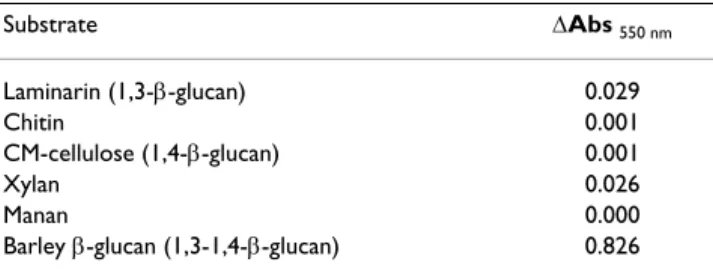
Enzyme specificity
The R. microsporus purified β-glucanase was tested for its ability to hydrolyze several other glucan substrates. As may be seen in table 2, only the barley β-glucan was effi-ciently hydrolyzed, as indicated by the much higher net absorbance. In comparison to the activity against the 1,3-1,4-β-glucan, very low or no activity at all was shown by the enzyme against the substrates laminarin (1,3-β-glu-can) and CM-cellulose (soluble 1,4-β-glu(1,3-β-glu-can), indicating clearly that the enzyme may be taken as a member of the EC 3.2.1.73 enzyme category.
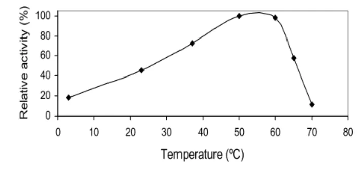
Effect of pH and temperature optima
The effect of pH and temperature on the activity of the purified 1,3-1,4-β-glucanase from Rhizopus microsporus
var. microsporus is shown in figures 4 and 5, respectively.
At 50°C, the enzyme showed substantial activity in the pH range of from 2 to 6. Maximal activity was recorded in the range of from 4 to 5. No enzyme activity was detected at pH higher than 6 (Figure 4). At pH 5.0, the purified enzyme was substantially active in the temperature range
Table 2: Hydrolysis of glucan substrates by the R. microsporus purified β-glucanase. ∆Abs 550 nm represents the net absorbance of the reaction mixture after incubation for 0.5 h with the enzyme at 50°C. Substrate ∆Abs 550 nm Laminarin (1,3-β-glucan) 0.029 Chitin 0.001 CM-cellulose (1,4-β-glucan) 0.001 Xylan 0.026 Manan 0.000
Barley β-glucan (1,3-1,4-β-glucan) 0.826
Ion exchange (SP-Sepharose column) chromatography of the concentrated culture filtrate of Rhizopus microsporus var. micro-sporus grown in liquid medium containing 0.5% chitin
Figure 2
Ion exchange (SP-Sepharose column) chromatography of the concentrated culture filtrate of Rhizopus microsporus var.
micro-sporus grown in liquid medium containing 0.5% chitin.
0
0,01
0,02
0,03
0,04
0,05
0,06
0,07
0,08
0,09
0,1
1
8
15
22
29
36
43
50
57
64
71
78
85
92
Fractions
A
b
s
(
280
nm
)
0
0,5
1
1,5
2
2,5
A
c
ti
v
ity
(
U
.m
L
-1)
Na
C
l(M
)
Abs (280nm)
Activity
NaCl(M)
PGI
PGII
from 20°C to 65°C. Maximal activity was detected at 50°C and 60°C, indicating that the optimal temperature for glucan hydrolysis is 55°C (Figure 5). The optima pH and temperature values determined for the purified 1,3-1,4-β-glucanase from R. microsporus var. microsporus were similar to those determined for 1,3-1,4-β-glucanases from several other fungi and bacteria [2]. In addition, these val-ues are comparable to those presented by enzymes cur-rently being used in the brewing industry [11,2]. The purified 1,3-1,4-β-glucanase retained 100% and 87% of its activity after incubation for 2 h and 24 h, respectively, at 50°C. The half-lives of the enzyme at the temperatures of 60°C and 70°C were found to be 10 min and 1 min, respectively. At 50°C, the half-life was 72 h (data not shown). For hydrolysis of β-glucan by a novel 1,3-1,4-β-glucanase produced by Bacillus halodurans C-125, the pH optimum was between 6 and 8, and the temperature
opti-mum was 60°C. After 2 h incubation at 50°C and 60°C, the residual activity remained 100% and 50%, respec-tively. The enzymatic activity was abolished after 3 min incubation at 70°C. The optimum temperature for hydrolysis of lichenan by a 1,3-1,4-β-glucanase from
Bacteroides succinogenes at ph 6.0 was 50°C [33].
Effect of metal ions
The effect of several ions on the activity of the purified 1,3-1,4-β-glucanase produced by R. microsporus var.
micro-sporus is shown in Table 3. The enzyme was sensitive to
copper and fairly sensitive to zinc and manganese, but insensitive to magnesium, calcium and aluminum (Table 3). Glucanases produced by Rhizopus oryzae [29], Bacillus
clausii [30], Bacillus halodurans [32] and Trichoderma har-zianum [31] show similar sensitivity to the divalent metal
ion copper.
SDS-PAGE (A) and MALDI-TOF mass spectrometry (B) analysis of the purified 1,3-1,4-β-glucanase from Rhizopus microsporus var.microsporus. A: line 1, molecular weight markers; line 2, PGI protein fraction; line 3, PGII protein fraction
Figure 3
SDS-PAGE (A) and MALDI-TOF mass spectrometry (B) analysis of the purified 1,3-1,4-β-glucanase from Rhizopus microsporus
var. microsporus. A: line 1, molecular weight markers; line 2, PGI protein fraction; line 3, PGII protein fraction. Figure 3
Table 1: Summary of the purification protocol of the 1,3-1,4-β-glucanase produced by Rhizopus microsporus var. microsporus.
Steps Total Protein(mg) Total Activity(U) Specific activity(U.mg-1) Purification (-fold) Yield(%)
Concentrated crude extract 1.574 0.228 0.197 1 100.000
Sephacryl S-100 eluate 0.235 0.227 0.966 4.915 99.561
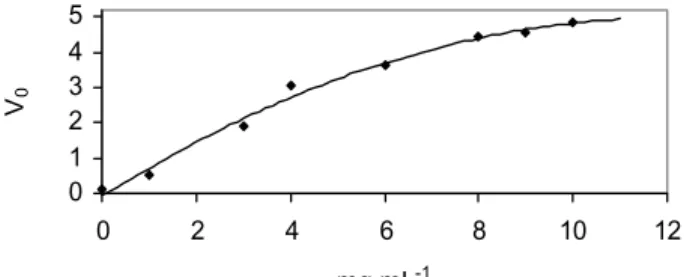
Kinetic Parameters
The purified 1,3-1,4-β-glucanase produced by R.
micro-sporus var. micromicro-sporus hydrolyzed 1,3 – 1,4-β-glucan in a
Michaelis-Menten fashion (Figure 7). Kinetic parameters were calculated using a Michaelis-Menten plot with a non-linear regression data analysis program [10]. Values of 19.8 mg.mL-1, 12.7s-1 and 16.5 U.mL-1 were determined
for Km, Kcat and Vmax, respectively. Km values of 1.2 – 1.5 mg.mL-1 for hydrolysis of barley β-glucan and 0.8 – 2
mg.mL-1 for lichenan were reported for the
1,3-1,4-β-glu-canase produced by Bacillus sp [2]. Values of 1,296 ± 51, 2.50 ± 0.09, and 518 were reported for Kcat (s-1), Km
(mg.mL-1) and Kcat/Km (s-1.M-1) respectively, for
hydrol-ysis of lichenan by a 1,3-1,4-β-glucanase produced by
Bacteroides succinogenes. [33].
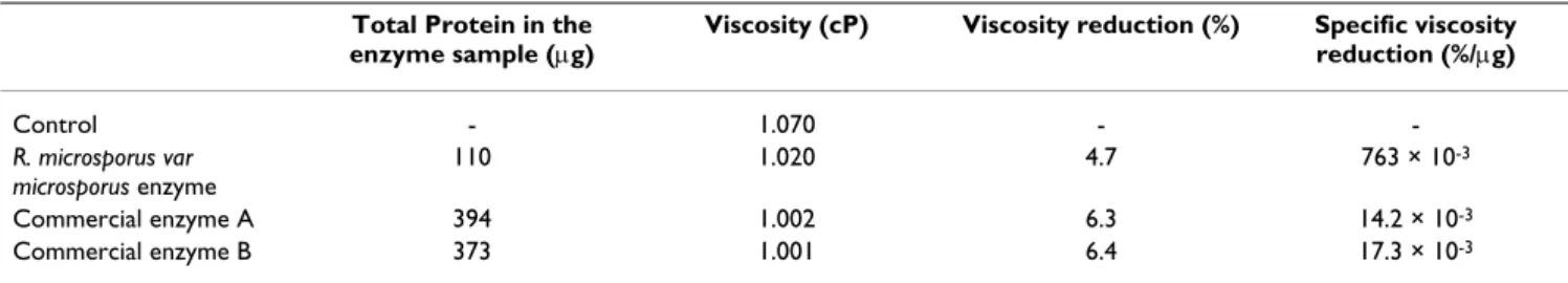
Capillary Viscosimetry and Filtration rate
The specific filtration rate and specific viscosity rate of the mash after incubation with the 1,3-1,4-β-glucanase from
R. microsporus var microsporus were compared with those
values calculated for two commercial β-glucanases cur-rently used in the brewing industry. The results are shown in Tables 4 and 5. Even at lower enzyme concentration, the 1,3-1,4-β-glucanase from R. microsporus var microsporus caused a higher reduction in the filtration rate (20.4%) of the mash (table 4). Similar results were obtained for the specific viscosity of the brewer's mash after treatment with the three β-glucanases (table 5).
Conclusion
The zygomycete Rhizopus microsporus var. microsporus pro-duced a 1,3-1,4-β-D-glucan 4-glucanhydrolase (EC 3.2.1.73) which could hydrolyze β-D-glucan substrate containing both 1,3- and 1,4-bonds. Its molecular mass as determined by both electrophoresis and mass spectrome-try (MALDI-TOF) was about 33.7 kDa. Its optimum pH and temperature were found to be in the ranges of 4–5 and 50–60°C, respectively. Kinetic analysis and its capac-ity to reduce both the viscoscapac-ity of the brewer mash and the
Thermostability of the purified 1,3-1,4-β-glucanase from Rhiz-opus microsporus var. microsporus, at temperatures of 50°C
(●), 60°C (❍) and 70°C (▲), at pH 5.0
Figure 6
Thermostability of the purified 1,3-1,4-β-glucanase from
Rhiz-opus microsporus var. microsporus, at temperatures of 50°C
(●), 60°C (❍) and 70°C (▲), at pH 5.0. 0 10 20 30 40 50 60 70 80 90 100 0 5 10 15 20 25 30 35 40 45 50 55 Time (minutes) R el ati ve a cti vi ty ( % ) Figure 6
Effect of temperature on the activity of the purified 1,3-1,4-β-glucanase from Rhizopus microsporus var. microsporus, at pH 5.0
Figure 5
Effect of temperature on the activity of the purified 1,3-1,4-β-glucanase from Rhizopus microsporus var. microsporus, at pH 5.0. 0 20 40 60 80 100 0 10 20 30 40 50 60 70 80 Temperature (ºC) R e la ti v e a c ti v ity ( % ) Figure 5
Effect of pH on the activity of the purified 1,3-1,4-β-glucanase from Rhizopus microsporus var.microsporus, at 50°C
Figure 4
Effect of pH on the activity of the purified 1,3-1,4-β-glucanase from Rhizopus microsporus var. microsporus, at 50°C.
0 20 40 60 80 100 0 1 2 3 4 5 6 7 8 9 10 pH
R
e
la
tiv
e
a
c
tiv
ity
(
%
)
Figure 4Table 3: Effect of metal ions on the activity of the purified 1,3-1,4-β-glucanase from Rhizopus microsporus var. microsporus.
Ion Residual activity (%)
Control 100 Cu+2 (12 mM) 0.3 Mg+2 (12 mM) 95.2 Fe+3 (12 mM) 89.6 Zn+2 (12 mM) 65.0 Mn+2 (12 mM) 62.3 Ca+2 (12 mM) 105.9 Al+3 (12 mM) 109.8
filtration time, indicate the possibility to use this enzyme in the brewing industry.
Methods
Chemicals
Barley 1,3-1,4-β-glucan, chitin, CM-cellulose, manan, xylan, laminarin, molecular mass standard proteins and sodium dodecyl sulfate (SDS) were from Sigma Chemical Co., USA. Sephacryl S-100 and SP-Sepharose were from Pharmacia-LKT, Sweden. All other chemicals were of ana-lytical grade.
Organism and enzyme production
The aerobic zygomycete microfungus Rhizopus microsporus
var. microsporus was isolated from a malt silo. The fungus
was maintained at 4°C, after growing for 48 hours in TLE modified solid medium [(0.5% chitin, 0.2% KH2PO4, 0.14%(NH4)2SO4, 0.03% MgSO4.7H2O, 0.0152% CaCl2, 0.02% glucose, 1.0 mL of 0.01% trace elements solutions (Fe+2, Mn+2, Zn+2, Co+2), 0.003% bactopeptone, 0.003%
urea, and 2% agar, pH 6.8)], at 40°C.
For enzyme production, one liter Erlemeyer flasks con-taining 250 mL of the liquid medium (TLE with no agar), were inoculated with 150 cm2 blocks of solid medium
taken from 2-day old R. microsporus var. microsporus cul-tures. Liquid cultures were then incubated for 24 hours with agitation (120 rpm) at 40°C. The culture superna-tants were then separated from the mycelium by filtration, using filter paper. The supernatants were then freeze-dried and used either for enzyme assay or enzyme purification as described in the following sections.
Enzyme assay
1,3-1,4-β-glucanase activity was assayed by the reducing-sugar method [6] with β-1,3-β-1,4-glucan as the substrate. The assay system consisted of 50 µL of 1% (wt/vol) β-glu-can dissolved in 100 mM sodium acetate buffer, pH 5.0, and 50 µL enzyme sample. The reaction was allowed to proceed for 30 min at 50°C, and was then stopped by the addition of 300 µL dinitrosalicylate reagent [6], and 5 min of boiling. The absorbance of the reaction mixture was determined at 550 nm using a Perkin Elmer mod. Lambda 11/Bio spectrophotometer. The amount of reducing sugar produced was determined using a curve constructed with glucose as standard. One unit of enzyme was defined as the amount of protein necessary to produce one µmol of reducing sugars.min-1.
The assays for xylanase, cellulase, 1,3-β-glucanase and mananase were performed as for 1,3-1,4-β-glucanase, except for the use of the substrates carboximetilcellulose, laminarin and manan, respectively. For chitinase the enzyme system consisted of 100 µL of enzyme sample, regenerated chitin 0,5% in 50 mM sodium acetate buffer, pH 5.2 [35]. The reaction was allowed to run for 12h at 37°C and stopped by addition of dinitrosalycilic reagent. The amount of reducing sugar produced was quantified using a standard curve constructed with glucose.
Purifications of the 1,3-1,4-β-glucanase from Rhizopus
microsporus var. microsporus
The supernatants from cultures of R. microsporus var.
microsporus grown in liquid medium containing β-glucan
were concentrated by ultrafiltration [(Amicon system; 10 k-Da cut-off membrane (PM10)]. Aliquots of concen-trated β-glucanase were loaded on a Sephacryl S-100 gel column (2.5 × 40 cm), equilibrated and eluted with 50 mM sodium acetate buffer, pH 5.0. Elution was
per-Table 4: Total protein in the enzyme samples, filtration time, filtration time reduction and specific filtration time reduction of the brewer's mash not treated or treated with enzymes.
Total Protein in the enzyme sample (µg)
Filtration time (seconds)
Filtration time reduction (%)
Specific filtration time reduction (%/µg) Control - 274 - -R. microsporus var microsporus enzyme 11.0 218 20.4 1854.5 × 10-3 Commercial enzyme A 394 195 29.9 75.9 × 10⁻³ Commercial enzyme B 373 176 35.8 95.8 × 10⁻³
Hydrolysis (µmol·min-1·mL-1) of β-glucan by the purified
1,3-1,4-β-glucanase from Rhizopus microsporus var. microsporus, in the presence of different concentrations of 1,3-1,4-β-glucan Figure 7
Hydrolysis (µmol·min-1·mL-1) of β-glucan by the purified
1,3-1,4-β-glucanase from Rhizopus microsporus var. microsporus, in the presence of different concentrations of 1,3-1,4-β-glucan.
0 1 2 3 4 5 0 2 4 6 8 10 12 mg.mL-1 V0 Figure 7
formed at a flow rate of 24 mL.h-1, and fractions of 4.0 mL
were collected. Active fractions were pooled and applied on a SP-Sepharose ion-exchange column (3.0 × 15 cm), previously equilibrated and eluted with 50 mM sodium acetate buffer, pH 5.0, and further eluted with a linear gra-dient formed with 100 mL of the acetate buffer and 100 mL of the same buffer containing 1 M NaCl. Elution was carried out at a flow rate of 24 mL.h-1, and fractions of 4
mL were collected. The resulting active fractions were pooled and dialyzed overnight against distilled water at 4°C, concentrated by ultrafiltration as above, and stored at -20°C until their use.
Protein determination
Protein was determined by the Bradford method [7], with bovine serum albumin as standard.
Electrophoresis
Enzyme samples were examined by electrophoresis under denaturing conditions in polyacrylamide slab gels (SDS-PAGE) as described by Laemmli [8]. Protein bands in the gel were visualized by the silver staining method [9].
Mass spectrometry
1,3-1,4-β-glucanase was analyzed by matrix-assisted laser desorption ionization-time of flight (MALDI-TOF) mass spectrometry with a Reflex IV mass spectrometer (Bruker Daltonik, Bremen, Germany) in linear positive mode. The purified enzyme sample (50 µg) was dissolved in 50 µL of 0.1 % (v/v) TFA, from which 1 µL was mixed with 1 µL of a saturated matrix solution of sinapinic acid dissolved in 50% (v/v) acetonitrile and 0.1% (v/v) trifluoroacetic acid, and applied to the MALDI plate. BSA was used for external mass calibration.
Effects of ions
The effects of several metallic ions (Cu+2, Mg+2, Fe+3, Zn+2,
Ca+2, and Al+3) on the purified 1,3-1,4-β-glucanase were
tested measuring the activity of the enzyme at 50°C (see 1,3-1,4–β-glucanase assay) in the presence of the ions.
pH and temperature optima
The effect of temperature on the enzyme was carried out at the temperature range of from 4° to 70°C, at pH 5.0 (in 50 mM sodium acetate buffer). The optimum pH value was determined by monitoring the enzyme activity at 50°C at pH values from 3.0 to 9.0. The following buffers were used: pH 3.0 – 6.0, 50 mM sodium acetate; pH 7.0, 50 mM sodium phosphate; and pH 8.0 – 9.0, 50 mM tris-HCl.
Kinetic Parameters
For determination of kinetic parameters, the enzyme assays were performed at 50°C, using 1,3-1,4-β-glucan at concentrations varying from 0.05 to 2.0 % dissolved in 50 mM sodium acetate buffer, pH 5.0. Km, Kcat and Vmax values were obtained using a Michaelis-Menten plot with a non-linear regression data analysis program [10].
Preparation of the mash
12.5 g of malt was triturated in a hammer mill (MARO-TEC), drizzled into a sieve of 0.2 mm spacing, and dis-solved in 50 mL of sodium acetate buffer (100 mM, pH 5.5), pre-heated to 45°C. Reaction started with 1.0 mL of enzyme sample taken from chromatography on a Sephacryl S-100 column, and allowed to proceed for 30 min at 45°C, followed by other periods of 10 min at 50°C, 15 min at 60°C, 60 min at 70°C, and 5 min of boil-ing. The reaction was then stopped by the addition of 100 mL of cold water and immediate cooling in an ice-water bath at about 20°C.
Capillary viscosimetry
Decrease in mash viscosity was measured by capillary vis-cosimetry using an Oswald viscosimeter [12,20]. Samples of 30 mL of mash were filtered using filter paper and placed in a viscosimeter at 20°C. Mash viscosity in the absence of enzyme was used as a control. The specific vis-cosity rate was calculated using the following equations: µmash = (µwater × Tmash × ρmash)/(Twater × ρwater) (1)
Table 5: Total protein in the enzyme samples, viscosity, viscosity reduction and specific viscosity reduction of the brewer's mash not treated or treated with enzymes.
Total Protein in the enzyme sample (µg)
Viscosity (cP) Viscosity reduction (%) Specific viscosity
reduction (%/µg) Control - 1.070 - -R. microsporus var microsporus enzyme 110 1.020 4.7 763 × 10-3 Commercial enzyme A 394 1.002 6.3 14.2 × 10⁻³ Commercial enzyme B 373 1.001 6.4 17.3 × 10⁻³
∆µ = (µmash control – µmash) × 100/(µmash control) (2) ∆µφ = ∆µ/δ (3)
Where µ is the viscosity, T is the flow time, ∆µ is viscosity reduction, δ is total protein, ∆µφ is specific viscosity rate and ρ is the density.
Filtration rate
The filtration rate was determined by filtration of 50 mL of mash through a filter paper [12]. Filtration rate in the absence of enzyme was used as a control. The specific fil-tration rate was calculated using the following equations: ∆ψ = (ψmash control – ψmash) × 100/(ψmash control) (4)
∆ψφ = ∆ψ/δ (5)
Where ψ is the flow time, ∆ψ is the filtration time of 50 mL, δ is total protein and ∆ψφ is the specific filtration reduction.
Authors' contributions
KRSC conceived of the study and participated in its design, performed all experiments, data quantification and was involved in the literature search and data inter-pretation. RBC participated and supported the mass spec-trometry analysis with a Reflex IV mass spectrometer. CRF participated in the design and coordination of the study, as well as on its supervision, and helped to draft the man-uscript. All authors read and approved the final manu-script.
Acknowledgements
CRF acknowledge the scholarship awarded by CNPq (process: 305123/ 2005-0).
References
1. Stone BA, Clarke AE: Chemistry and Biology of 1,3-β-Glucans. Bundoora: La Trobe University Press;; 1992.
2. Planas A: Bacterial 1,3-1,4-β-glucanases: structure, function
and protein engineering. Biochimica et Biophysica Acta 2000, 1543:361-382.
3. Bamforth CW: β-Glucan and β-glucanases in malting and
brewing: practical aspects. Brewers Digest 1994, 69:12-16.
4. Fincher GB, Stone BA: Advances in cereal science and
technol-ogy. Volume 7. American Association of Cereal Chemists. St. Paul;
1986.
5. Godfrey T, West S: Industrial enzymology. New York : Stockton Press; 1996.
6. Miller GL: Use of dinitrosalycilic acid reagent for
determina-tion of reducing sugar. Analytical Chemistry 1959, 31:426-428.
7. Bradford MM: A rapid and sensitive method for the
quantita-tion of microgram quantities of protein utilizing the princi-ple of protein dye binding. Analitical Biochemical 1976, 72:248-254.
8. Laemmli UK: Cleavage of structural proteins during assembly
of head of bacteriophage-T4. Nature 1970, 227:680-685.
9. Blum H, Beier H, Gross B: Improved silver staining of plant
pro-teins, RNA and DNA in polyacrilamide gels. Electrophoresis
1987, 8:93-99.
10. Leatherbarrow RJ: Enzifitter: a non-linear regression data
anal-ysis program for the IBM PC. London: Biosoft; 1987:1-91.
11. McCleary BV, Nurthen E: Measurement of
(1–3)(1–4)-β-D-glu-can in malt, wort and beer. Journal of the Institute of Brewing 1986, 92:168-173.
12. McCleary BV, Shameer I, Glennieholmes M: Measurement of (1–
3),(1–4)-β-D-glucan. Methods in Enzymology 1988, 160:545-551.
13. Kettunen A, Hamalainen JJ, Stenholm K, Pietila K: A model for the
prediction of β-glucanase activity and beta-glucan concen-tration during mashing. Journal of Food Engineering 1996, 29:185-200.
14. Bamforth CW, Martin HL: The degradation of β-glucan during
malting and mashing – the role of β-glucanase. Journal of the Institute of Brewing 1983, 89:303-307.
15. McCarthy T, Hanniffy O, Lalor E, Savage AV, Tuohy MG: Evaluation
of three thermostable fungal endo-β-glucanases from Talaromyces emersonii for brewing and food applications. Process Biochemistry 2005, 40:1741-1748.
16. Bhat MK: Cellulases and related enzymes in biotechnology.
Biotechnology Advances 2000, 18:355-383.
17. Jayus , McDougall BM, Seviour RJ: Purification and
characteriza-tion of the (1>3)-β-glucanases from Acremonium sp IMI 383068. FEMS Microbiology Letters 2004, 230:259-264.
18. Scheffler A, Bamforth CW: Exogenous β-glucanases and
pen-tosanases and their impact on mashing. Enzymes and Microbial Technology 2005, 36:813-817.
19. Murray PG, Grassick A, Laffey CD, Cuffe MM, Higgins T, Savage AV, Planas A, Tuohy MG: Isolation and characterization of a
ther-mostable endo-β-glucanase active on 1,3-1,4-β-D-glucans from the aerobic fungus Talaromyces emersonii CBS 814.70. Enzyme and Microbial Technology 2001, 29:90-98.
20. Vlasenko EY, Ryan AI, Shoemaker CF, Shoemaker SP: The use of
capillary viscometry, reducing end-group analysis, and size exclusion chromatography combined with multi-angle laser light scattering to characterize endo-1,4-β-D-glucanases on carboxymethylcellulose : A comparative evaluation of the three methods. Enzyme amd Microbial Technology 1998, 23:350-359.
21. Gan Q, Howell JA, Field RW, England R, Bird MR, O'Shaughnessy CL, MeKechinie MT: Beer clarification by microfiltration – product
quality control and fractionation of particles and macromol-ecules. Journal of Membrane Science 2001, 194:185-196.
22. Wang JM, Zhang GP, Chen JX, Wu FB: The changes of β-glucan
content and β-glucanase activity in barley before and after malting and their relationships to malt qualities. Food Chemis-try 2004, 86:223-228.
23. Wen TN, Chen JL, Lee SH, Yang NS, Shyur LF: A Truncated Fibro-bacter succinogenes 1,3-1,4-β-D-Glucanase with Improved
Enzymatic Activity and Thermotolerance. Fibrobacter succino-genes 2005, 44:9197-9205.
24. Lusk LT, Duncombe GR, Kay SB, Navarro A, Ryder D: Barley
β-glu-can and beer foam stability. Journal of the Ameriβ-glu-can Society of Brew-ing Chemists 2001, 59:183-186.
25. Edney MJ, LaBerge DE, Langrell DE: Relationships among the
β-glucan contents of barley, malt, malt congress extract, and beer. Journal of the American Society of Brewing Chemists 1998, 56:164-168.
26. Almin KE, Eriksson KE: Enzymic degradation of polymers .I.
vis-cometric method for determination of enzymic activity. Bio-chimica Et Biophysica ACTA 1967, 139:238-248.
27. Lima LHC, Ulhoa CJ, Fernandes AP, Felix CR: Purification of a
chi-tinase from Trichoderma sp. and its action on Sclerotium rolfsii and Rhizoctonia solani cell walls. Journal of General and Applied Microbiology 1997, 43:31-37.
28. McCarthy TC, Lalor E, Hanniffy O, Savage AV, Tuohy MG:
Compar-ison of wild-type and UV-mutant beta-glucanase-producing strains of Talaromyces emersonii with potential in brewing applications. Journal of Industrial Microbiology & Biotechnology 2005, 32:125-134.
29. Murashima K, Nishimura T, Nakamura Y, Koga J, Moriya T, Sumida N, Yaguchi T, Kono T: Purification and characterization of new
endo-1,4-β-D-glucanases from Rhizopus oryzae. Enzyme and Microbial Technology 2002, 30:319-326.
30. Miyanishi N, Hamada N, Kobayashi T, Imada C, Watanabe E:
Purifi-cation and characterization of a novel extracellular β-1,3-glucanase produced by Bacillus clausii NM-1 isolated from ezo abalone Haliotis discus hannai. Journal of Bioscience and Bio-engineering 2003, 95:45-51.
Publish with BioMed Central and every scientist can read your work free of charge
"BioMed Central will be the most significant development for disseminating the results of biomedical researc h in our lifetime."
Sir Paul Nurse, Cancer Research UK Your research papers will be:
available free of charge to the entire biomedical community peer reviewed and published immediately upon acceptance cited in PubMed and archived on PubMed Central yours — you keep the copyright
Submit your manuscript here:
http://www.biomedcentral.com/info/publishing_adv.asp
BioMedcentral
31. Rana DS, Theodore K, Naidu GSN, Panda T: Stability and kinetics
of β-1,3-glucanse from Trichoderma harzianum. Process Bio-chemistry 2003, 39:149-155.
32. Akita M, Kayatama K, Hatada Y, Ito S, Horikoshi K: A novel
β-glu-canase gene from Bacillus halodurans C-125. FEMS Microbiology Letters 2005, 248:9-15.
33. Erfle JD, Teather RM, Wood PJ, Irvin JE: Purification and
proper-ties of a 1,3-1,4-β-D-glucanase (lichenase, 1,3-1,4-β-D-glucan 4-glucanohydrolase, EC-3.2.1.73) from Bacteroides
succino-genes cloned in Escherichia coli. Biochemical Journal 1988,
255:833-841.
34. Schimming S, Schawarz WH, Staudenbauer WL: Properties of a
thermoactive β-1,3-1,4-glucanase (lichenase) from
Clostrid-ium thermocellum expressed in Escherichia coli. Biochemical and
Biophysical Research Communications 1991, 177:447-452.
35. Molano J, Duram A, Cabib E: A rapid and sensitive assay for