TRANSMISSÃO CONGÊNITA EM CABRAS REINFECTADAS COM
Toxoplasma gondii
CONGENITAL TRANSMISSION IN REINFECTED GOATS WITH Toxoplasma
gondii
SILVA, Helenara Machado da1,*; PEREIRA, Marina Mendonça1; OLIVEIRA, Thays Ávila de1; ALMEIDA, Henrique Meiroz de Souza1; GARCIA, João Luís2; LANGONI,
Hélio3; PEREIRA, Virgínia Bodelão Richini3; BRESCIANI, Kátia Denise Saraiva4; SOARES, Vando Edésio5; COSTA, Avimar José da1
1UNESP, FCAV, CPPAR, Depto. Patologia Veterinária, Jaboticabal, SP, Brazil. 2UEL, CCA, Depto. Medicina Veterinária Preventiva, Londrina, PR, Brazil.
3UNESP, FMVZ, Depto. Higiene Veterinária e Saúde Pública, Botucatu, SP, Brazil. 4UNESP, FMVA, Araçatuba, SP, Brazil, Depto. Apoio, Produção e Saúde Animal. 5Universidade Camilo Castelo Branco, Descalvado, SP, Brazil.
*Corresponding author: email address: helenarasilva@yahoo.com.br, phone: +55 16 3203-9300.
RESUMO
Avaliou-se o potencial de transmissão congênita em cabras experimentalmente reinfectadas com Toxoplasma gondii, em três estágios gestacionais (inicial, intermediário e final). Das 25 fêmeas não gestantes negativas para T. gondii, 20 foram inoculadas oralmente com 2,5x10³ oocistos de T. gondii cepa ME49. Destas, 15 fêmeas gestantes cronicamente infectadas foram reinoculadas, via oral, com 2,5x10³ oocistos T.
gondii cepa VEG. Cinco grupos experimentais foram formados (n=5): I, II e III
(reinoculações nos estágios gestacionais inicial, intermediário e final, respectivamente), IV (inoculação) e V (não inoculação). Exames clínicos e sorológicos (IgG RIFI [reação de imunofluorescência indireta]) em diferentes dias de avaliação, e bioensaio e PCR foram realizados em todos os animais. Nas cabras infectadas com T. gondii foram observados um pico de 40,2°C (IV) aos nove, soroconversão (IgG ≥64) aos 21 e estabilização (IgG <1024) aos 119 dias pós inoculação. Nas cabras reinfectadas com T.
gondii ocorreu um aumento nos títulos de IgG (≥1024) aos 28 (I) , 7 (II) e 3 (III) dias
pós-reinoculação. Durante o parto foram observados apenas nos grupos reinfectados: distocia, deformidades corporais, natimortalidade e fraqueza, e anticorpos IgG
anti-Toxoplasma foram detectados em todas e em algumas crias das cabras reinfectadas e
infectadas, respectivamente. Parasitismo tissular por T. gondii foi diagnosticado por bioensaio e PCR em cabras infectadas e reinfectadas e em sua prole. A toxoplasmose congênita foi possível em caprinos cronicamente infectados e reinfectados com T.
gondii. A infecção primária com T. gondii não protegeu as cabras prenhes contra a
doença congênita resultante de reinfecção toxoplásmica, em diferentes estágios de gestação (inicial, intermediário e final).
Palavras-chave: cabra, cepa VEG, reinfecção toxoplásmica, toxoplasmose congênita ABSTRACT
This study evaluated the potential of congenital transmission in goats experimentally infected and reinfected with Toxoplasma gondii, in three gestational stages (initial,
intermediate and final). Of the 25 non-pregnant females negative for T. gondii, 20 were orally inoculated with 2.5 x 10³ T. gondii ME49 oocysts. Of these, 15 pregnant females chronically infected were reinoculated, via oral, with 2.5 x 10³ T. gondii VEG oocysts. Five experimental groups were formed (n=5): I, II and III (reinoculations in the initial, intermediate and final gestational stage, respectively), IV (inoculation) and V (no inoculation). Clinical and serological exams (IgG IFAT [indirect immunofluorescence antibody test]) in different days of evaluation, and bioassay and PCR were performed in all goats. In the infected goats with T. gondii a peak of 40.2°C (IV) at nine, seroconversion (IgG≥64) at 21 and stabilization (IgG<1024) at 119 days post-inoculation were observed. In the reinfected goats with T. gondii occurred an increase in IgG titers (≥1,024) at 28 (I), 7 (II) and 3 (III) days post-reinoculation. During kidding were observed only in the reinfected groups: dystocia, malformation body, stillbirth and weakness, and IgG anti-Toxoplasma were detected in all and in some offsprings of the reinfected and infected goats, respectively. Tissue parasitism by T. gondii was diagnosed by bioassay and PCR in infected and reinfected goats and in their offspring. The congenital toxoplasmosis was possible in goats chronically infected and reinfected with T. gondii. The primary infection with T. gondii did not protect the pregnant goats against congenital disease resulting from toxoplasmic reinfection, in different gestational stages (initial, intermediate and final).
INTRODUCTION
Goats are commonly infected with Toxoplasma gondii, and the consequences of this parasitism directly affect the reproductive system. The transplacental transmission of T. gondii is a important route of infection and the increase in body temperature is often observed in animals infected with T. gondii (MAMIDI et al., 2002; ABOUZEID ET AL., 2010).
Several studies have described reproductive disorders, such as placental lesions and congenital transmission (DUBEY et al., 1980; ABOUZEID et al., 2010) and sexual transmission (SANTANA et al., 2013), as a result of toxoplasmic infection. However, studies concerning the consequences of reinfection of goats by T. gondii are still lacking, especially when there is a risk of animals coming into contact with different strains of the parasite. Immune protection conferred by one strain of T. gondii can be breached by reinfection with a strain belonging to another genotype (DAO et al., 2001). This can not be confused with reacutization of toxoplasmosis in animals.
Murine models have been extensively used to study toxoplasmic reinfections among individuals with a primary T. gondii infection (ARAÚJO et al., 1997; DAO et al., 2001; DZITKO et al., 2006; ELBEZ-RUBINSTEIN et al., 2009). These authors recommend further studies about reinfection with T. gondii on other animal models because this finding is extremely relevant for human and veterinary medicine. In one case, BRESCIANI et al. (2009) demonstrated transplacental transmission of T. gondii in reinfected female dogs. Thus, our objective was to evaluate the potential of congenital transmission in goats experimentally infected and reinfected with Toxoplasma gondii, in three gestational stages (initial, intermediate and final).
MATERIAL AND METHODS
The experiment was performed at the division of small ruminants at the “Centro de Pesquisas em Sanidade Animal (CPPAR)”, of “Faculdade de Ciências Agrárias e Veterinárias (FCAV)”, of “Universidade Estadual Paulista "Julio de Mesquita Filho" (UNESP)”, Jaboticabal Campus, São Paulo State, Brazil. The experimental period was from February 2010 to June 2011.
The study project was approved by the Ethics Committee on Animal Use (Comissão de Ética no Uso de Animais - CEUA), FCAV / UNESP (Protocol no. 010192), in June 2009.
Two Boer bucks (18 months and four years old) and 25 crossbred (Boer x Saanen) non-pregnant females of reproductive age (18 months to three years), seronegative for T. gondii, Neospora caninum, Brucella e Leptospira, were selected and remained in quarantine for three months. The animals were initially submitted to serological exams: indirect immunofluorescence antibody test (IFAT) for the detection of antibodies against T. gondii (CAMARGO, 1964) and N. caninum (CONRAD et al., 1993), acidified plate antigen test (ALTON et al., 1988) and microscopy serum agglutination test (COLE et al., 1973) were performed for the diagnosis of Brucella and
Leptospira, respectively. The females were kept in collective stalls, and the bucks were
housed individually. Quality food and drinking water were provided ad libitum.
We opted for a heterologous challenge to distinguish of reacutization of toxoplasmosis, and strains ME49 (type II) and VEG (type III) were used in inoculation and reinoculation with fresh inoculum of T. gondii oocysts, respectively. The oocysts T.
gondii (ME49 and VEG strains) used in the challenges of the animals were kindly
provided by Prof. João Luís Garcia (UEL, Brazil).
Immediately prior to the inoculation with T. gondii oocysts on day zero (D0), the 25 non-pregnant females were randomly distributed to the five experimental groups (Table 1), with five animals each (n=5) and transferred to five distinct stalls. Twenty non-pregnant females were orally inoculated with 2.5 x 10³ T. gondii ME49 oocysts belonging to groups: I, II, III and IV and the five remaining females were kept as negative control group (V - no inoculation).
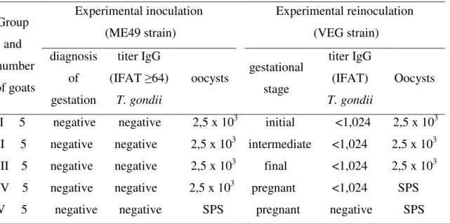
Table 1. Experimental design of goats that were non-inoculated, inoculated and reinoculated orally with Toxoplasma gondii oocysts.
Group and number of goats Experimental inoculation (ME49 strain) Experimental reinoculation (VEG strain) diagnosis of gestation titer IgG (IFAT ≥64) T. gondii oocysts gestational stage titer IgG (IFAT) T. gondii Oocysts I 5 negative negative 2,5 x 10³ initial <1,024 2,5 x 10³ II 5 negative negative 2,5 x 10³ intermediate <1,024 2,5 x 10³ III 5 negative negative 2,5 x 10³ final <1,024 2,5 x 10³ IV 5 negative negative 2,5 x 10³ pregnant <1,024 SPS V 5 negative negative SPS pregnant negative SPS
IFAT: indirect immunofluorescence assay test. SPS: sterile physiological solution.
The reproductive management started when all the inoculated females with T.
gondii ME49 oocysts of groups I, II, III and IV had titers IgG stabilized (IFAT<1,024).
Hormone treatment was used for the induction and synchronization of ovulation (MAIA, 1997) to allow for mating to occur at a fixed time for all the females of the five experimental groups. At 35 days post-mating, the females underwent transabdominal ultrasonography to confirm the pregnancy. After of the confirmed the pregnancy, the ultrasound screenings were performed every 15 days until the end of gestation.
Fifteen pregnant females that had been previously infected (ME49 strain) belonging to groups I, II and III and had IgG titers stabilized (IFAT<1,024) were reinoculated orally with 2.5 x 10³ T. gondii VEG oocysts (Table 1). In these groups, the experimental reinoculation were performed at different gestational stages (days of gestation), being: I – initial (40 days of gestion), II – intermediate (80 days of gestation) and III final (120 days of gestation). The remaining pregnant females comprised the positive (IV - infected) and negative (V - uninfected) control groups.
Before and after natural mating, the males were examined for the presence of antibodies anti-T. gondii, N. caninum, Brucella and Leptospira.
On days 0 (prior to inoculation), three, six, nine, 15, 21 and every seven days post-inoculation (DPI), the animals underwent clinical tests (heart and respiratory rates and rectal temperature), and blood samples (10 mL/goat) were collected by jugular venipuncture using vacuum tubes without an anticoagulant.
For the experimental reinfections, the females underwent clinical tests and blood sampling at three days post-reinoculation (DPR) and then weekly until the end of gestation.
Serum anti-Toxoplasma IgG antibodies were measured by the IFAT (CAMARGO, 1964). The samples were considered positive when the IgG titers were ≥64 (FIGUEIREDO et al., 2001). The slides used for the IFAT were prepared with antigens from the RH strain (Sabin, 1941).
The IFAT (CONRAD et al., 1993), buffered acidified plate antigen test (ALTON et al., 1988) and microscopy serum agglutination test (COLE et al., 1973) were performed for the diagnosis of N. caninum, Brucella and Leptospira, respectively. 2.4 T. gondii detection in the tissues of goats and offspring
All births were monitored by a veterinarian. After kidding, blood samples were collected from the goats (jugular venipuncture) and their offspring (heart blood and had not received colostrum) to perform the IFAT (CAMARGO 1964). Then, the animals were euthanized (AVMA, 2007) and necropsied to collect different tissues to T. gondii detection by bioassay and polymerase chain reaction (PCR).
The following tissue samples were collected from the adult goats and their offspring: central nervous system (CNS), lungs, heart, liver, spleen, kidneys and skeletal muscle, and only of the adult goats: ovaries, uterus and placenta. Each tissue was harvested in triplicate (2 cm³).
For bioassay, there was a pooled of tissues to goats and other to offspring, and the isolation of T. gondii in the tissues was performed according to the methodology proposed by DUBEY (1998). In 50mL aliquot of each pooled of tissue was added autoclaved buffer solution (pH 7.2) and antibiotic. Subsequently, 0.3mL of aliquot
diluted of each pooled of tissues of the goats and other of the offspring was inoculated intraperitoneally in mice, being six mice/ pooled of tissues. The mice were monitored daily for six weeks (COSTA et al., 1977) for clinical signs indicative of toxoplasmosis. Moreover, anti-Toxoplasma IgG antibodies (IFAT ≥32, CAMARGO, 1964) and brain cysts (DUBEY et al., 1998) were investigated in the mice.
For PCR, all the tissues collected from the goats and their offspring were processed separately and stored (-20°C) for later analysis recommended by FUENTES et al. (1996). The extraction of T. gondii DNA from animal tissue samples was performed with the Illustra TM Genomic Prep Cells & Tissue Mini Spin kit (GE Healthcare, USA). The use this kit involved the following steps: homogenization of animal tissue, lysis (incubated 56ºC for 1h, with frequent mixing (15min) by inversion), purification (ultra pure sterile water), wash and dry, elution and genomic DNA ready for downstream application.
Primers TOX4 (5'CGCTGCAGGGAGGAAGACGAAAGTTG'3) and TOX5 (5'CGCTGCAGACACAGTGCATCTGGATT'3) were used to amplify a 529-bp fragment (HOMAN et al., 2000). The reactions (final volume of 25µl) were prepared in microtubes containing the following reagents: 10mM Tris HCl pH 8.0, 50 mM KCl, 1.5 mM MgCl2, 0.2 mM dNTP, 10 ρmol of each primer, 0.2 units of Taq DNA polymerase and 10ng of DNA template. Amplification of DNA from parasites were performed over 35 cycles in EP Mastercycler Gradient thermocycler (Eppendorf), using the following cycling conditions: 7min at 94ºC for denaturation in cycle one, followed by 35 cycles on 60s at 94ºC for denaturation, 60s at 60ºC for annealing and 60s at 72ºC for extension, cycle 35 was followed by a final extension of 10min at 72ºC. The amplicon was analyzed by electrophoresis on a 1.5% agarose gel with SYBR® safe DNA gel stain (Invitrogen) and than stained with ethidium bromide and recorded in a Gel Documentation System (UVP, USA).
RESULTS
After inoculation with T. gondii ME49 oocysts, the goats belonging at groups I, II, III and IV had alterations in rectal temperature, this did not was observed in the goats
of the group V (no-inoculation). Hyperthermia was verified at 3º and 9º days post-inoculation (DPI) in all groups infected with T. gondii, being observed in the group IV a peak of the 40.2°C in the 9º DPI. In the 21º DPI were detected anti-Toxoplasma IgG antibodies (IFAT≥64) in all infected females with T. gondii which reached mean titers serum IgG as high as 16,384 (Table 2). In the 63º DPI, the IgG titers decreased (IFAT≥1,024), and in the 105º DPI the titers were lower than 1,024 (Table 2). The stabilization of the IgG titers below 1,024 was observed in all the females infected with
T. gondii in the 119º DPI (Table 2), when started the reproductive management in all
the experimental females.
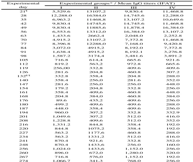
Table 2. Mean IgG antibody titers (indirect immunofluorescence assay test ‒ IFAT) of infected and reinfected goats with Toxoplasma gondii.
I II III IV 21¶ 5,529.6 13107,2 9,830.4 11,468.8 28 12,288.0 16384,0 14,745.6 12,288.0 35 6,963.2 11468,8 13,107.2 10,649.6 42 9,830.4 14745,6 14,745.6 11,468.8 49 9,830.4 11885,6 11,878.4 16,384.0 56 6,553.6 13312,0 16,384.0 13,107.2 63 1,433.6 2662,4 2,048.0 2,252.8 70 4,915.2 13107,2 7,372.8 11,468.8 77 8,396.8 12288,0 7,168.0 9,420.8 84 3,072.0 4915,2 8,192.0 7,372.8 91 1,638.4 4915,2 8,192.1 3,276.8 98 1,587.2 3174,4 3,481.6 3,891.2 105 716.8 614,4 665.6 921.6 112 819.2 563,2 972.8 665.6 119 256,00 332,8 409.6 409.6 126 281.6 204,8 460.8 307.2 132¶ ¶ 332.8 358,4 204.8 288.0 140 358.4 256,0 281.6 352.0 147 281.6 256,0 256.0 448.0 154 179.2 204,8 332.8 256.0 161 358.4 409,6 460.8 448.0 168 204.8 384,0 460.8 384.0 176 89.6 435,2 409.6 256.0 180 499.2 409,6 409.6 288.0 187 448.0 358,4 409.6 256.0 194 960.0 307,2 332.8 332.9 201 1,049.6 307,2 512.0 416.0 208 1,228.8 409,6 512.0 352.0 216 1,331.2 844,8 358.4 192.0 220 844.8 1075,2 358.4 192.0 227 563.2 1177,6 460.8 288.0 234 563.2 1331,2 512.0 416.0 241 870.4 921,6 332.8 352.0 248 870.4 1433,6 256.0 160.0 256 1,024.0 1433,6 1,152.0 256.0 260 896.0 672,0 1,280.0 320.0 267 716.8 576,0 1,152.0 192.0 274 1,066.7 341,3 768.0 256.0
Experimental groups* / Mean IgG titers (IFAT) Experimental
¶ Daysof seroconversion and ¶ ¶ of mating.
*Reinoculation (VEG strain): initial (I), intermediate (II) and final (III) gestational stage and inoculation (ME49 strain) - positive control (IV) with T. gondii oocysts.
The females of group V showed no anti-Toxoplasma IgG antibodies throughout the experimental period. It is worth noting that the bucks used in the present study were not infected with T. gondii, N. caninum, Brucella and Leptospira after natural mounting. After reinoculation with T. gondii VEG oocysts (groups I, II and III), goats at different gestational stages (initial, intermediate and final) showed a relevant increase in IgG titers (IFAT≥1,024, Table 2), being at 28 (I), 7 (II) and 3 (III) days post-reinoculation. In the goats of group IV (only inoculation with T. gondii ME49 oocysts), the IgG titers remained below 1,024 throughout the gestation period (Table 2).
Dystocia and clinical disorders in offsprings (malformation body, stillbirth and weakness) were diagnosed by a veterinarian, only during the kidding of the reinfected groups with T. gondii VEG strain ( groups I, II and III). The problems affected 57.1% (I), 75.0% (II) and 16.7% (III) of the offspring of goats reinfected with T. gondii in the initial, intermediate and final gestational stage, respectively (Table 3). Furthermore, all the offspring of the reinfected goats (VEG strain) and some offspring of the infected goats (ME49 strain, group IV) exhibited anti-Toxoplasma IgG antibodies (IFAT ≥64; Table 3).
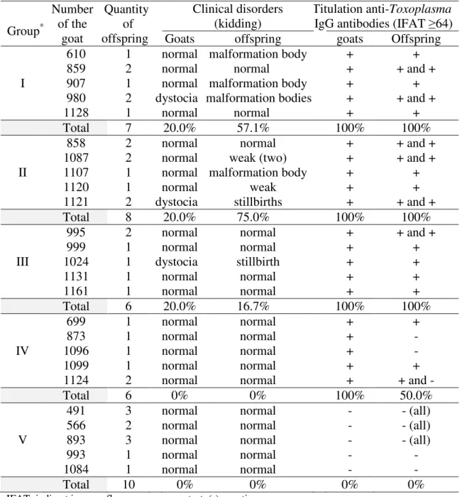
Table 3. Percentage (%) of clinical disorders during the kidding and positive titulation (+) of anti-Toxoplasma IgG antibodies in non-infected, infected and reinfected goats with T. gondii and their offspring.
Group* Number of the goat Quantity of offspring Clinical disorders (kidding) Titulation anti-Toxoplasma IgG antibodies (IFAT ≥64) Goats offspring goats Offspring I
610 1 normal malformation body + +
859 2 normal normal + + and +
907 1 normal malformation body + +
980 2 dystocia malformation bodies + + and +
1128 1 normal normal + +
Total 7 20.0% 57.1% 100% 100%
II
858 2 normal normal + + and +
1087 2 normal weak (two) + + and +
1107 1 normal malformation body + +
1120 1 normal weak + +
1121 2 dystocia stillbirths + + and +
Total 8 20.0% 75.0% 100% 100%
III
995 2 normal normal + + and +
999 1 normal normal + + 1024 1 dystocia stillbirth + + 1131 1 normal normal + + 1161 1 normal normal + + Total 6 20.0% 16.7% 100% 100% IV 699 1 normal normal + + 873 1 normal normal + - 1096 1 normal normal + - 1099 1 normal normal + +
1124 2 normal normal + + and -
Total 6 0% 0% 100% 50.0%
V
491 3 normal normal - - (all)
566 2 normal normal - - (all)
893 3 normal normal - - (all)
993 1 normal normal - -
1084 1 normal normal - -
Total 10 0% 0% 0% 0%
IFAT: indirect immunofluorescence assay test. (-) negative.
*Reinoculation (VEG strain): initial (I), intermediate (II) and final (III) gestational stage, inoculation (ME49 strain) – positive control (IV) and no inoculation – negative control (V) with T. gondii oocysts.
Notably, no goats received anthelmintic treatment throughout the experiment, and the ultrasound screenings did not reveal fetal abnormalities.
The bioassay (Table 4) and PCR (Table 5) results confirmed the presence of T.
gondii in tissue samples of the infected (ME49 strain) and reinfected (VEG strain) goats
and their offspring. This was not diagnosed in any tissue of goats and offspring belonging to GV. T. gondii tissue parasitism was confirmed by positive serology (IFAT-IgG ≥32) of the mice inoculated with the pool of tissue collected from infected (ME49 strain) and reinfected (VEG strain) goats and their offspring, and in some cases parasitism was also confirmed by the detection of bradyzoite-containing brain cysts (Table 4).
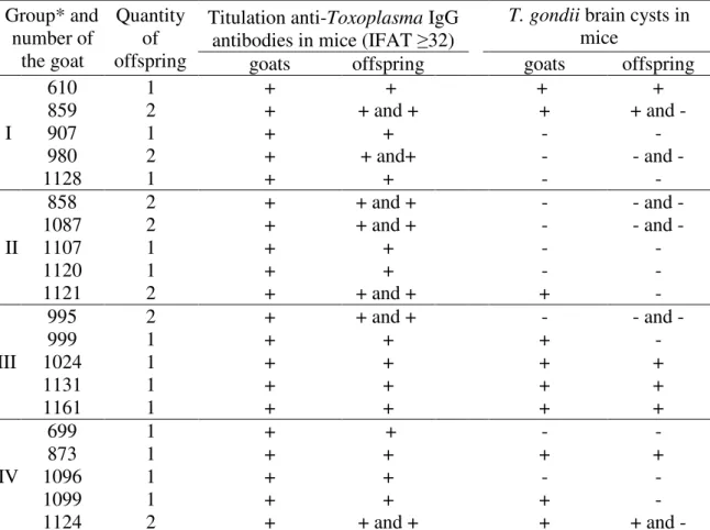
Table 4. Results of the bioassay using mice that were inoculated with tissue fragments of infected and reinfected goats with Toxoplasma gondii and their offspring.
Group* and number of the goat Quantity of offspring
Titulation anti-Toxoplasma IgG antibodies in mice (IFAT ≥32)
T. gondii brain cysts in
mice
goats offspring goats offspring
I 610 1 + + + + 859 2 + + and + + + and - 907 1 + + - - 980 2 + + and+ - - and - 1128 1 + + - - II 858 2 + + and + - - and - 1087 2 + + and + - - and - 1107 1 + + - - 1120 1 + + - - 1121 2 + + and + + - III 995 2 + + and + - - and - 999 1 + + + - 1024 1 + + + + 1131 1 + + + + 1161 1 + + + + IV 699 1 + + - - 873 1 + + + + 1096 1 + + - - 1099 1 + + + - 1124 2 + + and + + + and -
*Reinoculation (VEG strain): initial (I), intermediate (II) and final (III) gestational stage and inoculation (ME49 strain) – positive control (IV) with T. gondii oocysts.
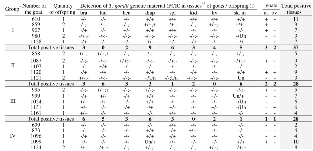
T. gondii DNA was detected by PCR in the tissues of the infected (ME49 strain)
and reinfected (VEG strain) goats and their offspring. The diaphragm was the most infected tissue with T. gondii DNA and GI animals had the highest tissue presence of genetic material of this protozoan (Table 5). The quantity of tissue with T. gondii genetic material varied in the offspring of the goats infected (spleen, diaphragm and skeletal muscle) and reinfected with T. gondii (Table 5): diaphragm, brain, spleen and skeletal muscle (initial stage of gestation); lungs and skeletal muscle (intermediate stage of gestation); and brain, lungs and heart (final stage of gestation).
Table 5. Results positive (+) and negative (-) of the polymerase chain reaction (PCR) to detect of T. gondii genetic material from tissues 1
samples of the infected and reinfected goats with T. gondii and their offspring. 2
Group Number of * the goat of offspring Quantity Detection of T. gondii genetic material (PCR)in tissues
** of goats / offspring(;) goats
(only) Total positive tissues
bra lun hea diap spl kid liv sk. m ut ov
I 610 1 -/- -/- -/- +/+ +/+ +/+ +/+ +/+ + - 11 859 2 -/-;- -/-;- -/-;- +/+;+ -/+;- -/-;- +/+;- +/+;- + - 9 907 1 -/+ -/- +/- +/+ +/+ -/- -/- -/- + - 7 980 2 -/+;- -/-;- -/-;- -/+;- -/-;- -/-;- -/-;- -/Un - + 3 1128 1 -/+ -/- +/- +/- +/- -/+ -/- -/+ - + 7
Total positive tissues 3 0 2 9 6 3 4 5 3 2 37
II 858 2 +/-;- +/+;+ -/-;- -/-;- -/-;- -/-;- -/-;- +/-;- - - 5 1087 2 -/-;- -/-;- +/+;+ -/-;- -/+;- -/-;- -/-;- +/+;+ + + 9 1107 1 -/- +/+ -/- -/- -/- -/- -/- -/- - - 2 1120 1 -/+ -/+ -/- +/+ -/- -/+ -/- +/+ + + 9 1121 2 +/-;- -/-;- -/-;- +/Un -/-;Un -/+;- -/-;- Un - - 3
Total positive tissues 3 6 3 3 1 2 0 6 2 2 28
III 995 2 -/-;- +/+;+ -/-;- +/-;- -/-;- -/-;- -/-;- -/-;- + - 5 999 1 -/+ +/- -/+ +/+ -/- -/- +/- Un/+ - - 7 1024 1 +/+ -/+ +/- +/+ -/- -/- -/- -/Un - - 6 1131 1 +/- -/- -/+ -/+ +/- -/- +/- -/Un - + 6 1161 1 +/+ -/- -/- -/- +/+ -/- -/- -/- - - 4
Total positive tissues 6 5 3 6 3 0 2 1 1 1 28
IV 699 1 -/- -/- -/- -/- +/+ -/- -/- -/- - - 2 873 1 -/- -/- -/- +/+ -/+ +/-;- -/- -/- - - 4 1096 1 -/+ -/- -/- +/+ -/+ -/- -/- -/- - - 4 1099 1 +/- -/- -/- Un/+ +/+ +/- +/- +/+ + + 10 1124 2 -/+;- -/+;+ -/-;- +/-;- -/-;- -/-;- +/+;- -/+;+ - - 8
Total positive tissues 3 2 0 6 6 2 3 4 1 1 28
Total general 15 13 8 24 16 7 9 16 7 6 121
*Reinoculation (VEG strain): initial (I), intermediate (II) and final (III) gestational stage and inoculation (ME49 strain) - positive control (IV) with T. gondii oocysts.
3
**bra=brain, lun=lungs, hea=heart, diap=diaphragm, spl=spleen, kid=kidneys, liv=liver, sk. m= skeletal muscle, ut=uterus, ov=ovaries. (Un) unrealized.
DISCUSSION
The clinical, serological and tissue parasitism results achieved in this study showed distinct situations between the toxoplasmic infection and reinfection and also in the congenital transmission of T. gondii in goats, exposed to different strains of the T.
gondii (ME 49 and VEG strain). The toxoplasmosis in goats and congenital
transmission by T. gondii were confirmed by presence of anti-Toxoplasma IgG antibodies and tissue parasitism by T. gondii, in goats infected (ME49 strain) and reinfected (VEG strain) with T. gondii and their offsprings. These diagnosis were performed by IFAT, bioassay and PCR, techniques commonly used in Veterinary Medicine (ABOUZEID et al., 2010; WIENGCHAROEN et al., 2011; GARCIA et al., 2012).
In the toxoplasmic infection, all the females had temperature increase post-inoculation with T. gondii ME49 oocyts, and the anti-Toxoplasma IgG antibodies detected in these females after of the seroconversion remained high for several days. Only when the IgG titers had stabilized in the infected goats with T. gondii (ME49 strain), indicating chronic toxoplasmosis (DUBEY and KIRKBRIDE, 1989), was performed the reproductive management in all experimental females. This served to rule out any chance of the animals experiencing acute infection (ME49 strain) at the time of fertilization and experimental reinoculation (VEG strain) with T. gondii, in different gestational stages (initial, intermediate and final).
The goats reinfected with T. gondii VEG strain, in different gestational stages, showed a relevant increase in IgG titers post-reinoculation with T. gondii oocysts, indicative of acute toxoplasmosis (DUBEY and KIRKBRIDE, 1989) and only during kidding, all monitored by a veterinarian, were diagnosed clinical disorders in the animals. This suggests that the protection conferred by one strain of T. gondii may have been breached by reinfection with a strain belonging to another genotype (DAO et al., 2001). In other hand, the infected group with T. gondii ME49 strain (only inoculation) showed low levels of IgG, indicative of chronic toxoplasmosis (DUBEY and KIRKBRIDE, 1989), throughout pregnancy with the births of all the normal animals, but not free of T. gondii.
The presence the IgG antibodies anti-Toxoplasma and genetic material of the T.
gondii in the offspring of the goats infected and reinfected with T. gondii evidenced a
possible contact with T. gondii. Once the placenta of ruminants (synepitheliochorial) do not allow the passage of maternal antibodies to the fetus (WOODING, 1992; AGERHOLM et al., 2006; BROADDUS et al., 2009) and the offspring not received colostrum, preventing the passive transfer of maternal immunoglobulins (O’BRIEN and SHERMAN, 1993). In addition, the distribution of genetic material T. gondii varied in the tissues of offspring of infected and reinfected goats with T. gondii, these results are unprecedented in the reviewed literature.
Based on exposures above and whereas this was a pioneer study, we can infer that in the reinfected goats with T. gondii the infectivity and the transplacental passage of this parasite was not limited only to one of the strains used in this study (ME49 and VEG strains), and that the toxoplasmic reinfection with congenital disease is a potential risk. Furthermore, in chronically infected goats with T. gondii (ME49 strain) the transplacental infection of T. gondii without reacutization and without congenital disease not can be excluded. We will believe that new studies about toxoplasmic reinfection in animals will be necessary even because in natural conditions, nothing prevents that this problem already is happening. Once there are high prevalence of
anti-T. gondii antibodies in goat herds and exact timescale and duration of each phase of T. gondii infection hardly are established in the field.
CONCLUSIONS
The congenital toxoplasmosis was possible in goats chronically infected and reinfected with T. gondii.
The primary infection with T. gondii did not protect the pregnant goats against congenital disease resulting from toxoplasmic reinfection, in different gestational stages (initial, intermediate and final).
ACKNOWLEDGEMENTS
We thank the Fundação de Amparo à Pesquisa do Estado de São Paulo – FAPESP (Process: 2009/11401-0) for the financial support provided for this study.
REFERENCES
ABOUZEID, N.Z., AMER, H.A., BARAKAT, T.M., SELIM, A.M., EL BALKEMEY, F.A., 2010. Toxoplasmosis in naturally and experimentally infected goats. J. Am. Sc. 11, 122-129.
AGERHOLM, J.S., AALBAEK, B., FOG-LARSEN, A.M., BOYE, M., HOLM, E., JENSEN, T.K., LINDHARDT, T., LARSEN, L.E., BUXTON, D., 2006. Veterinary and medical aspects of abortion in Danish sheep. Acta Path. Micro. Im. A. 114,146-152. ALTON, G.G., JONES, L.M., ANGUS, R.D., VERGER, J.M., 1988. Techniques for
the brucellosis laboratory. Paris: Institut National de la Recherche Agronomique.
ARAÚJO, F., SLIFER, T., KIM, S., 1997. Chronic infection with T. gondii does not prevent acute disease or colonisation of the brain with tissue cysts following reinfection with different strains of the parasite. J. Parasitol. 83,521-522.
AVMA Guidelines on Euthanasia (Formerly Report of the AVMA Panel on
Euthanasia), 2007. American Veterinary Medical Association.
BRESCIANI, K.D.S., COSTA, A.J., TONIOLLO, G., LUVIZOTTO, M.C.R., KANAMURA, C., MORAES, F.R., PERRI, S.H.V., GENNARI, S.M., 2009. Transplacental transmission of Toxoplasma gondii in reinfected pregnant female canines. Parasitol. Res. 104, 1213-1217.
BROADDUS, C.C., LAMM, C.G., KAPIL, S., DAWSON, L., HOLYOAK, G.R., 2009. Bovine Viral Diarrhea Virus abortion in goats housed with persistently infected cattle.
Vet. Pathol. 46, 45-53.
CAMARGO, M.E., 1964. Improved technique of indirect immunofluorescence for serological diagnosis of toxoplasmosis. Rev. I. Med. Trop. 6, 117-118.
COLE, JR., SULZER, C.R., PULSSELY, P.R., 1973. Improved microtechinique for the leptospiral microscopic aglutination. Appl. Microbiol. 25, 976-980.
CONRAD, P.A., SVERLOW, K., ANDERSON, M., ROWE, J., BONDURANT, R., TUTER, G., BREITMEYER, R., PALMER, C., THURMOND, M., ARDANS, A., DUBEY, J., DUHAMEL, G., BARR, B, 1993. Detection of serum antibody responses in cattle with natural or experimental Neospora infections. J.Vet. Diagn. Invest. 5, 572-578.
COSTA, A.J., ARAÚJO, F.G., COSTA, J.O., LIMA, J.D., NASCIMENTO E., 1977. Experimental infection of bovines with oocysts of Toxoplasma gondii. J. Parasitol. 63, 212-218.
DAO, A., FORTIER, B., PLENAT, F., DUBREMETZ, J.F., 2001. Successful reinfection of chronically infected mice by a different Toxoplasma gondii genotype. Int.
J. Parasitol. 31, 63–65.
DUBEY, J.P., SHARMA, S.P., LOPES, C.W., WILLIAMS, J.F., WILLIANS, C.S., WEISBRODE, S.E., 1980. Caprine toxoplasmosis: abortion, clinical signs, anda distribution of Toxoplasma in tissues of goats fed Toxoplasma gondii oocysts. Am. J.
Vet. Res. 41, 1072-1076.
DUBEY, J.P., KIRKBRIDE, C.A., 1989. Enzootic toxoplasmosis in sheep in north-central United States. J. Parasitol. 75, 673-676.
DUBEY, J.P., 1998. Refinement of pepsin digestion method for isolation of
Toxoplasma gondii frominfected tissues. Vet. Parasitol. 74, 75-77.
DZITKO, K., STACZEK, P., GATKOWSKA, J., DLUGONSKA, H., 2006.
Toxoplasma gondii: serological recognition of reinfection. Exp. Parasitol. 112, 134–
137.
ELBEZ-RUBINSTEIN, A., AJZENBERG, D., DARDÉ, ML., COHEN, R., DUMÈTRE, A., YERA, H., GONDON, E., JANAUD, J.C., THULLIEZ, P., 2009. Congenital toxoplasmosis and reinfection during pregnancy: case report, strain characterization, experimental model of reinfection, and review. J. Infec. Dis. 199, 280-285.
FIGUEIREDO, J.F., SILVA, A.O., CABRAL, D.D., MINEO, J.R., 2001. Seroprevalence of Toxoplasma gondii infection in goats by the indirect haemagglutination, immunofluorescence and immunoenzymatic test in the region of Uberlândia. Brazil. Mem. Inst. Oswaldo Cruz 96, 687–692.
FUENTES, S.I., RODRIGUEZ, M., DOMINGO, C.J., FERNANDO, C.C., JUNCOSA, T., ALVAR, J., 1996. Urine sample used for congenital toxoplasmosis diagnosis by PCR. J. Clin. Microbiol. 03, 2368-2372.
GARCIA, J.L., MARQUES, F.A.C., VIDOTTO, O., NAVARRO, I.T., MARTINS, G.F., ZULPO, D.L., CUNHA, I.A.L., TARODA, A., CARDIM, S.T., EWALD, M.P.C., 2012. Sero-occurrence of anti-Toxoplasma gondii antibodies and vertical transmission in slaughtered beef cows (Bos indicus). Semina Cienc. Agrar. 33, 1095-1102.
HOMAN, W.L., VERCAMMEN, M., DE BRAEKELLER, J., VERSCHUEREN, H, 2000. Identification of a 200- to 300-fold repetitive 529 bp DNA fragment in
Toxoplasma gondii, and its use for diagnostic and quantitative PCR. I. J. Parasitol. 30,
69-75.
MAIA, M.S., 1997. Manual de Inseminação artificial em Caprinos e Ovinos. Sebrae/RN.
MAMIDI, A., DESIMONE, J.A., POMERANTZ, R.J., 2002. Central nervous system infectious in individuals with HIV-1 infection. J. Neurovirol. 08, 158-167.
O’BRIEN, J.P., SHERMAN, D.M., 1993. Serum immunoglobulin concentrations of newborn goat kids and subsequent kid survival through weaning. Small Ruminant Res. 11, 71-77.
SABIN, A.B., 1941. Toxoplasmic encephalitis in children. J. Am. Vet. Med. Ass. 116, 801-807.
SANTANA, L.F., ROSSI, G.A.M., GASPAR, R.C., PINTO, W.M.R., OLIVEIRA, G.P., COSTA, A.J., 2013. Evidence of sexual transmission of Toxoplasma gondii in goats. Small Ruminant Res. 115, 130-133.
WIENGCHAROEN, J., THOMPSON, R.C., NAKTHONG, C., RATTANAKORN, P., SUKTHANA, Y., 2011. Transplacental transmission in cattle: is Toxoplasma gondii less potent than Neospora caninum? Parasitol. Res. 108, 1235-1241.
WOODING, F.B.P., 1992. Current topic: the synepitheliochorial placenta of ruminants:binucleate cell fusions and hormone production. Placenta, 13, 101-13.