www.rsbmt.org.br ̶ www.scielo.br/rsbmt
373
Short Communication
Address to : Dra. Carolina da Cunha Correia. Rua Francisco Alves 326, Ilha do Leite, 50070-490 Recife, PE, Brasil.
Phone: 55 81 3216-3131
e-mail: carolina.dra@gmail.com
Received 13 June 2011
Accepted 11 November 2011
Revista da Sociedade Brasileira de Medicina Tropical 46(3):373-376, May-Jun, 2013 http://dx.doi.org/10.1590/0037-8682-1202-2013
Features to validate cerebral toxoplasmosis
Carolina da Cunha Correia[1], Heloísa Ramos Lacerda Melo[2],
Vláudia Maria Assis Costa[3] and Alessandra Mertens Brainer[4]
[1]. Serviço de Neurologia, Universidade de Pernambuco, Recife, PE. [2]. Serviço de infectologia, Hospital das Clínicas, Universidade Federal de Pernambuco, Recife, PE. [3]. Departamento de Medicina Tropical, Universidade Federal de Pernambuco, Recife, PE. [4]. Serviço de Radiologa, Universidade de Pernambuco, Recife, PE.
ABSTRACT
Introduction: Neurotoxoplasmosis (NT) sometimes manifests unusual characteristics. Methods: We analyzed 85 patients with
NT and AIDS according to clinical, cerebrospinal fl uid, cranial magnetic resonance, and polymerase chain reaction (PCR)
characteristics. Results: In 8.5%, focal neurological defi cits were absent and 16.4% had single cerebral lesions. Increased sensitivity of PCR for Toxoplasma gondii DNA in the central nervous system was associated with pleocytosis and presence of >4 encephalic lesions. Conclusions: Patients with NT may present without focal neurological defi cit and NT may occur with presence of a single
cerebral lesion. Greater numbers of lesions and greater cellularity in cerebrospinal fl uid improve the sensitivity of PCR to T gondii. Keywords: AIDS. Cerebral toxoplasmosis. Diagnosis.
Neurotoxoplasmosis (NT) is the most frequent opportunistic infection of the central nervous system (CNS) among individuals with the acquired immunodeficiency syndrome (AIDS). It indicates severe immunodeficiency and, if it remains untreated, it may lead to death1.
The number of cases of NT in Brazil and around the world has declined since 1996, the year highly active antiretroviral therapy (HAART) was introduced. Nonetheless, it is still the most common opportunistic infection of the CNS in Brazil and
the third most common AIDS-defi ning disease in São Paulo2.
In countries with a high seroprevalence of toxoplasmosis, the incidence of NT has been estimated to be between 30% and 40% among AIDS patients who are not receiving prophylaxis3. Even
though NT is frequent neurological manifestation, pleomorphic presentations of the infection are not uncommon, and differential diagnoses must include other neurological infections and tumor diseases.
The aim of the present study was to describe important features of NT that might contribute to recognition of the disease, thereby avoiding delayed diagnoses. The descriptive cross-sectional study was conducted between February 2006 and December 2008, at 2 referral centers for neurological diseases in the City of Recife, State of Pernambuco, Brazil, the Hospital
da Restauração and the Oswaldo Cruz University Hospital. Eighty-fi ve AIDS patients were included in the study. They
all also had a diagnosis of NT that had been determined using
the criteria of the Centers for Disease Control and Prevention (CDC)4. These criteria include: I) presence of a recent
neurological abnormality consistent with intracranial disease; II) evidence of a cerebral lesion with a mass effect viewed using cranial tomography or magnetic resonance imaging (MRI);
III) positive serological fi ndings of Toxoplasma gondii; and IV) a therapeutic response to toxoplasmosis treatment. Aliquots
of cerebrospinal fl uid (CSF) were subjected to real-time and
PCR analysis for detection of the b1 gene of T. gondii.
The patients were characterized by variables relating to
gender, age, and presence of focal neurological defi cits. We defi ned the therapeutic response as good in cases in which
there was a clinical and radiologic improvement with regression of neurological signs and symptoms following institution of therapy, which consisted of sulfadiazine in association with pyrimethamine or clindamycin, both administered together with folinic acid.
Patient follow up was maintained for an average of 90 days. This study was approved by the Research Ethics Committee for studies involving human beings, and was given the protocol number 00430.102.172/05.
There were 43 men and 42 women evaluated, with a mean age of 35.8 ± 0.98 years. The time for neurological symptoms to become established ranged from 1 to 90 days, with a median of 14 days. At the time of diagnosis of cerebral toxoplasmosis, 65.8% of the patients were known to have been HIV-positive.
Among the most frequent manifestations, we observed hemiparesis in 75 (88.2.%) cases, headache in 76 (89.4%), and fever in 46 (54.1%).
Thirty-nine (45.9%) patients presented with convulsive crises. Analysis of the presence of seizures in patients with up to 4 encephalic lesions in relation to a group with more than
4 lesions did not show any statistically signifi cant difference
www.rsbmt.org.br ̶ www.scielo.br/rsbmt
374
Correia CC et al - Features to validate cerebral toxoplasmosis
TABLE 1 - Cellularity of cerebrospinal fl uid and number of encephalic lesions with PCR results from cerebrospinal fl uid.
PCR in cerebrospinal fl uid
positive (n = 29) negative (n = 53)
Variable n % n % p value
Cellularity of CSF (cells/mm³)
<4 3 10.3 19 35.8 0.013
≥4 26 89.7 34 64.2
Lesions seen MRI (n) (n = 23) (n = 46)
<4 6 26.1 25 54.3 0.026
≥4 17 73.9 21 45.7
PCR: polymerase chain reaction; CSF: cerebrospinal fl uid; MRI: magnetic resonance imaging; χ2 test; p=0.05 (α=5%).
on lesion location showed that patients with cortical lesions had a greater rate of seizures than did a group with deep lesions, and
this difference was statistically signifi cant (p = 0.02).
Ten (8.5%) patients did not present with any focal neurological signs, and among this group, the initial manifestation consisted predominantly of onset of convulsive crisis.
With regard to level of consciousness, 71 (83.5%) patients were alert. However, among this group, 47% presented with states of confusion on admission to hospital.
Defi cits of cranial nerve pairs were observed in 12 (14.1%)
patients, with CN III most (66.6%) frequently affected. CD4 evaluations revealed a mean count of 66.6 cells/mm³, with 93%-94% of the cases serologically positive for
anti-Toxoplasma IgG.
Eighty-three samples of cerebrospinal fl uid were evaluated,
and pleocytosis was observed in 52 (62.6%) cases, with a mean cell density of 27.1 cells/mm³. The predominant pattern was
lymphomononuclear. High levels of cerebrospinal fl uid proteins
were also present in 85.3% of the cases, with a mean protein concentration of 104.4mg/dL.
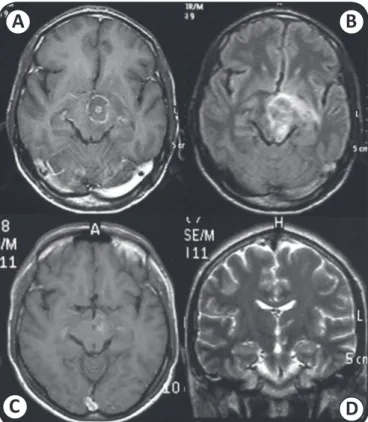
Cranial magnetic resonance imaging (MRI) was performed in 69 cases. Cases with 4 or more encephalic lesions predominated, accounting for 55.1% of all cases. There were 14 (16.4%) patients with single cerebral lesions (Figures 1 and 2).
On analysis of results of real-time PCR seeking T. gondii DNA
in the CSF, with regard to cell density in the fl uid, 89.7% of samples
with more than 4 cells/mm³ also had positive PCR results, whereas 35.8% of specimens with up to 3 cells/mm³ had negative PCR
results. This difference was statistically signifi cant (p = 0.013).
Analysis of the CSF PCR results in relation to the number of encephalic lesions (as identified by MRI) showed that 54.3% of patients with a maximum of 3 lesions had negative PCR, whereas 73.9% of the group with 4 or more intracranial lesions had positive results from PCR. This difference was also
statistically signifi cant (p = 0.026) (Table 1).
Clinical reassessments of 78 patients were performed, and
64 (82.1%) had improvement defi ned as full regression or recovery
from the signs and symptoms. There were 8 (10.3%) deaths, and
the PCR results from the cerebrospinal fl uid were positive in
A
C
B
D
FIGURE 1 - Mesencephalic lesion with a central target. (A) and (B), sequences before treatment; (C) and (D), sequences after treatment. Note reduction of the lesion and the cerebral edema. Images:author’s photos.
all 8 cases. Necropsies were only performed in 2 of these cases, and parasitic pseudocysts of T. gondii were observed in both. It was not possible to obtain data on the evolution of the other cases.
With regard to demographic characteristics, we did not fi nd
any predominance of the disease according to gender. Some studies have indicated that NT occurs predominantly among males5,6, but without demonstrating statistical signifi cance for
such an association7,8. The mean age of patients in our study
was similar to that of other studies1,6.
www.rsbmt.org.br ̶ www.scielo.br/rsbmt
375 Rev Soc Bras Med Trop 46(3):373-376, May-Jun, 2013
have also been reported in other series9-11, with emphasis not
only on focal defi cits but also with attention drawn to headaches
as an independent variable contributing towards the diagnosis of NT among individuals with AIDS5.
There was signifi cant rate of convulsive crisis (45.9% of the cases), and this was similar to the fi ndings in most other series12.
The seizures occurred during the acute phase of NT, and it was noteworthy that the number of lesions was not associated with the seizure rate, but the presence of cortical lesions decreased the seizure threshold. Another point to mention is that among the patients who were known to have been HIV-positive at the time of NT diagnosis, 62.5% also had experienced previous symptoms, such as chronic diarrhea, weight loss, or histories of pulmonary tuberculosis, that pointed towards the presence
of immunodefi ciency, but which were not given their value at
the time of initial medical care.
Although the CDC clinical criteria for inferring NT among AIDS patients are not pathognomonic, they are accepted by the
scientifi c community. However, absence of these criteria should
not be a factor in ruling out this diagnosis given that in our series there were 10 patients without any focal neurological signs, plus a small percentage of individuals who were serologically negative for anti-Toxoplasma IgG, who could have been judged
by these criteria to be presenting other diseases.
From a laboratory point of view, traditional methods
generally have not provided defi ning diagnostic information.
Serological detection of anti-Toxoplasma IgG antibodies in
FIGURE 2 - Parietal lesion, next to the meningeal plane. (A) and (B), sequences before treatment; (C) and (D), sequences after treatment. Note complete disappearance of the lesion and edema. Images: author’s photos.
A
C
B
D
immunosuppressed individuals serves as an indicator of chronic
infection by the parasite, but it does not help to defi ne active
disease13. Studies that have evaluated intrathecal synthesis of
IgG have shown low sensitivity and consequently little clinical utility13,14. In routine cerebrospinal fl uid analysis, fi ndings of
moderate pleocytosis with elevated protein concentrations are
nonspecifi c13, but increased cell counts may contribute to greater
sensitivity of PCR results, as we found in a previous study15
in which we reported on clinical variables that infl uenced and
thereby improved the performance of the method.
The present study showed that single lesions on MRI were not uncommon in individuals with NT (16.4%). However, this
fi nding has yet to be extensively explored with regard to its signifi cance as a diagnostic clue.
In conclusion, additional clinical and imaging variables, especially alongside results from real-time PCR, must be
identifi ed to improve the diagnosis of cerebral toxoplasmosis.
REFERENCES
The authors declare that there is no confl ict of interest. CONFLICT OF INTEREST
1. Nissapatorn V, Lee C, Quek KF, Leong CL, Mahmud R, Abdullah KA. Toxoplasmosis in HIV/AIDS patients: a current situation. J Infect Dis Jpn 2004; 57:160-165.
2. Camara VD, Tavares W, Ribeiro M, Dumas M. Manifestações neurológicas de toxoplasmose em AIDS. J Bras Doenças Sex Transm 2003; 15:46-50.
3. Antinori A, Larussa D, Cingolani A, Lorenzini P, Bossolasco S, Finazzi MG, et al. Prevalence, associated factors, and prognostic determinants of AIDS-related toxoplasmic encephalitis in the era of advanced highly active antiretroviral therapy. Clin Infect Dis 2004; 39:1681-1691.
4. Centers for Disease Control (CDC). 1993 revised classifi cation system for HIV infection and expanded surveillance case definition for AIDS among adolescents and adults. MMWR Recomm Rep 1992; 41(RR-17):1-19.
5. Raffi F, Aboulker JP, Michelet C, Reliquet V, Pelloux H, Huart, et al. A prospective study of criteria for the diagnosis of toxoplasmic encephalitis in 186 AIDS patients. AIDS 1997; 11:177-184.
6. Hernández-González E, Zamora F, Barnès J, Bender-del Busto J, Rodríguez Delgado F, Millan-Marcelo JC. Manifestaciones clínicas de la toxoplasmosis cerebral en pacientes cubanos con Sida. Rev Neurol 2002; 34:618-621.
7. Richards FO, Kovacs JA, Luft BJ. Preventing toxoplasmic encephalitis in persons infected with human immunodefi ciency virus. Clin Infec Dis 1995; 21(supl I):49-56.
8. Belanger F, Derouin F, Grangeot-Keros L, Meyer L. Incidence and risk factors for toxoplasmosis in a cohort of human immunodefi ciency vírus-infected patients. Clin Infec Dis 1999; 28:575-581.
9. Khan AN, Turnbull I, Al-Okaili R, MacDonald S. Imaging in CNS Toxoplasmosis [Internet]. Medscape’s full drug & disease; [Updated 2011 May, 25; Cited 2008 June 10]. Available from: www.emedicine.com/radio/ topic703.htm/.
10. Skiest DJ. Focal neurological disease in patients with acquired immunodefi ciency syndrome. Clin Infect Dis 2002; 34:103-115. 11. Montoya JG. Laboratory diagnosis of Toxoplasma gondii infection and
www.rsbmt.org.br ̶ www.scielo.br/rsbmt
376
12. Kellinghaus C, Engbring C, Kovac S, Möddel G, Boesebeck F, Fischera M, et al. Frequency of seizure and epilepsy in neurological HIV- infected patients. Seizure 2008; 17:27-33.
13. Borges AS, Figueiredo JFC. Detecção de imunoglobulinas IgG, IgM e IgA anti-Toxoplasma Gondii no soro, líquor e saliva de pacientes com Síndrome de Imunodefi ciência Adquirida e Neurotoplasmose. Arq Neuropsiquiatr 2004; 62:1033-1037.
14. Potasman I, Resnick L, Luft BJ, Remington J. Intrathecal production of antibodies against Toxoplasma gondii in patients with toxoplasmic encephalitis and the acquired immunodefi ciency syndrome (AIDS). Ann Inter Med 1988; 108:49-51.
15. Correia CC, Melo HR, Costa VM. Influence of neurotoxoplasmosis characteristics on real-time PCR sensitivity among AIDS patients in Brazil. Trans R Soc Trop Med Hyg 2010; 104:24-28.