David Alexandre Micael Pereira
METABOLITE PROFILING OF MARINE ORGANISMS : MODULATION OF ER-STRESS IN CANCER AND INFLAMMATION
Tese do 3º Ciclo de Estudos Conducente ao Grau de Doutor em Ciências Farmacêuticas
Especialidade de Fitoquímica e Farmacognosia
Trabalho realizado sob a orientação da
Professora Doutora Paula Cristina Branquinho de Andrade
e co-orientação da
Professora Doutora Natércia Aurora Teixeira
Formação Avançada, comparticipada pelo Fundo Social Europeu e por fundos nacionais do MCTES
IN TERESSAD O QU E A TAL SE COMPROMETE.
Dos trabalhos descritos nesta dissertação resultaram as seguintes publicações:
Pu blicaçõ e s e m re vistas in de xadas n o Jo u rn al Citatio n Re po rts da ISI W e b o f Kn o w le dge :
1. Federico Ferreres, David M. Pe re ira, Angel Gil-Izquierdo, Patrícia Valentão, J oão Botelho, Teresa Mouga, Paula B. Andrade. Metabolite profiling of cytotoxic carotenoids from the echinoderm M arthasterias glacialis (spiny sea-star). J ournal of Separation
Science, 2010, 33, 2250-2257.
2 . Pe re ira, D .M., Vinholes, J ., Correia-da-Silva, G., Valentão, P., Teixeira, N., Andrade, P.B. Fatty acids in marine organisms: In the pursuit of bioactive agents. Current Pharmaceutical Analysis, 2011, 7, 108-119.
3 . D avid M. Pe re ira, J uliana Vinholes, Paula Guedes de Pinho, Patrícia Valentão, Teresa Mouga, Natércia Teixeira, Paula B. Andrade. A gas chromatography-mass spectrometry multi-target method for the analysis of three classes of metabolites in marine organisms. Talanta, 2012, 100, 391-400.
4 . Lilian R. B. Mariutti, D avid M. Pe re ira, Adriana Zerlotti Mercadante, Patrícia Valentão, Natércia Teixeira, Paula B. Andrade. Further insights on the carotenoid profile of the echinoderm M arthasterias glacialis. Marine Drugs, 2012, 10, 1498-1510.
5. D avid M. Pe re ira, Patrícia Valentão, Natércia Teixeira, Paula B. Andrade. Amino acids, fatty acids and sterols profile of some marine organisms from Portuguese waters. Food Chemistry, 2013, 141, 2412-2417.
6.
D avid M. Pe re ira, Georgina Correia-da-Silva, Patrícia Valentão, Teresa Mouga, Natércia Teixeira, Paula B. Andrade.GC-MS/MS Lipidomic Profiling of the
Echinoderm
Marthasterias glacialis and Screening for Bioactivity Against Human
Cancer and Non-cancer Cell Lines.
Submitted.7.
D avid M. Pe re ira, Georgina Correia-da-Silva, Patrícia Valentão, Natércia Teixeira, Paula B. Andrade.The anti-inflammatory effect of unsaturated fatty acids and
8 . D avid M. Pe re ira, Georgina Correia-da-Silva, Patrícia Valentão, Natércia Teixeira, Paula B. Andrade. Palmitic acid and ergosta-7,22-dien-3-ol contribute to the ER-stress-mediated apoptotic effect and cell cycle arrest of an extract from Marthasterias glacialis L. in neuroblastoma cells. Submitted.
Capítu lo s de livro s
1. D avid M. Pe re ira, Georgina Correia da Silva, Patrícia Valentão, Natércia Teixeira, Paula B. Andrade. Marine Metabolomics in Cancer Chemotherapy. In OMICS:
Biomedical Prospective and applications", CRC Press. Edited by Debmalya Barh. pp. 377-398, 2011.
2. D avid M. Pe re ira, Patrícia Valentão, Paula B. Andrade. Lessons from the Sea: Distribution, SAR and Molecular Mechanisms of Anti-inflammatory Drugs from Marine Organisms. In Studies in natural products chemistry, vol. 40. Edited
byAtta-ur-Rahman (Ed.). Elsevier Science Publishers, Amsterdam, The Netherlands, 2013, pp. 205-228, 2013.
Re s u m o s pu blicado s e m re vis tas in de xadas n o Jo u rn al Citatio n Re po rts da ISI W e b o f Kn o w le dge :
1. D . Pe re ira, G. Correia-da-Silva, P. Valentão, N. Teixeira, P. B. Andrade. Biological properties of a lipophilic extract of sea-star M arthasterias glacialis L. upon several
cancer cell lines. The FEBS J ournal, 279 (Supplement 1), 2012.
2 . D . Pe re ira, G. Correia-da-Silva, P. Valentao, N. Teixeira, P. B. Andrade. Modulation of Cox-2 and iNOS expression in macrophages by a lipophylic extract of the sea-star
M arthasterias glacialis: cooperative effect of fatty acids and ergosta-7,22-dien-3-ol.
The FEBS J ournal, 280 (Supplment 1), 2013.
re spe ctivas Co m is s õ e s Cie n tíficas e ficaram re gistadas n o s livro s de actas
Co m u n icaçõ e s o rais
1. D avid M. Pe re ira, Federico Ferreres, Angel Gil-Izquierdo, Patrícia Valentão, Paula B. Andrade. Bioguided HPLC-PAD-APCI-MS metabolite profiling of cytotoxic carotenoids from the marine echinoderm M arthastherias glacialis (spiny sea-star). 3rd Meeting of
Young Researchers of University of Porto. Porto, 17th-19th February 2010, Portugal.
2 . D avid M. Pe re ira, Patrícia Valentão, Federico Ferreres, Angel Gil-Izquierdo, Georgina Correia da Silva, Natércia Teixeira, Paula B. Andrade. Marine organisms as a source of bioactive molecules: A case study with M arthasterias glacialis (spiny
sea-star). International Meeting On Marine Resources, Peniche, 16th-18th November, 2010,
Portugal.
3 . D avid M. Pe re ira, J uliana Vinholes, Patrícia Valentão, Natércia Teixeira, Paula B. Andrade. A GC-MS multi-target method for the simultaneous analysis of three classes of metabolites in marine organisms. 3rd Portuguese Young Chemists Meeting. Porto, 11th May, 2012, Portugal
Co m u n icaçõ e s s o b a fo rm a de pain e l
1. D avid M. Pe re ira, Federico Ferreres, Angel Gil-Izquierdo, Patrícia Valentão, Teresa
Mouga, Paula B. Andrade. HPLC-PAD-APCI-MS analysis of carotenoids: a case study with the marine equinoderm M arthasterias glacialis. 2nd Portuguese Meeting of Young
Chemists, Aveiro, 21st-23rd April, 2010, Portugal.
2 . D avid M. Pe re ira, Federico Ferreres, Angel Gil-Izquierdo, Patrícia Valentão, Paula B. Andrade. Marthasterias glacialis: cytotoxic carotenoids metabolite profiling by
HPLC-PAD-APCI-MS. Metabolomics 2010, Amsterdam, 27th J une-1st J uly, 2010, The
Netherlands.
3 . D avid Pe re ira, G. Correia-da-Silva, P. Valentão, T. Mouga, N. Teixeira, P. B. Andrade. Biological effects of the carotenoid extract of Marthasterias glacialis (spiny
4 . D . Pe re ira, G. Correia-da-Silva, P. Valentão, N. Teixeira, P. B. Andrade. Biological properties of a lipophilic extract of sea-star M arthasterias glacialis L. upon several
cancer cell lines. Young Scientist Program (FEBS/ IUBMB), Cádiz, 1st-4th September,
2012, Spain.
5. D . Pe re ira, G. Correia-da-Silva, P. Valentão, N. Teixeira, P. B. Andrade. Biological properties of a lipophilic extract of sea-star M arthasterias glacialis L. upon several
cancer cell lines. 22nd IUBMB and 27th FEBS Congress. Seville, 4th-9th September, 2012,
Spain.
6.
D . Pe re ira, G. Correia-da-Silva, P. Valentão, N. Teixeira, P. B. Andrade. Modulation of COX-2 and iNOS expression in macrophages by a lipophylic extract of the sea-starM arthasterias glacialis: cooperative effect of fatty acids and ergosta-7,22-dien-3-ol.
37
thFEBS Congress. St. Petersburg, 6
th-11
thJuly, 2013, Russia.
7. D . Pe re ira, G. Correia-da-Silva, P. Valentão, N. Teixeira, P. B. Andrade. The synergic anti-inflammatory effect of fatty acids and ergosta-7,22-dien-3-ol from M . glacialis
involves prevention of CHOP pathway-mediated ER-stress and NF-κB activation. EMBO Young Scientists' Forum, Lisbon, 15th-16th J uly, 2013, Portugal.
O autor declara que participou ativamente na recolha e estudo do material incluído em todos os trabalhos, tendo redigido os textos com a colaboração dos restantes coautores.
Uma das maiores alegrias em atingir a meta que um Curso de Doutoramento constitui é a constatação do caminho percorrido, relembrando e agradecendo às pessoas sem as quais o percurso não teria sido possível:
À Profª Doutora Paula Andrade, por ter acreditado e apostado em mim quando era um aluno do 2º Ano do Mestrado Integrado em Ciências Farmacêuticas, aceitando-me como aluno de investigação no Laboratório de Farmacognosia. Agradeço-lhe a paixão incutida pela pela áreas da Farmacognosia e da Química, bem como pela actividade científica em que me iniciou e na qual representa para mim uma referência e exemplo a seguir, tendo a minha total admiração. Não tenho hoje qualquer dúvida que o contacto e convivência que tivemos nos primeiros anos de trabalho conjunto marcaram de forma profunda e irreversível a construção da minha personalidade, da minha identidade científica e da minha postura face à Ciência. E ainda bem. Agradeço ainda à Dra Paula, motor do Laboratório de Farmacognosia, por todo o percurso científico que me possibilitou percorrer e que culminou no currículo científico que hoje tenho e cujas origens e raízes nunca esquecerei. O que hoje tenho devo-o principalmente a si. Por isto, e muito mais, muito obrigado.
À Prof Doutora Natércia Teixeira, co-orientadora desta dissertação o meu agradecimento e admiração por ter sempre a porta aberta e disponibilidade para me receber, conversar, discutir e pensar. Agradeço-lhe por ser sempre uma fonte de serenidade, calma, reflexão e amizade. Estou igualmente muito grato por me ter recebido e dados as boas vindas no Laboratório de Bioquímica e pelo facto de, desde o primeiro momento, me ter feito sentir parte da equipa. Muito obrigado.
À Prof. Doutora Georgina Correia-da-Silva, por todo o apoio prestado e ajuda na execução da tese, pela sua paixão e fascínio contagiantes pela Ciência e pela sua curiosidade inata, com a qual me identifico. Agradeço-lhe profundamente por todo o apoio dado na realização desta tese e por me ter acolhido na sua equipa, bem como pelo seu incentivo constante a não tomar nenhum conhecimento como adquirido e a questionar continuamente tudo o que sabemos.
À Prof. Doutora Patrícia Valentão, pelos ensinamentos e paciência que sempre teve aquando da minha estadia no Laboratório de Farmacognosia ainda como aluno de
escrita e revisão dos trabalhos e submissão dos mesmos, tarefa para a qual muito contribui a sua capacidade de pensamento analítico e atenção aos pormenores verdadeiramente únicas e que muito admiro.
À Prof. Doutora Teresa Mouga, da Escola Superior de Turismo e Tecnologias do Mar de Peniche, pela colaboração e fornecimento das amostras indispensáveis à realização deste trabalho.
À Fátima Fernandes, Andreia Oliveira e Marcos Taveira do Laboratório de Farmacognosia, ao qual tanto devo, pelo percurso que partilhámos durante anos, pelos bons momentos que passámos e também pela ajuda e apoio fundamentais para ultrapassar os maus momentos. Obrigado a todos.
À Engª Carla Sousa, por ter sido a primeira pessoa com quem trabalhei quando me iniciei no mundo da investigação, o meu reconhecimento por todos os ensinamentos e paciência.
A todos os meus colegas do Laboratório de Bioquímica, pelo ambiente acolhedor, divertido e crítico que sempre me proporcionariam. Um agradecimento especial ao Henrique Nascimento e J oão Fernandes, meus amigos, sócios e cúmplices, pelo seu apoio e amizade constantes e por terem tornado a minha experiência no Laboratório de Bioquímica tão rica. À Cristina Amaral, pela sua amizade, camaradagem e ajuda com os trabalhos de citometria de fluxo desenvolvidos ao longo desta tese. Ao Bruno Fonseca, companheiro de congressos, de bancada e amigo. À Marta Almada, pela sua irreverência que sempre constitui uma lufada de ar fresco. Não posso também deixar de agradecer à Susana Rocha pela disponibilidade e apoio demonstrados sempre que procurei o seu conselho e ajuda na condução de procedimentos laboratoriais. Obrigado a todos.
Um agradecimento também a todo o pessoal técnico que tornou este trabalho possível, nomeadamente o Rui Gonçalves do Laboratório de Farmacognosia e ainda o Luís Daniel, Ana Paula e D. Casimira do Laboratório de Bioquímica.
Aos meus amigos, por todo o apoio que sempre me deram, por terem sempre acreditado em mim e acima de tudo por toda a paciência demonstrada ao longo dos últimos 8 anos em todas as situações em que não pude estar com eles tantas vezes como gostaria. Valeu a
A todos os meus Professores, pelo seu contributo para a minha formação científica e pelo apoio e curiosidade manifestados no decorrer desta dissertação.
À Cláudia Silva, pela sua amizade e presença na minha vida.
Um agradecimento especial aos meus pais, que desde sempre apoiaram e incentivaram a minha paixão pela Ciência, pelo apoio constante e por terem criado condições para que eu pudesse percorrer um percurso que agora culmina numa Curso de Doutoramento. Muito obrigado. Um agradecimento também ao meu irmão, por ser o meu maior fã. O sentimento é recíproco.
À Fundação para a Ciência e Tecnologia, pelo suporte financeiro da atribuição da bolsa de doutoramento
SFRH/BD/62663/2009
no âmbito POPH – QREN – Tipologia 4.1 – Formação Avançada, comparticipada pelo Fundo Social Europeu e por fundos nacionais do MCTES.Atualmente, a Natureza permanece uma fonte importante de moléculas com atividades biológica e interesse para a saúde humana.
Durante vários anos as plantas constituíram a única fonte de produtos naturais, no entanto este paradigma tem vindo a mudar em função da atenção dada a fontes alternativas de produtos naturais, tal como os organismos marinhos.
Neste trabalho foram conduzidos estudos de caracterização metabolómica em várias espécies de organismos marinhos obtidos em ecossistemas de Portugal, sendo que o equinoderme M arthasterias glacialis L. (estrela-do mar de espinhos) foi selecionada para
subsequentes estudos biológicos.
Inicialmente, um extrato nativo foi obtido a partir de M . glacialis e analisado
quando à sua composição em carotenóides. A análise por HPLC-DAD e APCI-LC-MS revelou vários isómeros destes compostos, tendo-se encontrado a astaxantina, zeaxantina e luteína como compostos mais relevantes. Esta fração do extrato mostrou exercer um forte efeito anti-proliferativo na linha celular cancerígena RBL-3H3 (leucemia basofílica de rato), enquanto que contra a linha celular não-cancerígena V79 (fibroblastos de pulmão de rato), a toxicidade foi marcadamente inferior.
De forma a caracterizar outros metabolitos com relevância biológica, foi desenvolvido um método de GC-MS/ MS para a análise simultânea de três classes de metabolitos: aminoácidos, ácidos gordos saturados/ insaturados e esteróis. Este método provou ser rápido, reprodutível e depende da extração dos metabolitos com etanol, evitando assim solventes orgânicos mais perigosos e poluentes. Bons parâmetros analíticos foram obtidos para os 40 compostos sob a análise, incluindo 15 aminoácidos, 16 ácidos gordos, 6 esteróis e 3 lupanos. Para validação da técnica foram utilizados extratos etanólicos de vários indivíduos de M . glacialis colhidos em diferentes pontos geográficos e
em diferentes meses do ano. Este método foi posteriormente aplicado a outros organismos marinhos, nomeadamente os equinodermes Paracentrotus liv idus Lamarck
(ouriço-do-mar), H olothuria forskali Chiaje (pepino-do-mar) e os moluscos gastrópodes Aply sia fasciata Poiret e Aply sia punctata Cuvier (lesmas-do-mar). Todas as espécies
apresentaram os aminoácidos estudados, com a exceção de H . forskali, onde não foi
possível encontrar glicina, prolina, trans-4-hidroxi-prolina ou fenilalanina e A. fasciata, a
qual não apresentou prolina. Com a excepção de A. fasciata, os ácidos gordos insaturados
eram os ácidos gordos predominantes, em particular os pertencentes à série omega-6. No
caso dos esteróis, os únicos compostos encontrados foram o colesterol, o β-sitoesterol e um derivado não identificado do colesterol. A espécie P. liv idus foi aquela que apresentou
maior quantidade de esteróis.
o esterol ergosta-7,22-dieno-3-ol e que foi usado nos estudos biológicos. Este extracto, bem como os seus compostos predominantes, foram avaliados pela sua atividade anti-inflamatória na linha de macrófagos de ratinho RAW 264.7, usando o LPS como estímulo pró-inflamatório e avaliando os níveis de expressão da COX-2, iNOS, IκB-α e CHOP. O composto ergosta-7,22-dieno-3-ol foi o composto mais potente, no entanto a atividade máxima foi atingida apenas quando todos os compostos foram utilizados em combinação. Os resultados demonstraram que a atividade anti-inflamatória do extrato e dos seus principais componentes envolve a prevenção da via de stress reticular mediado pela CHOP e ativação do NF-κB.
De seguida, foi avaliado o impacto deste extrato purificado na viabilidade, integridade membranar e densidade celular de 3 linhas celulares humanas cancerígenas (SH-SY5Y, neuroblastoma; MCF-7, cancro da mama recetor do estrogénio positivo; Caco-2, cancro do cólon) e 2 linhas humanas não-cancerígenas (HDF, fibroblastos da derme; HFF, fibroblastos do antebraço). Os resultados demonstraram a atividade distinta do extrato de acordo com a linha celular testada, sendo a linha SH-SY5Y a mais sensível. As células não-cancerígenas revelaram ser menos sensíveis que as cancerígenas. Estes resultados preliminares foram posteriormente confirmados ao estudar o mecanismo de ação responsável pelo efeito anticancerígeno encontrado. Foram utilizadas as linhas celulares MCF-7 e SH-SY5Y e a avaliação da síntese de DNA mostrou uma diminuição significativa, sendo a linha celular SH-SY5Y a mais suscetível. Observou-se paragem do ciclo celular na fase G0/ G1, um efeito atribuído ao esterol ergosta-7,22-dieno-3-ol. Estudos morfológicos mostraram que o extrato causava o aparecimento de vesículas citoplasmáticas de conteúdo lipídico e condensação de cromatina compatível com apoptose, tendo este processo de morte celular sido confirmado por avaliação da atividade da caspase-9 e da caspase-3/ 7. O ácido palmítico revelou ser o principal responsável por este efeito pro-apoptótico, o qual foi demonstrado ser independente da ceramida e envolvendo a ativação da via de stress do reticulo endoplasmático.
Com esta tese de doutoramento, a composição química de vários macroorganismos marinhos de Portugal foi elucidada. O equinoderme M . glacialis foi selecionado para
subsequentes estudos biológicos, tendo a sua atividade anticancerígena e anti-inflamatória demonstrada, estabelecendo assim este organismo como uma potencial fonte de moléculas bioativas com aplicação em várias patologias.
Palavras -ch ave : Marthasterias glacialis; Ácidos Gordos; Inflamação; Apoptose; Stress
reticular
Nowadays, Nature still remains a very important source of biologically active molecules with application in human health.
For many years, plants have been the only natural products being studied, however this paradigm is being shifted towards alternative source of natural products, of which marine organism are a good example.
In this work, metabolite profiling was conducted in several species of marine macroorganisms obtained in Portuguese ecosystems, with Marthasterias glacialis (spiny
sea star) being selected for further biological evaluation.
Initially, a crude extract obtained from M . glacialis was analyzed for its carotenoid
composition. By employing HPLC-DAD and APCI-LC-MS, identification and differentiation of carotenoids isomers was achieved, with astaxanthin, zeaxanthin and lutein being the most important compounds. This carotenoid-enriched fraction displayed a strong cell proliferation inhibition against the rat basophilic leukemia cell line RBL-2H3, while against non-cancer rat lung fibroblasts the effect was weaker.
In order to evaluate other biologically-relevant metabolites in M . glacialis, a
GC-MS/ MS method for the simultaneous analysis of three classes of metabolites, amino acids, saturated/ unsaturated fatty acids and sterols, was developed. This method was shown to be fast, reproducible and relied on extraction with ethanol without using organic solvents such as chloroform and ether. Good analytical parameters were obtained for the 40 compounds under analysis (15 amino acids, 16 fatty acids, 6 sterols and 3 lupanes). Crude ethanolic extracts of several individuals of M. glacialis, collected in different temporal and
geographical points, were used for validating this technique. This method was later used in other marine organisms from Portuguese waters, the echinoderms Paracentrotus liv idus
Lamarck (sea urchin) and H olothuria forskali Chiaje (sea cucumber) and the gastropod
mollusks Aply sia fasciata Poiret and Aply sia punctata Cuvier (sea hares). In general, all
species presented the amino acids covered by the method, with the exceptions of H. forskali, in which no glycine, proline, trans 4-hydroxy-proline or phenylalanine were
found, and of A. fasciata which did not contain proline. In what regards fatty acids, among
all species studied 14 compounds were detected and 12 could be quantified. Apart from A. fasciata, unsaturated fatty acids were predominant compounds in all species, with those
from the ω-6 series being in higher amounts than their ω -3 homologues. In the case of
sterols, the only compounds found were cholesterol, β-sitosterol and one unidentified cholesterol derivative. P. liv idus was the species with the highest sterol content.
After a deeper knowledge of the chemical composition of M . glacialis was achieved,
a purified lipophilic extract containing mainly palmitic, cis 11-eicosenoic and cis
and its components. The purified extract, as well as the predominant compounds cis
11-eicosenoic and cis 11,14-eicosadienoic acids and the unsaturated sterol
ergosta-7,22-dien-3-ol were evaluated for anti-inflammatory activity in the macrophage cell line RAW 264.7 challenged with LPS, as assessed by the expression levels of COX-2, iNOS, IκB-α and
CHOP. Ergosta-7,22-dien-3-ol was the most active compound, although maximum activity was obtained only when all compounds were tested in combination. In addition, the anti-inflammatory activity of the extract from M . glacialis and its compounds was shown to
involve prevention of CHOP pathway-mediated ER stress and NF-κB activation.
After the anti-inflammatory activity of the extract and its compounds was established, the anticancer activity of the same matrix was evaluated. Initial studies evaluated the impact of this purified extract (78-625 µg/ mL) on cell viability, density and membrane integrity of three human cancer cell lines (SH-SY5Y, human neuroblastoma; MCF-7, human oestrogen receptor-positive breast cancer; Caco-2, human colon cancer) and two human non-cancer cell lines (HDF, human dermal fibroblasts; HFF, human foreskin fibroblasts). Differential activity towards the cell lines was found, being SH-SY5Y cell line the most susceptible. No activity was found for Caco-2 cell line and the non-cancer cell lines revealed to be less susceptible than non-cancer ones. These preliminary results were further investigated by studying the mechanism of action behind this activity. MCF-7 and SHSY-5Y cell lines were selected and evaluation of DNA synthesis revealed that both cell lines were markedly affected in a concentration-dependent way, being SH-SY5Y cell line more susceptible. G0/ G1 cell cycle arrest was observed, an effect induced by the sterol ergosta-7,22-dien-3-ol present in the extract. Morphological studies showed that incubation with the extract caused the advent of lipid droplets and chromatin condensation, the latter compatible with apoptosis, which was confirmed by the evaluation of caspase-3/ 7 and -9 activities. Palmitic acid was the main compound responsible for this apoptotic effect by a ceramide-independent mechanism that involved activation of the CHOP-mediated ER stress pathway.
With the doctoral thesis presented herein, the chemical composition of several marine macroorganisms from Portuguese waters was established. The echinoderm M. glacialis was further studied and its anticancer and anti-inflammatory capacity was
demonstrated, thus establishing this organism as a potential source of bioactive molecules with application in several pathologies.
Ke yw o rds:M arthasterias glacialis; Fatty acids; Inflammation; Apoptosis; ER stress
Pu blicatio n s ... VII
Agrade cim e n to s ... X
Re s u m o ... XVII
Abs tract ... XXI
Th e s is in de x ... XXV
Figu re in de x ... XXVII
Abbre viatio n s an d s ym bo ls ... XXIX
PART I
1. IN TRODU CTION
1.1. N atu re as a s o u rce o f bio active m o le cule s ... 1 1.2. Fatty acids ... 3 1.2.1.Chemistry ... 3 1.2.2. Biosynthesis ... 6 1.2.3. Analysis ... 7 1.2.4. Biological properties ... 9 1.3. Ste ro ls ... 13 1.3.1. Chemistry ... 13 1.3.2. Biosynthesis ... 14 1.3.3. Analysis ... 17 1.3.4. Biological properties ... 17 1.4. Caro te n o ids ... 20 1.4.1. Chemistry ... 20 1.4.2. Biosynthesis ... 22 1.4.3. Analysis ... 23 1.4.4.Biological properties ... 26 1.5. In flam m atio n ... 27 1.5.1.Targets in inflammation ... 27 1.6 Can ce r ... 34 1.6.1. Targets in cancer ... 34 1.7. ER s tre s s in in flam m atio n an d apo pto s is ... 39
2.OBJECTIVES ... 45
PART II
3. RESU LTS ... 47
HPLC-PAD-atmospheric pressure chemical ionization-MS metabolite profiling of cytotoxic carotenoids from the echinoderm Marthasterias glacialis (spiny sea-star).
... 49
A gas chromatography– mass spectrometry multi-target method for the simultaneous analysis of three classes of metabolites in marine organisms ... 75
Amino acids, fatty acids and sterols profile of some marine organisms from
Portuguese waters ... 87 GC-MS/ MS Lipidomic Profiling of the Echinoderm M arthasterias glacialis and
Screening for Bioactivity Against Human Cancer and Non-cancer Cell Lines ... 95 The anti-inflammatory effect of unsaturated fatty acids and ergosta-7,22-dien-3-ol from the echinoderm M arthasterias glacialis involves prevention of CHOP
pathway-mediated ER-stress and NF-κB activation ... 107 Palmitic acid and ergosta-7,22-dien-3-ol contribute to the apoptotic effect and cell cycle arrest of an extract from M. glacialis in neuroblastoma cells ... 119
PART III
4. D ISCU SSION ... 137 4.1. Ch e m ical co m po s itio n o f M . g la cia li s ... 139 4.2. An ti-in flam m ato ry activity... 140 4.3. Pro -apo pto tic activity ... 143
PART IV
5. BIBLIOGRAPH Y ... 147
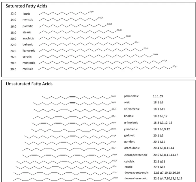
Figu re 1 -Lipid classes. ... 3
Figu re 2 -Most common saturated and unsaturated FA. ... 4
Figu re 3 -Non-methylene interrupted FA. ... 5
Figu re 4 -Methoxylated FA. ... 6
Figu re 5 - Biosynthesis of DHA. ...7
Figu re 6 - General structure and numeration of sterols. ... 13
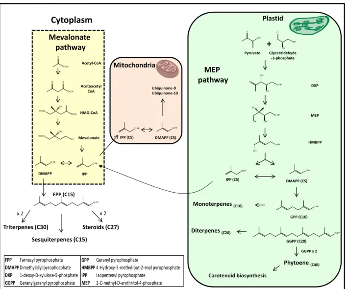
Figu re 7 - Major biosynthetic steps of the mevalonate / 1-deoxy-D-xylulose-5-phosphate pathways and their cellular compartments. ... 15
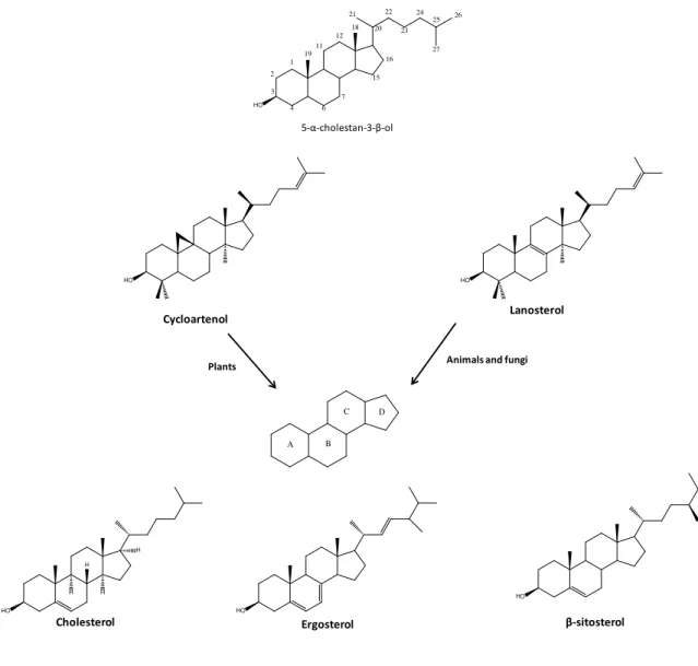
Figu re 8 - Origin of the sterol backbone in animals and plants. ... 16
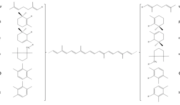
Figu re 9 - General chemical structure of carotenoids, including end-group classification. ... 21
Figu re 10 - Crocetin and its gentiobioside diester, crocin... 22
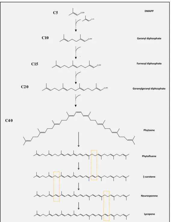
Figu re 11 - Biosynthesis of carotenoids from DMAPP. Major differences through the biosynthesis are highlighted in yellow.. ... 23
Figu re 12 - Spectrum and maxima of (A) lutein and (B) astaxanthin. In these compounds, the effect of a carbonyl group in conjugation with the polyene chain on the fine structure is visible. This functional group is present in astaxanthin and absent in lutein.. ... 25
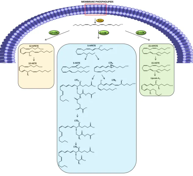
Figu re 13 - COX pathway. PLA2 hydrolyzes membrane phospholipids, originating
arachidonic acid, which is further metabolized by COX. Tissue-specific enzymes are responsible for the end-products formed.. ... 29
Figu re 14 - LOX pathway. PLA2 hydrolyzes membrane phospholipids, originating
arachidonic acid. According to the LOX isoform involved, different metabolites are obtained. ... 30
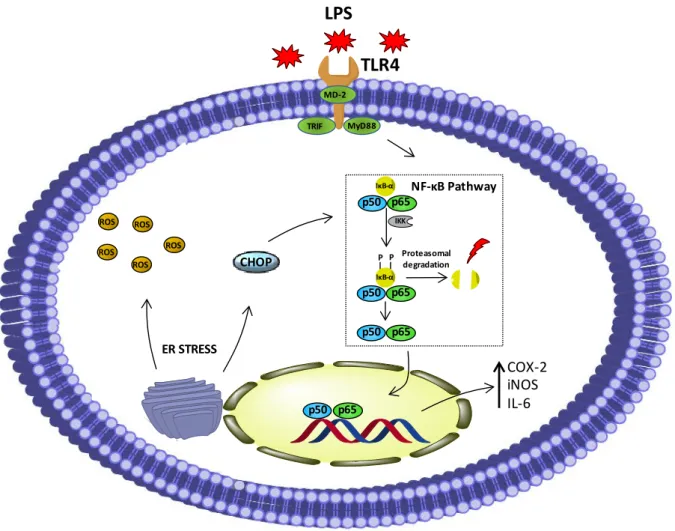
Figu re 15 - Representation of NF-κB activation. ... 32 Figu re 16 - Major proteins involved in ER stress sensing and signal transduction. ... 41
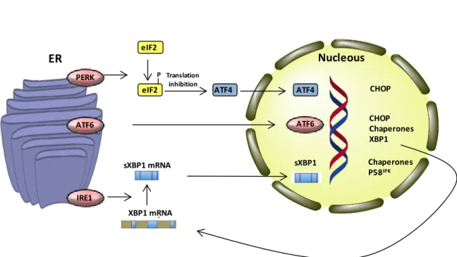
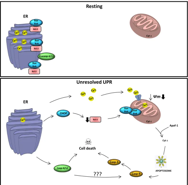
Figu re 17 - Major pathways connecting ER stress and apoptosis. ... 43
∆ψm Mitochondrial membrane potential
5-H ETE 5-Hydroxyeicosatetraenoic acid 5-H PETE 5-Hydroperoxyeicosatetraenoic acid
ACP Acyl carrier protein AIF Apoptosis inducing factor ALA α-Linolenic acid
Apaf-1 Apoptotic protease-activating factor 1 APCI Atmospheric pressure chemical ionization
APCI-MS Atmospheric pressure chemical ionization-mass spetrometry ATF6 Activating transcription factor 6
AN T Adenine nucleotide translocase
ATP Adenosine-5'-triphosphate Bak Bcl-2 antagonis killer 1 Bax Bcl-2 associated protein x Bcl-xL Bcl-2 long isoform
BH 4 Tetrahydrobiopterin BH T Butylated hydroxytoluene
C/ EBP CCAAT-enhancer-binding proteins Caco -2 Human colon adenocarcinoma cell line
CAT Catalase
CH OP C/ EBP homologous protein CLA Conjugated linoleic acid cPLA2 Cytosolic PLA2s
Co A Coenzyme A
COX Cyclooxigenases
COX-2 Cyclooxigenase 2 CypD Cyclophilin A
D AD Diode array detector D H A Docosahexaenoic acid
D ISC Death-inducing signalling complex D OXP 1-Deoxy-D-xylulose-5-phosphate D MAPP Dimethylallyl pyrophosphate
D MOX 4,4-Di-methyloxazoline D R Death receptor
ds RN A Double stranded ribonucleic acid
EPA Eicosapentaenoic acid
e N OS Endothelial nitric oxide synthase ER Endoplasmic reticulum
ERO1 Endoplasmic reticulum oxidoreductin 1
ERSE Endoplasmic reticulum stress responsive element
ESI Electrospray ionization
FA Fatty acids
FAD Flavon adenine dinucleotide
FADD Fas-associated death domain FAME Fatty acid methyl ester
FAK Focal adhesion kinase Fas L Fas ligand
FID Flame ionization detector FMN Flavin mononucleotide
GADD 153 DNA-damage inducible gene 153 GC Gas chromatography
GC-MS Gas chromatography-mass spectrometry GPrx Gluthatione peroxidase
GTP Guanosine-5'-triphosphate H D F Human dermal fibroblasts H FF Human foreskin fibroblasts
H IF Hypoxia inducing factor
H MG-Co A 3-Hydroxy-3-methylglutaryl-CoEnzyme A H PLC High pressure liquid chromatography IFN -γ Interferon gamma
IL Interleukin
IκB-α Nuclear factor kappa-light-chain-enhancer of activated B cells inhibitor alpha IKK IκB kinase complex
iPLA2 Calcium-independent PLA2
IPP Isopentenyl pyrophosphate iN OS Inducible nitric oxide synthase
IRE1α Inositol-requiring 1α
JN K c-J un N-terminal kinase
LA Linolenic acid
LC-MS Liquid chromatography-mass spectrometry
LPS Lipopolysaccharide LTA4 Leukotriene A4
LTB4 Leukotriene B4
LTC4 Leukotriene C4
LTD4 Leukotriene D4
LTE4 Leucotriene E4
MAP Microtubule-associated proteins MAPK Mitogen-activated protein kinases
MCF-7 Human estrogen receptor positive breast cancer cell line Mcl-1 Myeloid cell leukemia sequence 1
MEF Mouse embrionyc fibroblasts cell line MEP 2C-Methyl-D-erythritol-4-phosphate m iRN A Micro RNA
MS Mass spectrometry
MSTFA N-methyl-N-(trimethylsilyl)-trifluoroacetamide
MTA Microtubule targeting agents
MTT (3-(4,5-dimethylthiazol-2-yl)-2,5-diphenyltetrazolium bromide MU FA Mono unsaturated fatty acids
MyD 8 8 Myeloid differentiation factor 88 Mu LV Murine leukemia virus
N ADPH Nicotinamide adenine dinucleotide phosphate
N F-κB Nuclear factor kappa-light-chain-enhancer of activated B cells N MIFA Non-methylene-interrupted fatty acids
n N OS Neuronal nitric oxide synthase N O Nitric oxide
N OS Nitric oxide synthase
n N OS Neuronal nitric oxide synthase PAF Platelet-activating factor PD I Protein disulphide isomerase
PERK Double-stranded RNA-dependent protein kinase (PKR)-like ER kinase
PGD2 Prostaglandin D2
PGE2 Prostaglandin E2
PGG2 Prostaglandin G2
PGH2 Prostaglandin H2
PGI2 Prostaglandin I2
PMA Phorbol myristate acetate PP2 A Protein phosphatase 2A
PPAR Peroxisome proliferator-activated receptor PTP Permeability transition pores
PUFA Polyunsaturated fatty acids OMM Outer mitochondrial membrane Rn as e Ribonuclease
RN S Reactive nitrogen species ROS Reactive oxygen species
RP-H PLC Reversed phase high pressure liquid chromatography SH -SY5Y Human neuroblastoma cell line
SOD Superoxide dismutase SPE Solid phase extraction s PLA2 Secreted PLA2
TBARS Thiobarbituric acid reactive substances tBid Truncated Bid
TG Triglycerides
TIR Toll/ IL-1 receptor
TLC Thin layer chromatography TLR Toll-like receptor
TLR4 Toll-like receptor 4 TMS Trimethylsilyl
TN F-α Tumor necrosis factor α
TN FR Tumor necrosis factor receptor
TPA 12-O-Tetradecanoylphorbol-13-acetate
TXA2 Thromboxane A2
U PR Unfolded protein response U V Ultraviolet
VD A Vascular disrupting agents
VEGF Vascular endothelial growth factor
VEGFR Vascular endothelial growth factor receptor VEGFR2 Vascular endothelial growth factor receptor 2
VLCFA Very long chain fatty acids
VD AC Voltage-dependent anionic chanel W H O World Health Organization
1.1.
N atu re as a s o u rce o f bio active m o le cu le s
Even in the periods of pre-technical medicine, humans have always pursued to counter diseases with medicines. For thousands of years, natural products have been the sole option for treating a number of pathological conditions in the form of tinctures, powders and decocts, among others (1). This happens due to the fact that Nature is, and has been, an undeniable source of bioactive molecules. In recent History, the development of medicine and chemotherapy led to an obvious convergence to the use of isolated molecules within a formulation rather than chemically complex mixtures, thus providing us with molecules such as morphine, codeine, digitoxin and vincristine.
The interest in natural products is a consequence of the success of natural molecules as biologically active agents, which, in turn, arises from their complex chirality and ring systems and the number of heteroatoms and aromatic rings, thus allowing them to bind to complex proteins and other three-dimensional biological targets (2). For this reason, natural products have been addressed as “privileged structures”, as their molecular scaffolds are able to accommodate several pharmacophores, hence displaying multiple biological activities by fitting biological targets that are frequently conserved across species (3).
As it has been reviewed by Cragg and Newman in 2009, between 1981 and 2006, 66% of the 974 small molecule drugs introduced in clinical practice were synthetic. However, deeper insights revealed that 17% of these molecules contained a pharmacophore directly derived from natural products and 12% were derived from natural products. Thus, directly or indirectly, Nature remains a very important source of bioactive molecules (4). Nevertheless, it is undeniable that in the last 15 years there has been a decline in the approvals of Nature-derived molecules and many pharmaceutical industries have terminated their natural drug discovery pipeline (5). However, more than a threat this constitutes an opportunity, as the paradigm of drug discovery from natural products is changing with new hotspots of bioactivity under study and techniques for structure elucidation and target characterization.
Marine environment remains, to this day, the most diversified ecosystem on Earth and simultaneously the least studied. Marine organisms have to cope with the several challenges that marine life represents, including high pressures, low temperatures and light availability. In addition, many organisms have primitive immune systems and soft bodies and, for this reason, what they lack in physical defences is frequently balanced with remarkable chemical defences. In fact, organisms with lower physical defences, such as sponges and molluscs, are usually the ones with the most bioactive molecules. Marine natural products have proved to be an amazing source of chemical diversity and,
consequently, many sea-derived molecules were shown to exhibit a number of different pharmacological activities and are currently under clinical trials to treat various pathological conditions, such as cancer, inflammation and pain (6-9). Among the several classes of marine natural products described, the most relevant in a biological context are terpenes, alkaloids, peptides, fatty acids and sterols. In the next chapters, the chemical classes relevant for this thesis will be presented from a chemical, analytical, biosynthetic and biological point of view.
1.2 . Fatty acids
1.2.1. Chem istry
Fatty acids (FA) are a class of lipophilic molecules that can be found in all living organisms. They can occur in their free form or, alternatively, integrate more complex lipids, such as triglycerides (TG), phospholipids or glycolipids, as presented in Figu re 1. In addition, FA can be part of lipoproteins, lipopolysaccharides and alkaloids.
Figu re 1 – Lipid classes.
FA exhibit a very diversified chemistry, which results in several possible structures such as saturated/ unsaturated, branched and cyclic, which can, in turn, display distinct functional groups, such as hydroxyl, keto and epoxy, hence adding to the diversity of these molecules.
Classical nomenclature for FA indicates chain length, number and position of double bonds when present (example: 20:2 Δ5,11. In this example, there are 20 atoms of
carbon, with 2 double bonds, which are located in the position 5 and 11). The carbon
associated with the methyl end group is designated ω carbon and, accordingly,
Phospholipids Glycolipids Sphingolipids Glycerophospholipids Triacylglycerols Gl y ce ro
l Fatty acid
PO4 Alcohol
Glycosphingolipids Cerebrosides Gangliosides Sulfolipids Fatty acid Fatty acid G ly ce rol Fatty acid Fatty acid Monosaccharide S phi ng o si ne
CONH2 Fatty acid
Oligosaccharide S phi ng o si
ne CONH2 Fatty acid
S phi ng o si ne
CONH2 Fatty acid
PO4 Alcohol
Globosides Sialic Acid(s) Oligosaccharide S phi ng o si ne
CONH2 Fatty acid
unsaturated FA in which the position of the first double bond is located 3, 6 and 9 carbons
away from the ω carbon are named ω-3 (omega-3), ω-6 (omega-6) and ω-9 (omega-9).
Figu re 2 – Most common saturated and unsaturated fatty acids. Image obtained from (10).
In mammals, FA with a maximum chain length of 16-18 carbons constitute about 90% of total FA (11). FA consisting of 20 or more carbon atoms are referred as Very Long Chain Fatty Acids (VLCFA).
Saturated FA, as the name indicates, are molecules with no double bonds. These compounds usually have between 12 and 24 carbon atoms (12) (Figu re 2). On the other hand, when unsaturation is present the compounds are called either mono unsaturated fatty acids (MUFA) or polyunsaturated fatty acids (PUFA). In the case of the latter, double bonds are frequently in an non-conjugated arrangement, with —(CH=CHCH2)n—
4
CO2H
CO2H CO2H CO2H CO2H CO2H stearic
CO2H CO2H CO2H
CO2H
CO2H
CO2H CO2H
CO2H
CO2H
palmitic myristic lauric arachidic behenic lignoceric cerotic montanic melissic palmitoleic oleic cis-vaccenic linoleic α-linolenic γ-linolenic 12:0 14:0 16:0 18:0 20:0 22:0 24:0 26:0 28:0 30:0
16:1 Δ9
18:1Δ9
18:1 Δ11
18:2 Δ9,12
18:3 Δ9,12, 15
18:3 Δ6,9,12
CO2H gadoleic 20:1 Δ9
CO2H gondoic 20:1 Δ11 Saturated Fatty Acids
Unsaturated Fatty Acids
arachidonic 20:4 Δ5,8,11,14
eicosapentaenoic 20:5 Δ5,8,11,14,17
cetoleic 22:1 Δ11 CO2H
CO2H CO2H
erucic 22:1 Δ13
docosapentaenoic 22:5 Δ7,10,13,16,19 docosahexaenoic 22:6 Δ4,7,10,13,16,19 CO2H
CO2H
CO2H
repeating units (Figure 2), although some exceptions are known, as it will be presented later.
Figu re 3 – Non-methylene interrupted FA. Image obtained from (10).
The cis (Z) stereochemistry of the double bond causes a kink in the alkyl chain that
has consequences in the physical properties of the molecules, like fluidity, which is important in a biological context, namely in cellular membranes.
In the last few years, great advances have been made in the study of the chemistry of FA. In particular, the study of marine organisms has led to the discovery of new and less conventional classes of FA with remarkable chemistry, such as halogenated FA, acetylenic FA, non-methylene interrupted FA (NMIFA, Figu re 3) and methoxylated FA (Figu re 4). NMIFA are compounds that display unusual unsaturation features, namely double bonds with more than one methylene group between ethylenic bonds. These compounds are, in general, of marine origin and have been described in algae, mollusks and sponges (10, 13). A review on the chemistry of marine FA has been published recently (10).
Methoxylated FA are rare compounds, presenting limited distribution in Nature and described recently. These compounds are frequently found in cyanobacteria, bacteria and sponges (10, 14-16).
20:2 Δ5,11 20:2 Δ5,13
22:2 Δ7,13
22:2 Δ7,15
20:3 Δ5,11,14
20:4 Δ5,8,11,14
26:2 Δ5,9 30:3 Δ5,9,23
Non-methylene interrupted FA
HO2C
HO2C
HO2C
HO2C
HO2C
HO2C
HO2C
HO2C
Figu re 4 – Methoxylated FA. Image obtained from (10).
1.2 .2 . Bio s yn th e s is
Biosynthesis of FA, as it happens in most biological reactions, is not simply a reversal of its degradative pathway, of which β-oxidation is the most relevant. In general terms, FA biosynthesis takes place in cytosol and requires the action of fatty acid synthase, which comprises several enzymes pivotal to the synthesis. The building block of FA is acetic acid in the form of acetyl-S-Coenzyme A (CoA), which is exported from mitochondrion and acts as the direct initiator. Further elongation is assured by malonyl-CoA which, in turn, derives from acetyl-malonyl-CoA by the action of acetyl Co-A carboxylase.
Sequential elongation of the growing FA chain takes place by two units of carbon. Elongation stops when C14-C18 FA are obtained, typically in the C16:C18:C14 proportion of 7:1:2 (17). FA with higher carbon number, VLCFA, can be synthesized, however additional enzymes are required. In all cases, intermediates are covalently linked to the sulfhydryl groups of an acyl carrier protein (ACP). A four-step repeating cycle, which comprises condensation, reduction, dehydration and reduction once again, results in the addition of 2 carbons. While these steps are, in general, sufficient for saturated FA, biosynthesis of the unsaturated homologues requires additional steps, frequently microsomal dehydrogenation. A long-standing question has been whether or not multiple microsomal elongation systems exist in the cell with different saturation and/ or chain length specificities (18). The simplest ω-3 FA is α-linolenic acid (18:3 ω-3) and its biosynthesis differs according to the organism. In plants, α-linolenic acid is synthesized from linoleic acid (18:2 ω -6) v ia desaturation, catalysed by delta-15 desaturase.
Animals, however, do not possess this enzyme and, for this reason, cannot synthesize α-linolenic acid, which must be obtained from the diet. Despite this fact,
animals are still able to metabolize α-linolenic acid, mainly by the means of further desaturation and elongation. In particular, α-linolenic is an important source of eicosapentaenoic acid (EPA), as depicted in Figure 5.
2-methoxy-Hexadecanoic (16:0)
2-methoxy-5-hexadecenoic acid (16:1 Δ5)
2-methoxy-6-hexadecenoic acid (16:1 Δ6)
Methoxylated FA
HO2C OCH3 HO2C
OCH3 HO2C
OCH3
Figu re 5 – Biosynthesis of DHA.
When it comes to the biosynthesis of VLCFA, it is well accepted that the major site for biosynthesis of these molecules is the membranes of the endoplasmic reticulum (ER) (19).
1.2 .3 . An alysis
Ext r a ct i o n
Given the high chemical diversity in FA, extraction and analysis must be optimized to each class. In the case of free FA, soft and quick extraction procedures are required and they should minimize autoxidative degradation and formation of artefacts. Two reference methods are largely used for FA extraction, namely those of Folch (20) and of Bligh & Dyer (21), both of them based on the mixture of two immiscible solvents, one polar and one non-polar. In the case of the Bligh & Dyer method, lower final volumes are used (approximately 2 mL) and, for this reason, is considered a green method and adaptation of the Folch procedure. In this method, the fat fraction in the sample is extracted by the polar solvent mixture of chloroform, methanol and water, which gives a one-phase system. After
α-Linolenic acid
Eicosapentaenoic acid
CO2H
Docosahexaenoic acid
CO2H
Δ6-desaturase
Elongase
Δ5-desaturase
Elongase
Δ6-desaturase
β-oxidation
Stearidonic acid
Eicosatetraenoic acid
COOH
COOH COOH
extraction, the one-phase system is separated into chloroform and methanol/ water phases by addition of more chloroform and water. The non-polar phase, containing both neutral and polar lipids, can be further divided into the different lipid classes, by applying techniques like preparative thin-layer chromatography (TLC), high pressure liquid chromatography (HPLC) and silicic acid or alumina chromatography. Moreover, the extraction and enrichment of specific classes of FA can be also attained by several techniques: solid-phase extraction (SPE) for the extraction and purification of free FA (22), formation of silver ion complex for the enrichment in halogenated FA (23) and by the separation of molecules with different degrees of unsaturation by partial hydrogenation of their concentrates with hydrazine hydrate and subsequent isolation of the monoenoate products by argentation TLC (24).
D e r i v a t i z a t i o n a n d a n a ly s is
Nowadays, gas chromatography (GC) is one of the most widely used analytical techniques for the analysis of FA. Although GC can render very efficient separation of FA, one of its drawbacks resides in the need of a derivatization step prior to the analysis that improves the volatility of the analytes. Several derivatization agents can be used. The most frequent FA derivatized form is the fatty acid methyl ester (FAME), which requires a methylation reaction, sometimes called transesterification or transmethylation. Several different procedures can be found in literature, namely multistep or direct methylation methods.
In multistep methods, FA are extracted and subsequently methylated into FAMEs, which are then re-extracted and analysed by GC (24-29). Differently, in direct methylation methods, extraction and derivatization is combined in one single step, thus reducing some of the drawbacks of multistep methods, such as high volumes of solvents or loss of analyte (30).
Nowadays silylation is widely used as derivatization method for gas chromatography-mass spectrometry (GC-MS) metabolic profile studies, since it allows the determination of metabolites from different classes. Among silylation agents, N-methyl-N
-(trimethylsilyl)-trifluoroacetamide (MSTFA) has been increasingly used due to its ability to react with primary amines and amides, alcohols, phenols, carbohydrates and carboxylic groups (31, 32). Another type of derivatizing agents are nitrogen-bearing molecules, of which pyrrolidides, 4,4-di-methyloxazoline (DMOX) and piconyl ester derivatives are the most representative (33). Several reviews can be found in literature regarding each of these compounds (34-36), with each technique having its advantages and disadvantages. The technique of choice should be adapted to the type of sample and compounds under study. Piconyl esters frequently use mild temperatures, while DMOX derivatization
usually requires temperatures around 180 °C, which can be a problem in the case of some labile compounds. Also, the MS spectra of DMOX derivatives can be more informative regarding the location of the double bond in conjugated dienes, such as in conjugated linoleic acid (CLA) (36). Many other differences between the two methods are found in the above referred reviews.
1.2 .4 . Bio lo gical pro pe rtie s
Lipids in general, FA in particular, serve three major roles in an organism: they are structural components of biological membranes, provide energy reserves and serve as biologically active molecules exerting a wide range of functions.
FA are endogenous ligands of several nuclear receptors, which in part play important roles in controlling a number of metabolic pathways. Unsaturated FA with 18-20 carbon atoms are precursors of prostaglandins, thromboxanes and leucotrienes, which display several autocrine and paracrine effects and have a number of regulatory properties. These mediators will be discussed in Se ctio n 2 .4 when inflammation is addressed. FA with 20 and 22 carbon atoms are precursors of other biologically important molecules, such as non-classic eicosanoids, which include resolvins, lipoxins and neuroprotectins (37). For example, sphingolipids and glycerolipids are involved in DNA replication, cell recognition, signaling and transduction pathways and cell trafficking. In combination with diacylglycerols they can be involved in processes such as conformational changes of enzymes, cell division and apoptosis (38).
The number of biological properties attributed to FA and their derivatives has been increasing (8). There is a growing body of evidence that FA can be successful in the treatment of cancer, as it will be discussed later. Antimicrobial properties, not addressed herein, have also been extensively studied (39-41).
In the last few years we have assisted the rising importance of lipid rafts, membrane microdomains, in several biological processes, including infectious diseases, signal transduction and cytoskeleton reorganization (42-45). These microdomains are rich in cholesterol as well as saturated lipids such as sphingolipids. They also include a characteristic protein composition which can be important for signal transduction or even targeted membrane trafficking. In this regard, sterols have been increasingly regarded as pivotal for the formation of these structures (46, 47), as it will be presented later.
1.2 .4 .1. Fatty acids in in flam m atio n
The effect of FA on the onset and maintenance of inflammatory conditions is highly dependent on their chemistry. In this regard, it is important to highlight that saturated and unsaturated FA frequently exhibit opposing activities.
Several studies have shown that some saturated FA are able to induce the expression of pro-inflammatory markers such as cyclooxygenase-2 (COX-2), inducible nitric oxide synthase (iNOS) and interleukin-1α (IL-1α) as a consequence of the activation of Toll-like receptor 4 (TLR4). Among saturated FA, lauric and palmitic acids have been described as some of the most potent pro-inflammatory FA (48).
Differently, unsaturated FA are increasingly regarded as molecules that may exhibit anti-inflammatory properties, an effect that often results from several mechanisms
of action. Among FA or their derivatives, with anti-inflammatory properties, PUFAs are
particularly important (49).
Regarding FA necessary for the equilibrium of the human organism, with the exception of two compounds, linoleic acid (LA, C18:2 ω-6) (precursor of the ω-6 series of FA) and α-linolenic acid (ALA, C18:3 ω-3) (precursor of the ω-3 series of FA), all compounds are produced by humans.
Many inflammatory conditions result from an excessive production of pro-inflammatory mediators like eicosanoids, prostaglandin E2 (PGE2) and leukotriene B4
(LTB4). At this point, it is important to highlight that these molecules are synthesized in
the organism from the ω-6 FA arachidonic acid (C20:4 ω-6). If we consider that western diet results in high ω-6 and low ω-3 ratio of PUFA (50), it may be postulated that correction of these proportions, by increasing the consumption of ω-3 FA like EPA (C20:5
ω-3) and docosahexaenoic acid (DHA, C22:6 ω-3) may change this trend (51), as they may replace arachidonic acid as an eicosanoid substrate in cell membranes (52). In addition, several works have shown that the anti-inflammatory properties of FA can also occur downstream of phospholipase A2 (PLA2)-mediated production of arachidonic acid. In
particular, C22:6 ω-3, C20:5 ω-3, C20:4 ω-6, C18:2 ω-6 and C18:1 ω-9 have proved to be able to prevent saturated FA-induced increase in the expression levels of COX-2 at 5 µM (48). Another work evaluated the effect of a mixture containing high concentrations (200 µM) of oleic and palmitic acids in mouse peritoneal macrophages. Results showed that, in wild type (WT) macrophages, nuclear factor of kappa light polypeptide gene enhancer in B-cells inhibitor (IκB), a marker of Nuclear factor kappa-light-chain-enhancer of activated B cells (NF-κB) activation, was degraded, while in macrophages obtained from TLR4/
-animals no effect was noticed. A similar trend was found for other inflammatory markers, namely tumor necrosis factor α (TNF-α) and IL-6 mRNA (53).
In addition to NF-κB pathway inhibition, activation of peroxisome proliferator-activated receptors (PPAR) is referred as another mechanism for the anti-inflammatory activity of FA. Several PUFAs have been shown to activate PPAR (54-56), however the precise mechanism of cross-talk between this transcription factor and NF-κB is still not completely understood.
1.2 .4 .2 . Fatty acids in can ce r
The fact that populations of eskimos, which consume substantial amounts of long chain ω-3 polyunsaturated FA, EPA and DHA, exhibit lower incidence of cancer (57), launched the debate about the potential interest of these compounds as anti-carcinogenic molecules. Subsequent epidemiological studies showed an inverse relationship between blood levels of EPA and DHA and the risk of prostate (58) and colon (59, 60) cancers. These epidemiological data have been further confirmed by experimental models that showed the ability of some marine lipids to prevent the conversion of healthy cells to cancerous cells (61).
In a study by Hossain et al. (62) several FA (arachidonic acid, EPA and DHA) were tested for their ability to inhibit the growth and induce apoptosis in three colon cancer cell lines, HT-29, Caco-2 and DLD-1, both in their free form and as constituents of phospholipids. DHA was the most effective compound in the HT-29 cell line, especially when in the phospholipid form, with 100 µM causing a loss of 60 % in cell viability. The antioxidant butylated hydroxytoluene (BHT) was able to prevent DHA-mediated toxicity, suggesting that the mechanism of action of that FA may be related with the induction of oxidative stress. The finding of increased levels of thiobarbituric acid reactive substances (TBARS), a marker of lipid peroxidation, after incubation with DHA confirmed that hypothesis (62).
The effect of EPA on the human leukemia cell line HL60 was also evaluated (63). EPA inhibited cell growth, though in the econazole (Ec)-resistant HL60 clone E2R2 this compound was not so effective. Econazole is known to deplete calcium from the ER and to inhibit calcium influx in mammalian cells, thus leading to the activation of the unfolded protein response (UPR) and apoptosis. Gene expression analysis of HL60 cells revealed extensive changes in transcripts related to ER homeostasis, calcium homeostasis and cell cycle/ apoptosis (63).
In addition to the above mechanism of action, which frequently involve pro-apoptotic mechanisms, FA can also exert their anti-cancer effect via inhibition of
topoisomerase I, a key enzyme in the breaking and repair of DNA strands (64-66). While saturated FA seem to be devoid of interest as topoisomerase I inhibitors, several
unsaturated FA have proved to be active (64, 66, 67), including oleic acid (66), EPA (68, 69), very long chain (C26-C30) Δ5,9 FA (70) and phospholipids containing unsaturated FA
(65). FA with one double bond strongly inhibit this enzyme (66), an activity that is even higher in the case of conjugated FA (68). Palmitic acid has also been reported to be cytotoxic to several leukemic cell lines in the range of 10–15 μg/ml, an effect mainly
attributed to its ability to inhibit topoisomerase I (71).
1.3 . Ste ro ls
1.3 .1. Ch e m is try
Sterols are, typically, C27 steroid alcohols that are widespread in Nature, being synthesized by higher plants, algae, virtually all fungi and also vertebrates, although through different biosynthetic pathways. Curiously, insects are unable to synthesize these molecules and obtain them from diet. While sterols can exist in their free form, the presence of the C3 hydroxyl group turns esterification a possibility and, hence, sterols can be esterified with FA, hydroxycinnamic acids, hexoses or 6-fatty acyl hexoses.
From a chemical point of view, the general structure of sterols comprises a 4 ring system comprising cyclopenta[a]phenanthrene in trans junctures, methyl groups at C18
and C19 with β-stereochemistry and a carbon side chain at C17 (Figu re 6). In a general way, the hydroxyl group at C3 also displays β-stereochemistry. Saturated derivatives of sterols are known as stanols and they usually occur in trace amounts in plants, with the exception of cereal grains where they are present in considerable levels (12, 72).
Major sterols in plants include campesterol (24-methyl cholesterol), β-sitosterol (24-ethyl cholesterol) and stigmasterol (22-dehydro sitosterol) and they all have a double bond in position 5. They are similar to cholesterol, differing in the side chain. The additional unsaturation in the side-chain, trans Δ22, is trait common in plants sterols but
not in mammalian, as well as a one- or two-carbon substituent with variable stereochemistry in the side chain at C-24 which is conserved during metabolism.
Figu re 6 – General structure and num eration of sterols.
Ergosterol, a sterol with a β-24-methyl group, a transΔ22 double bond and another unsaturation in Δ7 is the predominant sterol found in fungi. If we take a structural criteria,
plant sterols can be divided into 4-desmethyl sterols (no substituent on C4), 4-α -monomethyl sterols and 4,4-dimethyl. The first family, in turn, can be categorized as Δ5, Δ7and Δ5,7, according to the position of double bonds in the B ring.
HO
1 2
3
4 6
19 11
7 12
18 20 22
23 24
25 21
27 26
15 16 17
8 9
10
13
14
1.3 .2 . Bio s yn th e s is
Plant sterols are biosynthesized v ia the isoprenoid pathway. In this pathway,
isoprene is not directly used in the biosynthesis, instead two C5 derivatives are possible: 3,3’-dimethylallyl diphosphate (DMAPP) and 3-isopentenyl diphosphate (IPP). For a long time, the mevalonate pathway, which starts with the production of acetoacetyl-CoA from two molecules of acetyl-CoA, was believed to be the sole and universal route leading to the production of IPP and DMAPP. Nowadays, this pathway is known to occur in a large number of animal species, eubacteria as well as in the cytosol and mitochondria of plants, fungi and some parasites. In fact, the pathway used can be highly dependent of the cellular compartment, as depicted in Figure 7.
An alternative pathway for IPP and DMAPP formation was described recently. This route, usually addressed as the non-mevalonate, 1-deoxy-D-xylulose-5-phosphate (DOXP) or 2-C-methyl-D-erythritol-4-phosphate (MEP) pathway, occurs in plant and algae chloroplasts, cyanobacteria and apicomplexan parasites (73, 74).
Mevalonic acid arises from the condensation of three molecules of acetyl-SCoA,
thus originating β-hydroxy-β-methylglutaryl-CoA (HMG-CoA). Hydrolysis and enzymatic reduction will eventually yield mevalonic acid.
Differently, in the 1-deoxy-D-xylulose-5-phosphate pathway, two products of glycolysis, pyruvic acid and glyceraldehyde are responsible for the synthesis of the intermediate 1-deoxy-D-xylulose-5-phosphate v ia the enzyme thiamine diphosphate.
In the case of sterols, mevalonate is the biosynthetic precursor. Biosynthesis of sterols differs between animals and plants. While in animals (75, 76) and fungi (77, 78) the tetracycle backbone is obtained from lanosterol, in the case of plants, the pentacycle cycloartenol is used instead. An additional enzyme, cycloeucalenol-obtusifoliol isomerase, opens the cyclopropyl ring, thus yielding a tetracycle moiety (79) (Figure 8).
Figu re 7
-
Major biosynthetic steps of the mevalonate / 1-deoxy-D-xylulose-5-phosphate pathways and their cellular compartments.In animals, lanosterol originates cholesterol in a process that requires the loss of three methyl groups, reduction of the carbon side-chain double bond and generation of a
Δ5,6 double bond instead of Δ8,9. The precise order by which the reactions take place are
thought to be species-dependent and cytochrome P450 enzymes participate in these reactions (12). Mevalonate pathway
Cytoplasm
Mitochondria Monoterpenes(C10) Steroids (C27) Phytoene(C40) Plastid MEP pathway Carotenoid biosynthesis Sesquiterpenes (C15) Triterpenes (C30) O-PP O-PP IPP IPP (C5) O-PP DMAPP (C5) O-PP DMAPP O-PP DMAPP (C5) O-PP IPP (C5) O-PP O-PP Diterpenes(C20) O-PP FPP (C15) GGPP (C20) GPP (C10)GGPP x 2 Mevalonate HMG-CoA DXP OH OH HOOC Acetyl-CoA OH O SCoA HOOC O O OH OH O OP Pyruvate Glyceraldehyde -3-phosphate
x 2 x 2
+
Ubiquinone-9 Ubiquinone-10 Acetoacetyl CoA O SCoA O O SCoA O-PP OH MEP OH OH O-PP O OH O-PP OH HO HMBPPFPP Farnesyl pyrophosphate GPP Geranyl pyrophosphate
DMAPPDimethylallyl pyrophosphate HMBPP4-Hydroxy-3-methyl-but-2-enyl pyrophosphate
DXP 1-deoxy-D-xylulose-5-phosphate IPP Isopentenyl pyrophosphate
GGPP Geranylgeranyl pyrophosphate MEP 2-C-methyl-D-erythritol-4-phosphate
Figu re 8 – Origin of the sterol backbone in animals and plants.
1.3 .3 . An alys is
Nowadays, most common techniques involve the extraction of the lipid fraction, saponification, extraction of non-saponifiables, derivatization, separation and detection by GC using a capillary quantification. Extraction is mainly performed by employing mixtures of different solvents, frequently chloroform-methanol, chloroform-methanol-water, hexane, methylene chloride or acetone (80). SPE using neutral alumina has also been reported (81, 82), as well as the combination of SPE with supercritical CO2 (83).
Traditionally, GC is the main technique for the separation of sterols and detection is conducted by either flame ionization detector (FID) (84) or mass spectrometry (MS) (85). The latter has been increasingly used as a powerful tool to the identification of many sterols given the extensive information provided by the mass fragmentation spectra. In
HO
H H
H H
Cholesterol β-sitosterol
HO HO
Cycloartenol
HO
Ergosterol
HO
A B
C D
Lanosterol
Animals and fungi Plants
HO
1 2
3
4 6
1911
7
12 18 20
22
23 24
25 21
27 26
15 16
5-α-cholestan-3-β-ol
these regards, GC analysis of trimethylsilyl (TMS) derivatives of sterols is a highly used technique for their study (80, 86, 87).
HPLC is also an option, in which case detection by ultraviolet (UV) (88, 89) or diode array detector (DAD) (90) is usually the most common option for detection due to their absorption maxima in the 190-230 nm range.
Recently, liquid chromatography-mass spectrometry (LC-MS) has been increasingly regarded as an excellent source for the analysis of sterols due to its low detection limits and the possibility of analyzing complex biological matrices. In this area, the widespread use of electrospray ionization (ESI) has been hindered by the difficulty of neutral sterols to be ionized and, therefore, achieve the sensitivity that can be obtained by using atmospheric pressure chemical ionization-mass spectrometry (MS) (91) or APCI-MS/ MS (92). Recent works have addressed these drawbacks in ESI and have successfully enhanced the efficiency by including charged moieties in the 3β-hydroxyl group v ia
derivatizing agents (93).
1.3 .4 . Bio lo gical pro pe rtie s
For the study of the biosynthesis and biological functions of sterols, the discovery of Arabidopsis thaliana L. sterol mutants has been fundamental, as has been reviewed
before (94, 95).
Sterols play a pivotal role in most organisms, mainly due to their function as membrane constituents. Membrane dynamics is essential for cell survival. A delicate balance between a highly fluid membrane with great permeability and a rigid membrane that yields any transfer across the bilayer impossible could only be achieved through millions of years of evolution. Sterols are one of the mechanisms by which eukaryotic cells are able to control and modulate the properties of biological membranes, such as fluidity. Even organisms that lack sterols, such as some bacteria, have developed surrogates that play the same role, in this case hopanoids like bacteriohopanetetrol (96, 97).
The barrier properties of cell membranes and other lipid bilayer are strongly affected by the qualitative and quantitative profile of sterols included, which are frequently addressed as membrane-active sterols. The knowledge that all membrane active-sterols share similar chemical and physical properties allowed the identification of the minimum requirements for membrane-active sterols, which includes a polar group in C3, virtually always an hydroxyl, a C17 side-chain similar to that of cholesterol, a flat fused ring system and low area (<40 A/ molecule) (98).