UNIVERSIDADE DA BEIRA INTERIOR
Ciências da Saúde
Regulação da Expressão de Moléculas MHC Classe I
em Linfócitos T em Divisão
André João Gabriel Esgalhado
Dissertação para obtenção do Grau de Mestre em
Ciências Biomédicas
(2º ciclo de estudos)
Orientador: Prof. Doutor Fernando Arosa
ii
Agradecimentos
Em primeiro lugar gostava de endereçar um grande obrigado ao meu orientador, Professor Doutor Fernando Arosa, pela oportunidade de trabalhar com ele nesta área, algo que que era há algum tempo do meu interesse. Agradeço-lhe a sua orientação científica, todo o apoio e ajuda que me deu, mas principalmente os conhecimentos que me transmitiu, tendo eu a plena consciência que ao logo deste tempo lhe devo a maior parte do que aprendi. Quero também agradecer à Professora Doutora Elsa Cardoso por toda ajuda que me deu a nível laboratorial, pelo que me ensinou e pela disponibilidade que sempre mostrou para me ajudar no que fosse preciso. Muitas horas passamos juntos no laboratório e muito aprendi sobre citometria de fluxo graças a ela. Um enorme obrigado aos dois!
Um especial obrigado aos meus pais, Américo e Maria do Carmo, por sempre terem acreditado em mim e por me terem dado tudo o que preciso para triunfar. Estiveram sempre do meu lado nos momentos mais difíceis e nunca me deixaram desistir, mostrando sempre o seu apoio incondicional. Agradecer também à minha família que sempre depositaram muita confiança em mim e me deram força para completar mais um ciclo da minha vida.
À Jéssica, minha namorada, não há palavras que possam descrever tudo o que ela fez e tem feito por mim, todo o apoio que me deu nos momentos verdadeiramente difíceis e por ter estado sempre ao meu lado. Foi uma das principais razões por nunca ter desistido de perseguir os meus sonhos e objetivos. Estar-lhe-ei eternamente grato!
Aos meus dois grandes amigos de infância, o Bruno e o João, que desde pequenos sempre acreditaram nas minhas capacidade e potencialidades, e que me estiveram disponíveis para mim sempre que precisei só tenho que agradecer e continuar a retribuir aos dois todo o carinho que demonstraram.
Agradecer aos meus amigos, Tiago, Hugo, Diogo, Joana, Ana, Catarina, Laura e Sofia por todos os excelentes momentos que passamos juntos, pelas grandes vivências, pela amizade e por sempre apoiarem as minhas opções e decisões. Obrigado por tudo!
Às minhas afilhadas, Catarina Chendo, Catarina Nascimento, Cláudia Martins, Joana Coelho e Joana Pereirinha, obrigado pela amizade, carinho e apoio que sempre demonstraram.
Aos meus colegas do CICS-UBI, muito obrigado pelo companheirismo, pelo bom ambiente de trabalho, pelas conversas e desabafos, assim como pelos momentos que passamos juntos.
iii
Por fim, agradecer ao Dr. Jorge Martínez do Serviço de Imunohemoterapia do Centro Hospital Cova da Beira pela seleção de doentes e fornecimento de flebotomias, assim como a todos os funcionários do mesmo serviço pela amabilidade e disponibilidade demonstrada ao longo deste percurso, em especial ao Sr. Rui Morgado. Agradecer à Dra. Teresa Cairo e ao seu pessoal do seu serviço do Instituto Português do Sangue e Transplantação de Coimbra (IPST-C) pelo envio de buffy-coats de dadores de sangue, assim como à Dra. Helena Silva do Centro Hospital Tondela-Viseu (CHTV) pelo fornecimento de amostras de sangue de pacientes de Policitemia Vera. Muito obrigado a todos, pois de outra maneira não seria possível a realização deste trabalho.
Um especial agradecimento ao Dr. Simon Powis (St. Andrews University, Glasgow, Scotland, UK) por carinhosamente ter fornecido o anticorpo HC-10.
iv
Resumo
As moléculas do Complexo Major de Histocompatibilidade (MHC) Classe I são glicoproteínas transmembranares compostas por uma cadeia pesada de 44-49 kDa, uma cadeia leve de 12 kDa denominada β2-microglobulina e um péptido de 9-11 aminoácidos. Estas moléculas apresentam péptidos que são principalmente originados de péptidos intracelulares a células T citotóxicas e são necessárias para a indução e desenvolvimento de respostas imunes adaptativas. As moléculas de MHC Classe I são expressas por todas as células nucleadas. Contudo, estas moléculas podem ser expressas numa conformação diferente, na qual as cadeias pesadas de MHC classe I não se encontram associadas com o péptido e/ou com a β2-microglobulina (denominados conformações abertas) que se podem associar com uma grande variedade de recetores expressos à superfície celular como por exemplo, o recetor de insulina, recetores de células Natural Killer (NK) e recetores de CD8, entre outros, modulando assim a sua ativação. Estas são também capazes de associar entre elas com implicações em doenças como a espondiloartrite. Estudos anteriores mostraram que estas conformações abertas são expressas na superfície celular de linfócitos ativados.
Neste trabalho, as células mononucleares do sangue periférico foram isoladas de amostras de sangue de pessoas saudáveis e de pacientes com excesso de ferro e eritrocitose, os glóbulos vermelhos lisados, as células marcada com CFSE, tendo-se adicionado diferentes estímulos capazes de induzir divisão celular e colocaram-se as células em cultura durante 6 dias. Depois deste período, as células foram marcadas como diferentes anticorpos e analisadas por citometria de fluxo. Células do sangue total de pacientes com Policitemia Vera foram isoladas de amostras de sangue de 3 mL. Esta doença carateriza-se por elevado número de glóbulos vermelhos, aumento o volume do sangue, tornando-o assim mais espesso, sendo que este excesso de glóbulos vermelhos pode levar a outras complicações. Os glóbulos vermelhos foram lisados, as células lavadas e marcadas com os anticorpos W6/32 e HC-10. As amostras foram analisadas por citometria de fluxo.
Os nossos resultados mostram claramente que os estímulos aplicados nas células resultaram em diferentes níveis de proliferação e que em linfócitos T em divisão existe uma tendência para a manutenção ou um ligeiro decréscimo na expressão de moléculas MHC classe I na sua conformação fechada, ao passo que as conformação abertas aumento, tendo isto sido
verificado tanto para linfócitos T CD4+ como para linfócitos T CD8+. No caso dos doentes de
Policitemia Vera, os resultados mostram que a expressão de moléculas MHC classe I na sua conformação fechada encontra-se aumenta nas três diferentes populações leucocitárias estudadas: linfócitos, monócitos e granulócitos. Em relação à expressão de moléculas MHC classe I na sua conformação aberta, a sua expressão encontrava-se aumenta nos monócitos e
v
granulócitos ao passo que no caso dos linfócitos se encontrava diminuída em relação aos controlos.
Os resultados obtidos neste estudo indicam que a proliferação e divisão de células T conduz a uma alteração na expressão de moléculas MHC classe I à superfície das células, resultando numa alteração que aparenta estar a levar a um equilíbrio entre as conformações fechadas e abertas, o que poderá ter várias implicações imunes e não-imunes, tendo em conta o tipo de associações a que as conformações abertas têm sido implicadas. Ainda assim, mais investigações são necessárias, sendo que existe a necessidade de aumentar o número de indivíduos estudados. O alargamento do tempo de cultura das células poderá levar a um melhor conhecimento sobre os mecanismos de expressão destas moléculas, visto que o número de divisões será maior, o que poderá mostrar qual a verdadeira tendência associada à expressão destas conformações de moléculas MHC classe I. No caso do estudo da expressão destas moléculas em pacientes com Policitemia Vera será necessário aumentar o número de controlos saudáveis, com uma idade superior a 65 anos, assim como monitorizar os doentes de modo a perceber possíveis alterações que estejam a ocorrer, visto que os dados apresentados correspondem apenas a resultados preliminares.
Palavras-chave
vi
Abstract
MHC Class I molecules are transmembrane glycoproteins composed by a 44-49 kDa heavy chain, a 12 kDa light chain named β2-microglobulin and a 9-11 amino acid peptide. These molecules present peptides originating manly from intracellular peptides to cytotoxic T cells and are mandatory for the induction and development of adaptive immune responses. MHC Class I molecules are expressed by all nucleated cells. However, these molecules can be expressed in a different conformation, where MHC class I heavy chains are not associated with peptide and/or β2-microglobulin (called open conformers) that can associate with a variety of cells surface receptors, resulting in modulation of their activation. Previous studies showed that open conformers are expressed at the cell surface of proliferating lymphoid cells. In this work, we isolated peripheral blood mononuclear cells, from blood samples of healthy patients and iron overload and erythrocytosis patients, lysed red blood cells, labeled cells with CFSE, added different stimulus and cultured the cells for 6 days. After this period of time cells were labeled with different antibodies and analyzed by flow cytometry. Whole blood cells from Polycythemia Vera patients were isolated from 3 mL blood samples, red blood cells were lysed and cells were labelled with W6/32 and HC-10 antibodies.
Our results clearly demonstrate that the stimulus applied to the cells resulted in different proliferation rates and that in dividing T cells there’s a tendency for the maintenance or a slightly decrease in the expression of closed MHC Class I molecules and an increase in the
expression of open conformers, for both CD4+ and CD8+ T lymphocytes. Regarding,
Polycythemia Vera patients, expression of closed MHC class I molecules is increased, while the levels of open conformers expression are increased in monocytes and granulocytes.
Keywords
vii
Table of Contents
Chapter 1 - Introduction 1
1.1. Brief Historical Perspective 1
1.2. MHC Class I Molecules: from structure to function 1
1.3. MHC Class I Cell Surface Expression : endocytosis and beyond 2
1.4. Non-immunological Functions of MHC Class I Molecules: guilty by
cis-association 3
1.5. MHC Class I Molecules on T Cells 4
Chapter 2 – Aims of the Study 5
Chapter 3 – Materials and Methods 6
3.1. Cells and Reagents 6
3.2. CFSE Labelling 6
3.3. Culture Conditions 6
3.4. Flow Cytometry 7
3.5. Statistical Analysis 7
Chapter 4 – Results 8
4.1. T Cell Proliferation under Different Activation Conditions 8
4.2. Monitoring the Expression of Closed/Open MHC Class I Conformers on
Proliferating/Dividing T Cells 9
4.3. Impact of Cell Division Number on the Level of Expression of MHC Class
I Conformers 12
4.4. Expression of Closed and Open MHC Class I Conformers on Leukocyte
Populations of Patients with Polycythemia Vera 13
Chapter 5 – Discussion 16
Chapter 6 – Conclusion 18
Chapter 7 – Future Perspectives 19
viii
List of Abbreviations
APC Antigen Presenting Cells
Β2m Beta-2 microglobulina
CFSE 5-(and -6)-Carboxyfluorescein diacetate succinimidyl ester
DC Dentritic Cell
EGF Epidermal Growth Factor
ERAD Endoplasmic Reticulum-associated degradation
FBSi Fetal Bovine Serum inactivated
HC Healthy Controls
HLA Human Leukocyte Antigen
IL Interleukin
MHC Major Histocompatibility Complex
NK Natural Killer
PHA Phytohemagglutinin
PBS Phosphate-Buffered Saline
PBMC Peripheral Blood Mononuclear Cells
PV Polycythemia Vera
RBC Red Blood Cells
1
Chapter 1 - Introduction
1.1. Brief Historical Perspective
The Major Histocompatibility Complex (MHC) is a chromosomal region containing genes that play key roles in the immune response. The MHC locus was identified as a result of transplantation studies in mice during the 1940-1960s, initially performed by Peter Gorer and subsequently by George D. Snell and Baruj Benacerraf, that lead to the identification of the group of genes responsible for transplant rejection that were designated Histocompatibility group 2 genes (thereafter H-2). The equivalent of the H-2 locus in humans was identified by Jean Dausset on leukocytes and designated Human Leukocyte Antigens (thereafter HLA) (1, 2). The MHC locus was shown to encode two classes of molecules: class I and class II. In 1970s, while studying the immune response of mice to viruses, Doherty and Zinkernagel demonstrated that the immune response mediated by T cells was restricted by the own MHC, thus introducing the concept of MHC restriction (3). On the following years the structure of the MHC class I and class II molecules was characterized and defined as classical and non-classical based on polymorphism and pattern of expression.
1.2. MHC Class I Molecules: from structure to function
Classical MHC class I molecules are composites of a 44-49 kDa α heavy chain, a 12 kDa light chain, called beta-2 microglobulin (β2m) and a 9-11 amino acid peptide (4-6). These composites are also designated as closed MHC-class I conformers. The heavy chain consists of an extracellular part constituted by three domains (α1, α2 and α3), a transmembrane part and a cytoplasmic tail containing tyrosine and serine motifs. While the α3 domain is conserved, the α1 and α2 domains are highly polymorphic and form a groove/cleft where the peptide binds. These composites are assembled in the endoplasmic reticulum (ER) with the help of a number of molecular chaperones, including calnexin, calreticulin ERp57 and TAP (7). The peptides that are part of the final structure of MHC class I molecules are generated in the cytoplasm, mainly from endogenous proteins, by the enzymatic activity of a multimeric complex called proteasome. The peptides are then transported to the ER by the transporters associated with antigen processing (TAP) a complex formed by two subunits, TAP1 and TAP2, were there bind to the peptide binding cleft formed by the α1 and α2 domains. Once the structure is stabilized it is transported to the plasma membrane, via the Golgi apparatus where the α-chain is glycosylated in the α1 domain, where its main known function is to present peptides to CD8+ T lymphocytes. MHC class I molecules that fail to assemble correctly and become misfolded, due to deficiency in any of the components involved, are
2
retrotranslocated into the cytosol for their subsequent degradation by the proteasome. The process involves the use of the Sec61 pore complex that removes unstable or accumulating misfolded proteins from the ER, a process called ER-associated degradation (ERAD) (8). Contrary to MHC class II molecules, which typically present peptides derived from exogenous proteins acquired by endocytosis or from internalized plasma membrane proteins, MHC class I molecules generally present peptide antigens derived from endogenously proteins. MHC class I molecules are expressed by the vast majority of cells, allowing circulating CD8+ cytotoxic T cells to survey them for possible infection or improper protein expression (9). Before becoming competent effector cells, T cells must first be primed against specific antigens by professional antigen presenting cells (thereafter APCs), namely mature dendritic cells (DCs). DCs express abundantly MHC class I and II molecules at the cell surface, akin to costimulatory and adhesion molecules and are thought to be the main “priming” cell in vivo (10). Besides presenting peptides derived from intracellular proteins, MHC class I molecules can also present peptides originated from exogenous antigens, a phenomenon called cross-presentation and that allows immune responses against tumor cells or extracellular pathogens.
1.3. MHC Class I Cell Surface Expression: endocytosis and
beyond
At the cell surface, MHC-class I molecules are subjected to continuous and spontaneous cycles of endocytosis. This process was initially described on T cells by the group of Benvenutto Pernis (11) and subsequently found to take place in other cell types (12, 13). This endocytosis is thought to be regulated by conserved motifs located in the cytoplasmic tail of the majority of MHC-class I alleles (14) and proposed to have important implications for peptide presentation and viral escape. On the other hand, cell surface MHC class I molecules are also subjected to proteolytic cleave by membrane proteases leading to shedding of soluble MHC-class I molecules which are thought to have immunoregulatory properties (15-18). Interestingly, these studies revealed that cell surface MHC class I molecules could loss the peptide and/or the light chain and become misfolded MHC class I heavy chains, also designated as β2m-free and peptide-free heavy chains. However, to differentiate these heavy chains from truly misfolded ones resident in the ER, they were later designated as open MHC-class I conformers, in the sense that they still keep an ordered structure and in opposition to the closed MHC-class I conformers (19). These open conformers are thought to be originated after endocytosis and have been described mostly in activated T cells and cell lines (20-22) thanks to the production of a monoclonal antibody that was specific for a linear aminoacid sequence containing Arg and Asn on positions 62 and 63 of the α1 domain, that is only detected in the absence of peptide (21).
3
Years later, these open MHC-class I conformers, more specifically open HLA-B27 heavy chains, were shown to self-associate and form homodimers (23). These finding brought about much interest on the biology of cell surface MHC-class I molecules and their relationship with disease beyond antigen presentation. In fact, these “unconventional” structures (HLA class I dimers) have been shown to interact in trans with a variety of receptors expressed by NK cells (24, 25), therefore regulating their function and with implications for autoimmune disorders, such as spondyloarthritis (26, 27).
On the other hand, a number of studies carried out in the 1980s and 1990s showed that cell surface MHC class I molecules could interact with a variety of receptors present on the same plasma membrane (cis interactions). Although these studies were controversial, they showed that by associating with receptors for hormones, such as the insulin receptors, MHC class I molecules were capable to fine-tune intracellular signaling (28), with implications for tumor cell survival and escape (29). More recently, this contextual framework has been revived, again in the context of cancer escape, by showing that expression of open MHC-class I conformers by medulloblastoma cells favor cancer progression due to interference with cell survival signals (30, 31).
1.4. Non-immunological Functions of MHC Class I Molecules:
guilty by cis-association
As already mentioned, MHC class I molecules have the ability to physically cis-interact with various receptors. One of the first interactions discovered and studied was with the insulin receptor. MHC class I molecules were found to physically interact with the insulin receptor in mouse liver membranes (32) and raised the hypothesis that MHC class I molecules could be a partner involved in the regulation of receptor-associated kinases and therefore cell growth and proliferation. Indeed, these studies showed that the MHC class I heavy chain and the insulin receptor are structurally associated in the cell membrane, due to replacement of β2m by the insulin receptor, which results in a higher affinity binding of insulin to its receptor (33). This interaction was shown to be involved in hormone signaling (34). MHC class I molecules, most likely open conformers, have also been shown interact with a variety of other receptors, including the epidermal growth factor (EGF) receptor, the interleukin (IL)-2 in low affinity receptor, with the CD8 receptor, among others, with implications for the signaling pathways mediated by these receptors (35-39).
This novel role of MHC-class I molecules, that is cis-associations with other receptors, has been also observed in another contexts as well as for non-classical MHC class I molecules, including HFE, HLA-F and M10. HFE has been shown to cis-interact with the transferrin receptor and regulates iron homeostasis (40, 41). HLA-F also cis-associates with open MHC-I conformers association, contributes to a novel and unrecognized model of antigen
4
presentation in on activated lymphocytes and monocytes which may significantly contribute to the regulation of the immune system mediated by these lymphocytes (42). Finally, M10 cis-interacts with pheromone receptors and regulates neuronal functions (43, 44). Importantly, classical MHC-class I molecules have also been shown to modulate neuronal signaling both through cis-interactions (45) and trans-interactions after shedding (46). Similar finding have been reported on the cis-associations between MHC-class I molecules and NK receptors (47).
1.5. MHC Class I Molecules on T Cells
Expression of open MHC class I conformers in activated, but not resting, T cells was first described more than 25 years ago (20-22). Although the molecular mechanisms responsible for the generation of these open conformers have not been completely elucidated, there is evidence that endocytosis and tyrosine phosphorylation of the cytoplasmic tail are involved (14, 15, 20, 48, 49). Activated T cells and cell lines were then shown to self-associate via unpaired cysteines resulting in the formation of disulphide-linked homodimers (23). These homodimers are part of larger clusters MHC class I molecules that have been described at the cell surface of activated T cells and other metabolically active cells by some authors (50-52). The physiological significance of these homotypic interactions has not been solved but could be related to early studies showing that crosslinking of cell surface MHC-class I molecules on activated T cells modulated signal transduction pathway (53-56).
Expression of open MHC-class I conformers by T cells in the context of chronic disorders is scarce or lacking. Indeed, there are only reports showing anomalies in the expression of these forms on peripheral blood cells types of patients with spondyloarthropathies (57-59). On the other hand, a number of studies have reported the existence of anomalies in the expression of open MHC-I class I conformers in solid tumors (30, 60). In this regard, it is important to refer that in patients with Polycythemia Vera (PV), a myeloproliferative malignancy there is a significant downregulation of several HLA class I genes, which may be of major importance for defective tumor immune surveillance (61). However, and to our knowledge, there are no studies examining the expression level of open MHC-class I conformers in blood cell populations in PV patients.
Searching the literature, few investigation have been made to characterize the expression of MHC-I molecules in PV patients. One of the few works found relating this matter, studied the expression of HLA class I and II genes in PV. Since no work could be found about the expression of MHC-I molecules in their closed and open conformation, we studied the expression of these molecules in lymphocytes, monocytes and granulocytes of PV patients.
5
Chapter 2 - Aims of the Study
Given our interest in studying the functional role of open MHC class I conformers present at the cell surface of activated T cells we went to characterize the expression of HC-10-reactive (i.e., open conformers) and W6/32-reactive (i.e., closed conformers) in T cells activated in vitro with combinations of stimuli that we have previously shown to induce different levels of T cell proliferation and survival (62, 63). In addition, we performed a throughout characterization of the expression of open and closed MHC-class I conformers on peripheral blood leukocyte populations (bulk lymphocytes, monocytes and granulocytes) from Polycythemia Vera patients. The specific objectives were as follows:
1. To monitor the expression of closed and open MHC class I conformers in dividing T cells
2. To characterize the expression of these conformers on three leukocytes population of Polycythemia Vera patients
6
Chapter 3 - Materials and Methods
3.1. Cells and Reagents
Phytohemagglutinin (PHA, from Phaseolus vulgaris), bovine serum albumin, trizma base, ammonium chloride, RPMI-1640 medium and the antibiotic-antimycotic solution were obtained from Sigma-Aldrich (Madrid, Spain). Lymphoprep was from STEMCELL Technologies (Genobre, France). Sodium azide was obtained from Amresco (Solon, USA). CellTrace Carboxyfluorescein diacetate succinimidyl ester (CFSE) Cell Proliferation Kit was from
eBioscience (San Diego, USA). W6/32 was from eBioscince (San Diego, USA), anti-CD4 and
CD8 antibodies were purchased to Immunotools (Germany), while HC-10 antibody was a special gift from Dr. Simon Powis (St. Andrews University, Glasgow, Scotland, UK).
Fresh human peripheral blood monocuclear cells were obtained from buffy coats and phlebotomy from hereditary haemochromatosis and polyglobulia patients and buffy-coats after centrifugation over Lymphoprep. Peripheral blood mononuclear cells (PBMC) were washed twice in Phosphate-Buffered Saline (PBS) and contaminating RBC lysed in red cell lysis
solution (10 mM Tris, 150 mM NH4CL, pH 7.4) for 10 minutes at 37ºC. RBC were collected from
the pellet region after Lymphoprep centrifugation and diluted 1:10 in RPMI supplemented with 2.5% Fetal Bovine Serum inactivated (FBSi) and 1% antibiotic-antimycotic. Whole Blood Cells (WBC) were isolated from 3 mL bloods samples of PV patients. RBC were lysed in red cell lysis solution (described above) for 10 minutes at 37ºC one time. Then, cells were washed twice in PBS.
3.2. CFSE Labelling
Ten million PBMCs were labelled with CFSE at a final concentration of 5 μM for 10 minutes at 37ºC, with occasional mixing. Then, cells were washed twice with Washing Buffer (PBS and 20% FBSi) and ressuspended in culture medium.
3.3. Culture Conditions
PBMCs (1.5x10⁶) were cultured in six-well plates, in a final volume of 5 mL for up to 6 days in
an incubator at 37ºC, 5% CO2 and 99% humidity. PBMCs were cultured in three different conditions: unstimulated, stimulated with PHA and stimulated with PHA plus RBC at a ratio of 1:10 in RPMI medium.
7
3.4. Flow Cytometry
Staining steps of resting and activated T cells for flow cytometry studies were performed at
4ºC for 45 minutes in staining buffer (PBS, 0.2% Bovine Serum Albumin, 0.1% NaN3) in 96-well
round-bottom microtiter plates with approximately 0.5x10⁶ cells per well. At the end of
culture, cells were harvested and washed with PBS. In cultures that received RBC, erythrocytes were lysed twice with red cell lysis solution and washed again with PBS. First, cells were labelled with W6/32 and HC-10 antibodies, followed by a Goat anti-Mouse secondary antibody. Finally, anti-CD4 and anti-CD8 antibodies were used. CFSE halving was used to determine rounds of cell divisions. For each sample at least 10000 viable lymphocytes were acquired using forward scatter/side scatter characteristics in a BD Biosciences Accuri C6 Flow Cytometer and analyzed using BD Accuri C6 software. For leukocyte populations of PV patients, Staining steps of resting and activated T cells for flow cytometry studies were performed at 4ºC for 45 minutes in staining buffer (PBS, 0.2% Bovine Serum Albumin, 0.1%
NaN3) in 96-well round-bottom microtiter plates with approximately 0.5x10⁶ cells per well.
Cells were labelled with W6/32 and HC-10 antibodies, followed by a Goat anti-Mouse Secondary antibody. For each sample, 15000 viable lymphocytes were acquired were acquired using forward scatter/side scatter characteristics in a BD Biosciences Accuri C6 Flow Cytometer and analyzed using BD Accuri C6 software.
3.5. Statistical Analysis
Statistical analysis was performed using Graph Pad Prism 6 software. Independent Student’s T-test was used to determine the differences between group means. Spearman’s correlation was used to assess the correlations between the proliferation index of T cells and the ratio of the expression between closed and open MHC class I conformers. Statistical significance was defined as P<0.05.
Proliferation index was calculated according to this formula: 1
8
Chapter 4 - Results
4.1. T Cell Proliferation under Different Activation Conditions
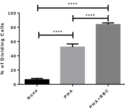
The objective of this study was to ascertain the existence of a possible relationship between the expression of MHC class I molecules at the cell surface and the extent of T cell division. For that objective we cultured PBMC in vitro with stimuli previously known to induce different levels of proliferation that is PHA and PHA plus red blood cells RBC). PBMC were cultured for 6 days in the absence of stimulus (NONE), with PHA or with PHA+RBC. At the end of the culture the percentage of dividing in each culture condition was calculated by adding the percentage of cells in the upper- and lower-left dot-plots of CFSE/W6/32 or HC-10 characteristics. As shown in Figure 1, T cells proliferated at different levels depending on the stimulus, being the culture condition with PHA+RBC much better than PHA (P<0.0001).
% o f D iv id in g C e ll s No ne PH A PH A+ RB C 0 2 0 4 0 6 0 8 0 1 0 0
****
****
****
Figure 1. Proliferation of PBMC. After isolation over Lymphoprep,
cells were labelled with CFSE and culture in three different conditions: None, PHA and PHA+RBC, for six days. At the end of this time period, cells were harvested, washed and cultures with RBC were lysed twice in RBC lysis solution. PBMC were labelled with antibodies previously referred and anlyzed by flow cytometry. The expression of MHC class I molecules was analysed in the lymphocyte region, according to FSC/SSC characteristics using BD Accuri C6. The percentage of proliferating lymphocytes in the different culture conditions was calculated by determining the fraction of cells with CFSE lower than the observed in unstimulated cultures. Student’s t-tests were performed. ****P<0.0001.
9
4.2. Monitoring the Expression of Closed/Open MHC Class I
Conformers on Proliferating/Dividing T Cells
Next, we wanted to analyze the expression of closed and open MHC class I conformers on T dividing Cells. For that purpose, we labelled PBMC with the dye CFSE to monitor cell division cycles and at the end of the culture, cells were labelled with W6/32 or HC-10 antibodies to
study expression of these conformational states on CD4+ and CD8+ T cells.
Figure 2 illustrates a representative experiment where the expression of closed (W6/32) and open (HC-10) MHC class I conformers along the proliferation process (determined by CFSE halving) is shown in the different culture conditions. The level of proliferation as well as the expression of open MHC class I conformers in unstimulated PBMC (NONE, left dot-plots) were very low. Unstimulated PBMC, however, showed expression of closed MHC class I conformers. In cultures of PBMC stimulated with PHA there was an apparent entrance into the cell cycle (middle dot-plots) akin to an increase in the expression of open MHC class I conformers. However, a large fraction of T cells did not divide. The level of expression of closed MHC class I conformers remained steady. In cultures of PBMC stimulated with PHA+RBC, the percentage of non-dividing T cells was very low with the majority of T cells entering the cell cycle and dividing up to six times (right dot-plots), thus corroborating the data shown in Figure 1. Interestingly, the expression of open MHC class I conformers increased further in the PHA+RBC culture conditions in comparison to PHA alone or to unstimulated cultures. In contrast, the expression of closed MHC class I conformers slightly decreased.
10
In view of these results, we wanted to ascertain whether there was a correlation between the levels of expression of MHC Class I conformers on dividing T cells at the end of the six days culture and the extent of proliferation. To that end, we performed a cross-relation analysis between the proliferation index (a measure of the extent of proliferation, see Material & Methods) and the ratio of the expression of the closed to open MHC class I conformers (as a measure of the oscillations in the expression of these conformers, see Materials and Methods)
in CD4+ and CD8+ T cells. As illustrated in Figure 3, there was an inverse relationship between
the extent of cell proliferation and the ratio of the expression of closed to open MHC class I
conformers, both in CD4+ and CD8+ T cells. These results indicate that the overall level of
open MHC class I conformers at the cell surface of dividing T cells increases, while the level of closed conformers is maintained or slightly decreases, at least in the culture conditions with PHA+RBC.
Figure 2. Proliferation of PBMC in response to different stimuli. After isolation over
Lymphoprep, cells were labelled with CFSE and culture in three different conditions: None, PHA and PHA+RBC, for six days. At the end of this time period, cells were harvested and cultures with RBC were lysed twice in RBC lysis solution. PBMC were labelled with antibodies previously referred and analyzed by flow cytometry. The expression of MHC class I molecules was analysed in the lymphocyte region, according to FSC/SSC characteristics. Dot-plots show CFSE halving in the different culture conditions versus expression closed (W6/32, upper dot-plots) and open (HC-10, lower dot-dot-plots) MHC class I conformers.
1 0 5 4 3 2 1 0 6 5 4 3 2 1 0
1 0 5 4 3 2 1 0 6 5 4 3 2 1 0
5.6% 94.4% 54.8 % 45.2% 88,4% 6.8% 0% 0% 0% 0% 4.7% 0.1% 0.4% 62.1% 51.1% 40.4% 88.6% 5.7% 5.7% 31.8% 5.8% 2.7% 5.4% 0.3%11 R a t io W 6 / 3 2 / H C - 1 0 P r o li fe r a ti o n I n d e x 0 2 0 4 0 6 0 0 5 1 0 1 5 2 0
r = - 0 .4
p = 0 .0 3
R a t io W 6 / 3 2 / H C - 1 0 P r o li fe r a ti o n I n d e x 0 2 0 4 0 6 0 0 5 1 0 1 5 2 0r = - 0 .5
p = 0 .0 0 5
A
B
Figure 3. Correlation between cell proliferation and expression of MHC class I conformers in T lymphocytes. After isolation over Lymphoprep, PBMC were labelled with CFSE and culture in three
different conditions: None, PHA and PHA+RBC, for six days. At the end of this time period, cells were harvested and cultures with RBC were lysed twice in RBC lysis solution. PBMC were labelled with W6/32, HC-10, and anti-CD4 and CD8 antibodies and analyzed by flow cytometry. The expression of MHC class I molecules was analysed in the lymphocyte region, according to FSC/SSC characteristics.Graph shows an inverse relationship between the extent of proliferation and the ratio of closed/open MHC Class I conformers (as determined by W6/32 and HC-10 expression levels) in CD4+ T lymphocytes (A) and in CD8+ T lymphocytes (B) in stimulated cultures. Coefficient
12
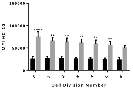
4.3. Impact of Cell Division Number on the Level of Expression
of MHC Class I Conformers
Since the use of CFSE allows to monitor the different cycles of cell division along the six-day culture (see Figure 2), we went to analyze the expression of closed and open MHC Class I conformers in non-dividing cells and in each division cycle. As shown in Figure 4, PBMC cultures stimulated with PHA did not show significant differences in the expression of closed MHC class I conformers. In marked contrast, in cultures of PBMC stimulated with PHA+RBC, in addition to higher expression levels when compared to cultures with PHA (P<0.05), there was an evident decrease in the expression of closed MHC class I conformers from cell division to cell division, that was statistically significant (r=-1 , P=0.0004 ).
A similar analysis for the expression of open MHC class I conformers revealed a similar pattern as for closed MHC class I conformers in cultures of PBMC stimulated with PHA. However, a marked decrease was observed between the 5th and 6th cell division cycle (see Figure 5). In PBMC cultures stimulated with PHA+RBC there was only a slight decrease in the level of open MHC class I conformers that did not reach statistical significance. Noteworthy, the level of expression of open MHC class I conformers in cultures with PHA+RBC were statistically significantly higher when compared to cultures with PHA (P<0.0001) in each division cycle but the last.
Figure 4. Expression of closed MHC class I conformers (W6/32) in each cell division of stimulated cultures. After isolation over Lymphoprep, cells were labelled with CFSE and culture in three
different conditions: None, PHA and PHA+RBC, for six days. At the end of this time period, cells were harvested and cultures with RBC were lysed twice in RBC lysis solution. PBMC were labelled with W6/32 and HC-10 antibodies and analyzed by flow cytometry. The expression of MHC class I molecules was analyzed in the lymphocyte region, according to FSC/SSC characteristics. Graphic shows the expression of closed MHC Class I conformers in non-dividing cells and each cell division. Expression levels in stimulated cultures are shown (Mean±SEM). It is also showed an inset correlation between the expression of closed MHC class I conformers ande the number of cell division (spearman’s Correlation *P<0.05 C e ll D iv is io n N u m b e r M F I W 6 /3 2 0 1 2 3 4 5 6 0 5 0 0 0 0 0 1 0 0 0 0 0 0 1 5 0 0 0 0 0 P H A P H A + R B C
*
W 6 /3 2 C o r r e la tio n N u m b e r o f C e ll D iv is io n s M F I W 6 /3 2 0 1 2 3 4 5 6 0 2 0 0 0 0 0 4 0 0 0 0 0 6 0 0 0 0 0 8 0 0 0 0 0 1 0 0 0 0 0 0 r= - 1 p = 0 .0 0 0 413
4.4. Expression of Closed and Open MHC Class I Conformers on
Leukocyte Populations of Patients with Polycythemia Vera
Since the main focus of this work was to monitor the expression of closed and open MHC Class I conformers in dividing T lymphocytes, we took advantage of having access to blood samples of Polycythemia Vera (PV) patients to analyze the expression of MHC class I molecules on the three main leukocyte populations; lymphocytes, monocytes and granulocytes (mostly neutrophils). Most of these patients are considered elderly (74±2.4) and a number of previous
studies have shown that T cell populations, namely CD8+ T cells, in the elderly have
undergone many cycles of cell division (64). Likewise, these PV patients are characterized by a chronic inflammatory status (65). Therefore, we went to characterize the expression of closed an open MHC class I molecules.
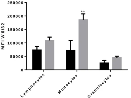
Total leukocytes were obtained from 3 mL blood samples after RBC lysis, as indicated in Material and Methods. Cells were then stained with W6/32 and HC-10 antibodies followed by secondary goat anti-mouse antibody. After staining, cells were acquired using a BD Accuri C6 and expression of closed and open MHC class I conformers was analyzed in the three major leukocyte populations shown in Figure 6.
C e ll D iv is io n N u m b e r M F I H C -1 0 0 1 2 3 4 5 6 0 5 0 0 0 0 1 0 0 0 0 0 1 5 0 0 0 0 P H A P H A + R B C
****
**
**
**
**
**
Figure 5. Expression of open MHC class I conformers (HC-10) in each cell division of stimulated cultures. After isolation over Lymphoprep, cells were labelled with CFSE and culture in three
different conditions: None, PHA and PHA+RBC, for six days. At the end of this time period, cells were harvested and cultures with RBC were lysed twice in RBC lysis solution. PBMC were labelled with W6/32 and HC-10 antibodies and analyzed by flow cytometry. The expression of MHC class I molecules was analyzed in the lymphocyte region, according to FSC/SSC characteristics. Graphic shows the expression of open MHC Class I conformers in non-dividing cells and each cell division. Expression levels in stimulated cultures are shown (Mean±SEM). *P<0.05
14
Figure 7 shows the expression (MFI, Mean±SEM) of closed MHC Class I conformers in the different leukocyte populations in patients and healthy controls (HC). Differences in the expression of both conformers between PV patients and HC were observed. However, only in monocytes the difference in the expression of W6/32-reactive conformers reached statistical significance (185699 in PV vs. 99238 in HC, P<0.001).
Figure 6. Leukocyte Populations studied in Polycythemia Vera patients. WBC
were isolated from 3 mL blood samples. RBC were lysed in REB lysis solution and washed twice in PBS. Then cells were labelled with W/6/32 and HC-10 antibodies for 30 minutes on ice followed by labelling with Goat anti-Mouse secondary antibody. Cells were acquired by flow cytometry in BD Accuri C6 and analyzed in BD Accuri C6 software. Study of closed and open MHC Class I conformers expression was performed in Polycythemia Vera patients in different leukocyte populations. R1 corresponds to the lymphocytes region, R2 to the monocytes region and R3 to granulocytes region.
15 M F I W 6 /3 2 Ly mp ho cy tes Mo no cy tes Gra nu loc yte s 0 5 0 0 0 0 1 0 0 0 0 0 1 5 0 0 0 0 2 0 0 0 0 0 2 5 0 0 0 0 H D P V
**
M F I H C -1 0 Ly mp ho cy tes Mo no cy tes Gra nu loc yte s 0 2 0 0 0 4 0 0 0 6 0 0 0 8 0 0 0 H D P VFigure 7. Expression of closed MHC Class I conformers (W6/32) in the different leukocyte populations of Polycythemia Vera patients and healthy controls. . WBC
were isolated from 3 mL blood samples. RBC were lysed in REB lysis solution and washed twice in PBS. Then cells were labelled with W/6/32 and HC-10 antibodies for 30 minutes on ice followed by labelling with Goat anti-Mouse secondary antibody. Cells were acquired by flow cytometry in BD Accuri C6 and analyzed in BD Accuri C6 software. Graphic shows the different expression levels of closed MHC Class I conformers (W6/32) between Polycythemia Vera patients (PV) and healthy controls (HD) in lymphocytes, monocytes and granulocytes (Mean±SEM). **P<0.001
Figure 8. Expression of closed MHC Class I conformers (W6/32) in the different leukocyte populations of Polycythemia Vera patients and healthy controls. . WBC
were isolated from 3 mL blood samples. RBC were lysed in REB lysis solution and washed twice in PBS. Then cells were labelled with W/6/32 and HC-10 antibodies for 30 minutes on ice followed by labelling with Goat anti-Mouse secondary antibody. Cells were acquired by flow cytometry in BD Accuri C6 and analyzed in BD Accuri C6 software. Graphic shows the different expression levels of open MHC Class I conformers (HC-10) between Polycythemia Vera patients (PV) and healthy controls (HD) in lymphocytes, monocytes and granulocytes (Mean±SEM). **P<0.001
16
Chapter 5 - Discussion
The main objective of this work was to monitor the expression of closed and open MHC Class I conformers in dividing T lymphocytes. For this purpose, T cells were stimulated with previously described stimulus known to induce different rates of proliferation.
Previous experimental studies showed that open MHC class I conformers are expressed at the cell surface of activated, but not resting, lymphoid cells (20-22, 48, 66). These studies were performed in various cell lines and in activated PBMC of diseased individuals. From this point, we went to investigate the expression of closed and open MHC class I conformers in activated PBMC, isolated from iron-overload/erythrocytosis patients and buffy-coats from healthy individuals, on dividing T cells. These were cultured for six days and different stimuli were applied to induce T cell proliferation. Expression of W6/32-reactive molecules and HC-10-reactive molecules follows different trends. In the present study, a correlation between the extent of proliferation and the decrease in the ratio closed to open MHC class I conformers
was found to occur for both CD4+ and CD8+ T lymphocytes, within the six-day culture, pointing
to an increase in the expression of open MHC class I conformers, while the expression of closed MHC class I conformers was maintained or slightly decreasing. A recent work (49), showed that open conformers are expressed at the cell surface of normal human T cells. Cells were activated for 5 days and the expression of open conformers was analyzed at different time points (days 0, 1, 3 and 5), showing an increase in the expression of open MHC class I conformers. On the other hand, in our work, expression of both closed and open MHC class I conformers was monitored in six-day cultures, where different stimulus were applied and the expression of these molecules was studied in each cell division cycle, extending the knowledge about this subject. When stimulated with PHA, PBMC expression of closed conformers increases after cell division, slightly decreasing after some division cycles, whereas the expression of open conformers is maintained from cell division to cell division, being higher than in non-dividing cells, in the first cell division cycles. Cell surface expression of open MHC class I conformers in activated T cells is associated with tyrosine phosphorylation, which may be responsible for the increase in open conformers expression, since shedding of closed MHC class I conformers is reduced, possibly leading to a closed to open equilibrium (49). However, PHA+RBC stimuli induced an evident decrease in the expression of closed MHC class I conformers from cell division to cell division, while expression of open MHC class I conformers slightly decreases in each cell division cycle. Furthermore, expression of both conformers was increased in PHA+RBC-stimulated cells, when compared to PHA-stimuli alone, which may be explained by the known ability of RBC to promote survival, growth and cell cycle progression of T lymphocytes (62, 63).
Regarding the expression of closed and open MHC Class I conformers in PV patients, the data presented represents only preliminary results. Expression of closed conformers is increased in
17
the three leukocyte populations of PV patients in comparison to HC, with statistically significance on monocytes. Although the expression of open conformers is also increased in monocytes and granulocytes of PV patients, the same cannot be said about lymphocytes. In this case, HC express higher levels of open MHC Class I conformers. A recently published paper (61), revealed that HLA genes are downregulated in myeloproliferative neoplasm, in which PV is included. One can speculate that since MHC class I molecules are being overexpressed in this patients, HLA genes may be repressed, in order to decrease the expression of these molecules. Since one of the features of PV patients is an uncontrolled production of RBC, higher expression of open MHC class I molecules may be related with it, allowing cells to divide more often and in higher rates. Nevertheless, it important to take in consideration that the number of HC used is very low, so few conclusions can be taken concerning this matter. More importantly, for our knowledge, this is the first work describing the expression of open MHC class I conformers in PV patients.
This work contributes to the knowledge about the expression of MHC class I conformers, in particular open conformers that have been implicated in a variety of immune and non-immune functions, reviewed in (19). As shown in this work, expression of open conformers increases with the extent of cell proliferation, contributing to a closed to open equilibrium. Considering the immune and non-immune functions of open MHC class I conformers, further studies addressing the formation of these molecules as well their ability to trans- and cis-associate with cell surface receptors are required, in order to disclose novel insights about a possible relation between open MHC class I conformers and several diseases.
18
Chapter 6 - Conclusion
In the present work we were able to monitor the expression of MHC class I conformers on dividing T cell for a short period of time (six-day cultures). The main finding is that the levels of expression of W6/32-reactive molecules and HC-10-reactive molecules at the cell surface of activated T cells follow different trends. This was more evident in PHA+RBC-stimulated cultures, where it is known that T cells divide much more and dye much less. Thus, closed MHC class I conformers appear to decrease their levels of expression with the increase of cell divisions, whereas open MHC class I conformers seem to maintain or slightly decrease their expression levels. Of note, expression of open conformers increases sharply after activation, being more evidence, once again, in cultures stimulated with PHA+RBC, in contrast to closed conformers.
This study comprises a preliminary characterization of the expression of closed and open MHC class I conformers in leukocyte populations of PV patients. Expression levels of W6/32-reactive molecules are increased in the three populations studied, whereas HC-10-W6/32-reactive molecules expression is increased in monocytes and granulocytes. The first steps were taken for a better understanding of the expression of MHC class I molecules, which may have implications for the disease development.
19
Chapter 7 - Future Perspectives
Further studies are required in order to confirm our results and to ameliorate our understanding about the expression of MHC class I conformers. Considering that we monitored the expression of these molecules in six-day cultures, it would be interesting to perform the same experiences in long-term cultures of PBMC from patients and healthy blood donors. A bigger time window would clarify what happens to the expression of W6/32-reactive molecules with an increase of the number of cell cycle division and if HC-10-reactive molecules would maintain or not, their levels of expression. Since the expression of open MHC class I conformers is increased at the cell surface of activated T cells, leading to a closed to open equilibrium, studies about the association of these molecules with other cell surface receptor and with themselves could contribute for a better understanding of several diseases that may be related with the non-immunological functions of open conformers. Furthermore, investigations addressing tyrosine phosphorylation of MHC class I molecules may help understand the influence of open MHC class I conformers in several, non-related, diseases. Regarding PV patients, it is very important to increase the number of HC with a mean age above 65 years so that our results can be confirmed, or not, improving our knowledge about
the disease. It would be interesting to extend our investigations to CD4+ and CD8+ T
lymphocytes, considering their importance to the immune system. Finally, follow-up studies should be performed so possible correlations between the expression of closed and open MHC class I conformers and the progression of the disease may be established.
20
References
1. Snell GD. The immunogenetics of tumor transplantation. Cancer research.
1952;12(8):543-6.
2. Dausset J. [The biological implications of the main histocompatibility system: the
HL-A system]. Comptes rendus des seances de la Societe de biologie et de ses filiales. 1974;168(2-3):160-72.
3. Doherty PC, Zinkernagel RM. Enhanced immunological surveillance in mice
heterozygous at the H-2 gene complex. Nature. 1975;256(5512):50-2.
4. Germain RN, Margulies DH. The biochemistry and cell biology of antigen processing
and presentation. Annual review of immunology. 1993;11:403-50.
5. Pamer E, Cresswell P. Mechanisms of MHC class I--restricted antigen processing.
Annual review of immunology. 1998;16:323-58.
6. Solheim JC. Class I MHC molecules: assembly and antigen presentation. Immunological
reviews. 1999;172:11-9.
7. Antoniou AN, Powis SJ, Elliott T. Assembly and export of MHC class I peptide ligands.
Current opinion in immunology. 2003;15(1):75-81.
8. Grotzke JE, Cresswell P. Are ERAD components involved in cross-presentation?
Molecular immunology. 2015;68(2 Pt A):112-5.
9. Ackerman AL, Cresswell P. Cellular mechanisms governing cross-presentation of
exogenous antigens. Nature immunology. 2004;5(7):678-84.
10. Mellman I, Steinman RM. Dendritic cells: specialized and regulated antigen processing
machines. Cell. 2001;106(3):255-8.
11. Tse DB, Pernis B. Spontaneous internalization of Class I major histocompatibility
complex molecules in T lymphoid cells. The Journal of experimental medicine. 1984;159(1):193-207.
12. Dasgupta JD, Cemach K, Dubey DP, Yunis EJ, Amos DB. The role of class I
histocompatibility antigens in the regulation of T-cell activation. Proceedings of the National Academy of Sciences of the United States of America. 1987;84(4):1094-8.
13. Dasgupta JD, Yunis EJ. Receptor-like role of HLA-class I antigens: regulation of T cell
activation. Journal of immunology. 1987;139(3):672-7.
14. Vega MA, Strominger JL. Constitutive endocytosis of HLA class I antigens requires a
specific portion of the intracytoplasmic tail that shares structural features with other endocytosed molecules. Proceedings of the National Academy of Sciences of the United States of America. 1989;86(8):2688-92.
15. Demaria S, Bushkin Y. Soluble HLA proteins with bound peptides are released from the
cell surface by the membrane metalloproteinase. Human immunology. 2000;61(12):1332-8.
16. Zavazava N. Soluble HLA class I molecules: biological significance and clinical
21
17. Ghio M, Contini P, Mazzei C, Brenci S, Filaci G, Indiveri F, et al. Soluble HLA class I
and Fas ligand molecules in blood components and their role in the immunomodulatory effects of blood transfusions. Leukemia & lymphoma. 2000;39(1-2):29-36.
18. Puppo F, Contini P, Ghio M, Brenci S, Scudeletti M, Filaci G, et al. Soluble human MHC
class I molecules induce soluble Fas ligand secretion and trigger apoptosis in activated CD8(+) Fas (CD95)(+) T lymphocytes. International immunology. 2000;12(2):195-203.
19. Arosa FA, Santos SG, Powis SJ. Open conformers: the hidden face of MHC-I molecules.
Trends in immunology. 2007;28(3):115-23.
20. Schnabl E, Stockinger H, Majdic O, Gaugitsch H, Lindley IJ, Maurer D, et al. Activated
human T lymphocytes express MHC class I heavy chains not associated with beta 2-microglobulin. The Journal of experimental medicine. 1990;171(5):1431-42.
21. Madrigal JA, Belich MP, Benjamin RJ, Little AM, Hildebrand WH, Mann DL, et al.
Molecular definition of a polymorphic antigen (LA45) of free HLA-A and -B heavy chains found on the surfaces of activated B and T cells. The Journal of experimental medicine. 1991;174(5):1085-95.
22. Demaria S, Schwab R, Bushkin Y. The origin and fate of beta 2m-free MHC class I
molecules induced on activated T cells. Cell Immunol. 1992;142(1):103-13.
23. Bird LA, Peh CA, Kollnberger S, Elliott T, McMichael AJ, Bowness P. Lymphoblastoid
cells express HLA-B27 homodimers both intracellularly and at the cell surface following endosomal recycling. European journal of immunology. 2003;33(3):748-59.
24. Gonen-Gross T, Achdout H, Arnon TI, Gazit R, Stern N, Horejsi V, et al. The
CD85J/Leukocyte Inhibitory Receptor-1 Distinguishes between Conformed and 2-Microglobulin-Free HLA-G Molecules. The Journal of Immunology. 2005;175(8):4866-74.
25. Benoit LA, Shannon J, Chamberlain JW, Miller RG. Influence of xenogeneic
beta2-microglobulin on functional recognition of H-2Kb by the NK cell inhibitory receptor Ly49C. Journal of immunology. 2005;175(6):3542-53.
26. Allen RL, Bowness P, McMichael A. The role of HLA-B27 in spondyloarthritis.
Immunogenetics. 1999;50(3-4):220-7.
27. Raine T, Brown D, Bowness P, Hill Gaston JS, Moffett A, Trowsdale J, et al. Consistent
patterns of expression of HLA class I free heavy chains in healthy individuals and raised expression in spondyloarthropathy patients point to physiological and pathological roles. Rheumatology. 2006;45(11):1338-44.
28. Edidin M. Function by association? MHC antigens and membrane receptor complexes.
Immunology today. 1988;9(7-8):218-9.
29. Fishman D, Elhyany S, Segal S. Non-immune functions of MHC class I glycoproteins in
normal and malignant cells. Folia biologica. 2004;50(2):35-42.
30. Smith C, Santi M, Rajan B, Rushing EJ, Choi MR, Rood BR, et al. A novel role of HLA
22
31. Smith C, Santi M, Rushing EJ, Cornelison R, MacDonald TJ, Vukmanovic S.
Characterization of signaling function and expression of HLA class I molecules in medulloblastoma. Journal of neuro-oncology. 2011;103(2):197-206.
32. Fehlmann M, Peyron JF, Samson M, Van Obberghen E, Brandenburg D, Brossette N.
Molecular association between major histocompatibility complex class I antigens and insulin receptors in mouse liver membranes. Proceedings of the National Academy of Sciences of the United States of America. 1985;82(24):8634-7.
33. Due C, Simonsen M, Olsson L. The major histocompatibility complex class I heavy
chain as a structural subunit of the human cell membrane insulin receptor: implications for the range of biological functions of histocompatibility antigens. Proceedings of the National Academy of Sciences of the United States of America. 1986;83(16):6007-11.
34. Samson M, Cousin JL, Fehlmann M. Cross-linking of insulin receptors to MHC antigens
in human B lymphocytes: evidence for selective molecular interactions. Journal of immunology. 1986;137(7):2293-8.
35. Schreiber AB, Schlessinger J, Edidin M. Interaction between major histocompatibility
complex antigens and epidermal growth factor receptors on human cells. The Journal of cell biology. 1984;98(2):725-31.
36. Harel-Bellan A, Krief P, Rimsky L, Farrar WL, Mishal Z. Flow cytometry resonance
energy transfer suggests an association between low-affinity interleukin 2 binding sites and HLA class I molecules. The Biochemical journal. 1990;268(1):35-40.
37. Blue ML, Craig KA, Anderson P, Branton KR, Jr., Schlossman SF. Evidence for specific
association between class I major histocompatibility antigens and the CD8 molecules of human suppressor/cytotoxic cells. Cell. 1988;54(3):413-21.
38. Bushkin Y, Demaria S, Le JM, Schwab R. Physical association between the CD8 and
HLA class I molecules on the surface of activated human T lymphocytes. Proceedings of the National Academy of Sciences of the United States of America. 1988;85(11):3985-9.
39. Jelonek MT, Classon BJ, Hudson PJ, Margulies DH. Direct binding of the MHC class I
molecule H-2Ld to CD8: interaction with the amino terminus of a mature cell surface protein. Journal of immunology. 1998;160(6):2809-14.
40. Bennett MJ, Lebron JA, Bjorkman PJ. Crystal structure of the hereditary
haemochromatosis protein HFE complexed with transferrin receptor. Nature. 2000;403(6765):46-53.
41. Lebron JA, Bjorkman PJ. The transferrin receptor binding site on HFE, the class I
MHC-related protein mutated in hereditary hemochromatosis. Journal of molecular biology. 1999;289(4):1109-18.
42. Goodridge JP, Lee N, Burian A, Pyo CW, Tykodi SS, Warren EH, et al. HLA-F and MHC-I
open conformers cooperate in a MHC-I antigen cross-presentation pathway. Journal of immunology. 2013;191(4):1567-77.
43. Olson R, Dulac C, Bjorkman PJ. MHC homologs in the nervous system--they haven't
23
44. Olson R, Huey-Tubman KE, Dulac C, Bjorkman PJ. Structure of a pheromone
receptor-associated MHC molecule with an open and empty groove. PLoS biology. 2005;3(8):e257.
45. Boulanger LM, Shatz CJ. Immune signalling in neural development, synaptic plasticity
and disease. Nature reviews Neuroscience. 2004;5(7):521-31.
46. Escande-Beillard N, Washburn L, Zekzer D, Wu ZP, Eitan S, Ivkovic S, et al. Neurons
preferentially respond to self-MHC class I allele products regardless of peptide presented. Journal of immunology. 2010;184(2):816-23.
47. Held W, Mariuzza RA. Cis interactions of immunoreceptors with MHC and non-MHC
ligands. Nature reviews Immunology. 2008;8(4):269-78.
48. Pickl WF, Holter W, Stockl J, Majdic O, Knapp W. Expression of beta
2-microglobulin-free HLA class I alpha-chains on activated T cells requires internalization of HLA class I heterodimers. Immunology. 1996;88(1):104-9.
49. Santos SG, Powis SJ, Arosa FA. Misfolding of major histocompatibility complex class I
molecules in activated T cells allows cis-interactions with receptors and signaling molecules and is associated with tyrosine phosphorylation. The Journal of biological chemistry. 2004;279(51):53062-70.
50. Matko J, Bushkin Y, Wei T, Edidin M. Clustering of class I HLA molecules on the
surfaces of activated and transformed human cells. Journal of immunology. 1994;152(7):3353-60.
51. Jenei A, Varga S, Bene L, Matyus L, Bodnar A, Bacso Z, et al. HLA class I and II
antigens are partially co-clustered in the plasma membrane of human lymphoblastoid cells. Proceedings of the National Academy of Sciences of the United States of America. 1997;94(14):7269-74.
52. Triantafilou K, Triantafilou M, Wilson KM, Fernandez N. Human major
histocompatibility molecules have the intrinsic ability to form homotypic associations. Human immunology. 2000;61(6):585-98.
53. Skov S, Odum N, Claesson MH. MHC class I signaling in T cells leads to tyrosine kinase
activity and PLC-gamma 1 phosphorylation. Journal of immunology. 1995;154(3):1167-76.
54. Skov S, Bregenholt S, Claesson MH. MHC class I ligation of human T cells activates the
ZAP70 and p56lck tyrosine kinases, leads to an alternative phenotype of the TCR/CD3 zeta-chain, and induces apoptosis. Journal of immunology. 1997;158(7):3189-96.
55. Skov S. Intracellular signal transduction mediated by ligation of MHC class I
molecules. Tissue antigens. 1998;51(3):215-23.
56. Pedersen AE, Skov S, Bregenholt S, Ruhwald M, Claesson MH. Signal transduction by
the major histocompatibility complex class I molecule. APMIS : acta pathologica, microbiologica, et immunologica Scandinavica. 1999;107(10):887-95.
57. Ding J, Feng Y, Zheng ZH, Li XY, Wu ZB, Zhu P. Increased expression of human
leucocyte antigen class I free heavy chains on monocytes of patients with spondyloarthritis and cells transfected with HLA-B27. International journal of immunogenetics. 2015;42(1):4-10.
24
58. Kollnberger S, Chan A, Sun MY, Chen LY, Wright C, di Gleria K, et al. Interaction of
HLA-B27 homodimers with KIR3DL1 and KIR3DL2, unlike HLA-B27 heterotrimers, is independent of the sequence of bound peptide. European journal of immunology. 2007;37(5):1313-22.
59. Wong-Baeza I, Ridley A, Shaw J, Hatano H, Rysnik O, McHugh K, et al. KIR3DL2 binds
to HLA-B27 dimers and free H chains more strongly than other HLA class I and promotes the expansion of T cells in ankylosing spondylitis. Journal of immunology. 2013;190(7):3216-24.
60. Perosa F, Luccarelli G, Prete M, Ferrone S, Dammacco F. Increased serum levels of
beta2m-free HLA class I heavy chain in multiple myeloma. British journal of haematology. 1999;106(4):987-94.
61. Skov V, Riley CH, Thomassen M, Larsen TS, Jensen MK, Bjerrum OW, et al. Whole
blood transcriptional profiling reveals significant down-regulation of human leukocyte antigen class I and II genes in essential thrombocythemia, polycythemia vera and myelofibrosis. Leukemia & lymphoma. 2013;54(10):2269-73.
62. Fonseca AM, Pereira CF, Porto G, Arosa FA. Red blood cells promote survival and cell
cycle progression of human peripheral blood T cells independently of CD58/LFA-3 and heme compounds. Cellular Immunology. 2003;224(1):17-28.
63. Antunes RF, Brandao C, Maia M, Arosa FA. Red blood cells release factors with growth
and survival bioactivities for normal and leukemic T cells. Immunology and cell biology. 2011;89(1):111-21.
64. Effros RB. Telomerase induction in T cells: a cure for aging and disease? Experimental
gerontology. 2007;42(5):416-20.
65. Mondet J, Hussein K, Mossuz P. Circulating Cytokine Levels as Markers of Inflammation
in Philadelphia Negative Myeloproliferative Neoplasms: Diagnostic and Prognostic Interest. Mediators of inflammation. 2015;2015:670580.
66. Demaria S, Schwab R, Gottesman SR, Bushkin Y. Soluble beta 2-microglobulin-free
class I heavy chains are released from the surface of activated and leukemia cells by a metalloprotease. The Journal of biological chemistry. 1994;269(9):6689-94.