Patterns of
MHC-G-Like
and
MHC-B
Diversification in New World Monkeys
Juan S. Lugo, Luis F. Cadavid*
Department of Biology and Institute of Genetics, Universidad Nacional de Colombia, Bogotá, Colombia
Abstract
The MHC class I (MHC-I) region in New World monkeys (Platyrrhini) has remained relatively understudied. To evaluate the diversification patterns and transcription behavior of MHC-I in Platyrrhini, we first analyzed public genomic sequences from theMHC-G-likesubregion inSaimiri boliviensis,Ateles geoffroyiandCallicebus moloch, and from theMHC-B subre-gion inSaimiri boliviensis. WhileS.boliviensisshowed multiple copies of bothMHC-G-like (10) and–B(15) loci,A.geoffroyiandC.molochhad only three and fourMHC-G-like genes, respectively, indicating that not all Platyrrhini species have expanded their MHC-I loci. We then sequencedMHC-G-likeand-BcDNAs from nine Platyrrhini species, recover-ing two to five unique cDNAs per individual for both loci classes. In twoSaguinusspecies, however, noMHC-BcDNAs were found. In phylogenetic trees,MHC-G-likecDNAs formed genus-specific clusters whereas theMHC-BcDNAs grouped by Platyrrhini families, sug-gesting a more rapid diversification of the former. Furthermore, cDNA sequencing in 12 capuchin monkeys showed that they transcribe at least fourMHC-G-likeand fiveMHC-B polymorphic genes, showing haplotypic diversity for gene copy number and signatures of positive natural selection at the peptide binding region. Finally, a quantitative index for MHC:KIR affinity was proposed and tested to predict putative interacting pairs. Altogether, our data indicate thati)MHC-I genes has expanded differentially among Platyrrhini species, ii) Callitrichinae(tamarins and marmosets)MHC-Bloci have limited or tissue-specific expression,iii) MHC-G-likegenes have diversified more rapidly thanMHC-Bgenes, andiv) the MHC-I diversity is generated mainly by genetic polymorphism and gene copy number variation, likely promoted by natural selection for ligand binding.
Introduction
Major Histocompatibility Complex (MHC) class I molecules present processed peptides to CD8+ T lymphocytes and interact with Natural Killer (NK) cell receptors to modulate immune responses directed against self-altered cells [1]. The hallmark of the MHC class I system is the high genetic polymorphism of its loci, which has been promoted by selective pressures from rapidly evolving pathogens to evade immune recognition [2]. In humans, there are six func-tional MHC class I genes,HLA-A, -B. -C, -E, -F, and -G, that, together with 11 MHC class I
a11111
OPEN ACCESS
Citation:Lugo JS, Cadavid LF (2015) Patterns of
MHC-G-LikeandMHC-BDiversification in New World Monkeys. PLoS ONE 10(6): e0131343. doi:10.1371/journal.pone.0131343
Editor:Tzen-Yuh Chiang, National Cheng-Kung University, TAIWAN
Received:February 4, 2015
Accepted:June 1, 2015
Published:June 29, 2015
Copyright:© 2015 Lugo, Cadavid. This is an open access article distributed under the terms of the
Creative Commons Attribution License, which permits unrestricted use, distribution, and reproduction in any medium, provided the original author and source are credited.
Data Availability Statement:All relevant data are within the paper and its Supporting Information files. cDNA sequences reported here are available from GenBank under accession numbers KM219682-KM219782.
Funding:The authors have no support or funding to report.
pseudogenes and several non-MHC genes and pseudogenes, constitute MHC class I region on chromosome 6p21 [3].HLA-A, -B, and -Cgenes are known as classical MHC class I genes and are characterized by their high polymorphism and expression on virtually all nucleated cells, whereasHLA-E, -F, and -Gare nonclassical MHC class I genes displaying limited polymor-phism and restricted tissue distribution [4]. The human MHC class I region is divided into three contiguous subgenomic sections, often referred to as the alpha block, located at the telo-meric end of the region and containingHLA-F, -G, and -A, the beta block, located at the cen-tromeric end of the region and containingHLA-Band -C, and the kappa block, at the central part of the region, containingHLA-E[5].
Functional MHC class I loci have remained relatively well conserved in Catarrhini primates (humans, apes and Old World monkeys) [6]. Species from theHominidaefamily (humans and great apes) have orthologous genes toHLA-A, -B, -C, -E. -F, and -G, althoughMHC-Cis pres-ent in only about 50% of orangutan (Pongo pygmeus)haplotypes [7]. TheHylobatidaefamily (gibbons or lesser apes) has at least threeMHC-Bgenes and a single copy of eachMHC-A, -E, and -Fgenes, but they lackMHC-CandMHC-Gloci [8]. Likewise, Old World monkeys (family Cercopithecidae) have multiple copies of theMHC-Aand-Bloci, lackMHC-C, have maintained MHC-E and—Fas single copy genes, andMHC-Gis a pseudogene [9]. Platyrrhini species
(New World monkeys) are distributed from Mexico to the North of Argentina and shared a common ancestor with Catarrhini some 35–45 million years ago (MYA) [10]. At least 15 Pla-tyrrhini genera have been recognized, which are grouped into three families:Pitheciidae, Ateli-dae, andCebidae[11]. Platyrrhini are typically known to express classical MHC class I loci more closely related toMHC-G[12,13], lacking orthologues ofMHC-AandMHC-Cloci, and containing a functionalMHC-E[14]. A genomic analysis of the MHC class I alpha block (MHC-F/G/A) in the Platyrrhini speciesCallithrix jacchus(common marmoset) revealed the presence of fiveMHC-G-likegenes, nineMHC-G-likepseudogenes, oneMHC-Fgene, and four MHC-Fpseudogenes [15]. The expansion of this region in the common marmoset has proba-bly occurred by repeated segmental duplications of units containing fourMHC-G-likeand one MHC-Fgenes [15]. In addition, genomic sequencing of the MHC class I beta block (MHC-B/ C) in this species showed the presence of ninebona fide MHC-Bgenes, five of which were puta-tively transcribed [16]. However, cDNA sequencing efforts to identifyMHC-Btranscripts in this species have given mixed results. In a population of 15 common marmosets, van der Wiel et al (2013) failed to identify anyMHC-Btranscript [17], although Cao et al (2015) found four uniqueMHC-Bsequences with one to three sequences per individual in a population of eight common marmosets [16]. This suggests thatMHC-Bloci have either low transcription levels that hinder their detection by conventional PCR-based techniques, and/or they have differen-tial tissue expression. Although the vast majority of classical MHC class I cDNAs reported in Platyrrhini are from theMHC-G-likeloci, cDNAs with high similarity toMHC-Bhave been identified in the owl monkey (Aotussp) [18], the saki monkey (Pithecia pithecia) [13], the white-bellied spider monkey (Ateles belzebuth)[13], and the brown-headed spider monkey (Ateles fusciceps). Furthermore, sequence and retroelement analyses of the 50-flanking regions of MHC class I genes, have provided evidence for the presence of various copies ofMHC-Bin the owl monkey, the capuchin monkey (Cebus apella), and the spider monkey [18].
In order to gain a better understanding of the MHC class I genes in Platyrrhini, we have investigated their diversification processes at three different levels,i) at the genomic level by analyzing the MHC class I gene organization in three Platyrrhini species using public sequence information,ii) at the transcriptomic level by cloning and sequencingMHC-G-likeand MHC-BcDNAs from 10 species representing two Platyrrhini families (CebidaeandAtelidae), andiii) at the gene level by studying the polymorphism ofMHC-G-likeandMHC-Bloci in a population of 12 white-fronted capuchin monkeys (Cebus albifrons).
Materials and Methods
Ethics Statement
We used samples from the following primate species: cotton-top tamarin (Saguinus oedipus), white-footed tamarin (Saguinus leucopus), squirrel monkey (Saimiri sciureus), tufted capuchin monkey (Cebus apella), white-headed capuchin monkey (Cebus capucinus), red howler mon-key (Alouatta seniculus), white-bellied spider monkey (Ateles belzebuth), brown spider monkey (Ateles hybridus), and common woolly monkey (Lagothrix lagotricha). Animals used to obtain blood samples were maintained at the Center for Rescue and Rehabilitation of Wild Fauna (URRAS) from the Universidad Nacional de Colombia0s Veterinary School. The Center is authorized by the Bogotá Secretary of the Environment following the Colombian legislation on the use of animals for research (law 84/1989, resolution 8430/1993), and it is regulated by the Bioethics Committee of the Universidad Nacional de Colombia0s Veterinary School, directed by Dr. Gloria C. Ramírez-Nieto ([email protected]) (Art. 22, Resolution 138/2011). Colombia’s Ministry of the Environment and Sustainable Development authorized the access to genetic resources for scientific research without commercial interests (contract No. 1 of 2012). The study was approved by the Center’s director, Dr. Claudia Brieva (cibrievar@unal. edu.co), as no Institutional Animal Care and Use Committee has been formally established at the University. For animal care monitoring, the results of the present study were reported to the Center’s Director, which in turn reports periodically to the Bioethics Committee. The mon-keys were temporarily maintained in cages of 1 x 1.5 x 2 meters with an average temperature of 22 degrees centigrade, and a relative humidity of 67%. The monkeys’diet was based on a supply of fruits, vegetables and a nutritional supplement including vitamins, minerals and proteins. Their environment was enriched by including visual barriers (to avoid social conflicts), feeding devices, some branches and vegetation, perches and a nest box. Trained veterinary personnel carried out all procedures requiring the handling of animals. Blood samples were obtained as part of routine animal care by the Center0s veterinarian (Dr. Robin Poches), from animals anes-thetized with Ketamine 20 mg/Kg or Tyletamine/Zolazepam (Zoletil) 4.4 mg/Kg. No animals were sacrificed during, or as a result of, this study.
MHC class I region genomic sequences
BLAST searches against the NCBI GenBank database using known MHC class I sequences were conducted to identify Platyrrhini bacterial artificial chromosomes (BACs) and next gener-ation sequence scaffolds containing MHC class I genes. Significant BLAST matches
(identity>80% and e-value<10−70) were found in the Bolivian squirrel monkey (Saimiri
boli-viensis), the spider monkey (Ateles geoffroyi), and the dusky titi monkey (Callicebus moloch). Data from the former two species were produced by the National Institutes of Health— Intra-mural Sequencing Center and released on April 2010. Data from the latter species were pro-duced by the Broad Institute as part of a genome project and released on October 2011. Gene predictions on the genomic sequences were carried out with the GeneScan tool (genes.mit.edu/ GENSCAN.html) and with the Augustus software (augustus.gobics.de/), and the identified genes were annotated by BLAST against the NCBI Reference Sequence Database (RefSeq,
www.ncbi.nlm.nih.gov/refseq). Genomic sequences were aligned with the Mauve multiple genome alignment software v2.3.1 [19].
Samples, cDNA cloning, and sequencing
white-footed tamarin (Saguinus leucopus), squirrel monkey (Saimiri sciureus), tufted capuchin mon-key (Cebus apella), white-headed capuchin monkey (Cebus capucinus), red howler monkey (Alouatta seniculus), white-bellied spider monkey (Ateles belzebuth), brown spider monkey (Ateles hybridus), and common woolly monkey (Lagothrix lagotricha). For polymorphism analyses, 12 unrelated white-fronted capuchin monkeys (Cebus albifrons) were also sampled. Total RNA was isolated from peripheral blood with the Trizol reagent (Invitrogen) and used for complementary DNA (cDNA) synthesis with the RevertAid First strand cDNA Synthesis kit (Fermentas). Full-length MHC class I cDNAs were amplified by PCR with generic primers designed for bothMHC-G-likeandMHC-B, as follows: forward primer5'-TAACGGTCMT GGMGCCCCGAA-3'(where M means A or C), and reverse primer5'-AATGAGAGACACATC AGAGCC-3'. The reaction contained 400μM dNTPs, 2 mM MgCl2, and 0.5 U of Taq DNA
polymerase (Invitrogen) in a volume of 25μl. Amplifications were carried out with an initial cycle of 5 min at 94°C, followed by 35 cycles of 94°C for 45 s, 62.6°C for 55 s, and 72°C for 65 s, with a final extension of 72°C for 10 min. PCR products were gel-purified with the PureLink kit (Life Technologies), cloned into the pGEM-T Easy vector (Promega), and at least 24 clones per individual were sequenced with the Sanger method at Macrogen (Korea). Reported sequences derived from at least three identical plasmid clones.
Sequence and evolutionary analysis
Genomic and cDNA sequences were aligned with the Muscle software [20] and the alignment were manually edited using the MEGA v5.2 software [21]. Sequence distances were estimated with the Maximum Composite Likelihood model [22] with standard errors estimated by boot-strap after 1,000 replicates using MEGA v5.2. Phylogenetic analyses were carried out with the Neighbor-Joining, Maximum Likelihood, and Bayesian approaches based on nucleotide substi-tution models estimated by the ModelTest software v3.7 [23]. Neighbor-joining and Maximum likelihood phylogenies were constructed with the MEGA v5.2 package [21] and their reliability was evaluated by bootstrap with 1,000 replicates. Bayesian phylogenies were inferred with MrBayes v3.2 [24,25] using 10,000,000 iterations in five chains. Natural selection acting on MHC class I genes was evaluated by codon-based selection analysis using the PAML v4.4 pack-age [26]. For this, the dN / dS ratio (ω) was estimated by maximum likelihood using the F3X4 model for codon frequencies. The neighbor-joining tree generated previously was used to esti-mate the tree topology likelihood comparing a null model that does not allowω>1 (M1) with three models that does (M2, M3, and M8). Comparisons were carried out by likelihood ratio tests and their significance was evaluated by comparing two times the likelihood difference between the models (2ΔL) with a Chi-square distribution with two degrees of freedom. Codons
withω>1 were identified through a Bayes Empirical Bayes approach [27] when at least two of the three selection models had a probability greater than 0.9.
Prediction of MHC-I:KIR interactions
and Rhesus monkey MHC:KIR interactions [29,31–34] in order to asses its predictability and to quantify the rate of false positives and false negatives.
Results
Genomic diversification of the MHC class I region in Platyrrhini
Sequence database searching using known MHC class I sequences resulted in the identification of BAC clone sequences and next generation sequencing scaffolds containing unpublished MHC class I genes from the Bolivian squirrel monkey (Saimiri boliviensis), the spider monkey (Ateles geoffroyi), and the dusky titi monkey (Callicebus moloch). The search of MHC class I sequences inS.boliviensisyielded eight non-overlapping next-generation sequence scaffolds, with the first six (NW_003943887.1, NW_003943835.1, NW_003943876.1, NW_003943863.1, NW_003943949.1 and NW_003943840.1) spanning the entireMHC-F/G/Aregion. This sub-genomic block was circumscribed at the telomeric end by the framework genesMOG(myelin oligodendrocyte glycoprotein) andZNF57(zinc finger protein-57) and at the centromeric end by framework genesETF1P1(eukaryotic peptide chain release factor subunit 1-like) and the non-coding RNAZNRD1-AS1(Fig 1A). The scaffolds contained sixMHC-G-likegenes (five partially sequenced), fourMHC-G-likepseudogenes, one partially sequencedMHC-Fgene, and 44 non-MHC genes, pseudogenes, and non-coding open reading frames (ORFs) (Fig 1A
andS1 Table). In addition, an MHC class I sequence (tentatively namedMHC-X) with no close similarity to known primate MHC class I genes was found between the centromeric framework genesETF1P1andZNRD1-AS1(Fig 1A). One of the six scaffolds spanning theMHC-F/G/A region (NW_003943840.1) extended beyond the alpha block into the kappa block (MHC-E) and contained this block0s telomeric framework geneRNF39(gene ring finger protein 39) and seven non-MHC genes also found in humans and in the common marmoset (S1 Fig). The fol-lowing scaffold (NW_003943826.1) spanned part of the MHC class I kappa block and had a sin-gleMHC-Egene (Sabo-E), together with 12 non-MHC genes and the centromeric framework genesCDSN(corneodesmosin),CCHR1(coiled-coil alpha-helical rod protein-1), andTCF19 (transcription factor 19) (S1 Fig). Finally, scaffolds NW_003943835.1 and NW_003943627.1 contained genes and pseudogenes homologous to those of the primate MHC class I beta block (MHC-B/C), with the framework genePOU5F1(encoding the POU domain, class 5, transcrip-tion factor 1) flanking the region at the telomeric end, and aMICgene (MHC class I chain related) at the centromeric end (Fig 1B). These two scaffolds contained 15MHC-Bgenes, two MHC-Bpseudogenes (Sabo-B8and-B12), and 24 non-MHC genes, pseudogenes, and non-cod-ing ORFs (Fig 1andS1 Table). Scaffold NW_003943835.1 was unusual in that it contained two MHC-G-likeand nineMHC-Bsequences, suggesting that the scaffold has been misassembled.
overlapped by a region of approximately 120 kb, resulting in a contig of approximately 240 kb with no internal gaps (Fig 1A). This region was homologous to the primate MHC class I alpha block, having the telomeric framework genesMOGandZNF57, but lacking centromeric frame-work genes, indicating that the region has not been completely covered. The contig contained two full-lengthMHC-G-likegenes (Camo-G1and-G3), twoMHC-G-likepseudogenes ( Camo-G2and-G4), oneMHC-Fgene (Camo-F1), and 18 MHC genes, pseudogenes, and non-coding ORFs (Fig 1AandS1 Table).
In order to have better insights on the diversification dynamics of the Platyrrhini MHC class I region, a Bayesian phylogenetic analysis was performed based on intronic sequences from the genes and pseudogenes presented above. Since introns evolve neutrally, their phylog-eny reflects closer the actual evolutionary history of the genes than that based on exons, as they are subjected to natural selection. The phylogenetic tree showed that PlatyrrhiniMHC-G-like, Fig 1. Comparison of the MHC class I region ofS.boliviensis,A.geoffroyi, andC.molochusing as reference that of humans [3] andC.jacchus[15,
16].A, MHC class I alpha block (MHC-A/G/F). B, MHC class I beta block (MHC-B/C). Red rectangles represent centromeric and telomeric framework genes, dark blue rectangles indicate putatively transcribed MHC class I genes, including partially sequenced genes (names underlined), and light blue rectangles depict MHC class I pseudogenes. BAC clone or scaffold accession numbers are indicated for each species. TheS.boliviensisscaffold NW_003943835.1 containedMHC-G-likeandMHC-Bsequences, likely reflecting an erroneous assembly, but for clarity, the genes were located in their respective regions. Non-MHC class I sequences annotated in these regions are shown inS1 Table.
doi:10.1371/journal.pone.0131343.g001
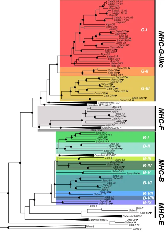
MHC-FandMHC-Bsequences clustered as a sister groups of their Catarrhini counterparts (Fig 2). PlatyrrhiniMHC-G-likesequences formed three well-differentiated clades (G-I, G-II, and G-III), where clade G-I contained the majority of the sequences from the analyzed species, clade G-II included threeC.jacchuspseudogenes and a partial sequence fromS.boliviensis (Sabo-G4), and clade G-III included only pseudogenes from the three species analyzed, along with genomic sequences fromC.jacchus, the squirrel monkey (Saimiri sciureus), and the cotton top tamarin (Saguinus oedipus) found in public databases (Fig 2). Within the clades most sequences cluster with paralogous genes, whereas orthologousMHC-G-likegenes were hypoth-esized only forSabo-G10/Sasc31,Aotr-G2/Sabo-G6/Atge-G1,Sabo-G9/Sasc-25, andCaja-G8/ Saoe-G13. This suggests that mostMHC-G-likeloci have diversified independently in Platyr-rhini taxa by a continuous process of gene duplication and inactivation or deletion, followed by sequence divergence. The pseudogeneMHC-Xin the centromeric end of the alpha block ofC. jacchus,A.geoffroyi, andC.moloch, formed a monophyletic group ancestral to all known pri-mate MHC class I loci (Fig 2).
PlatyrrhiniMHC-Fgenes and pseudogenes were organized as a sister group of Catarrhini sequences, with the exception of the pseudogeneCaja-F3that was outside of these two groups (Fig 2). The available data suggests that theMHC-Flocus has duplicated differentially among the Platyrrhini species after the divergence from Catarrhini, with at least one duplication event inA.geofroyiiand various events inC.jacchus. PlatyrrhiniMHC-Bsequences formed nine well-differentiated clades (I-IX), each with at least one sequence fromC.jacchusandS.boliviensis, except for clade B-V that contained a single sequence from eachS.boliviensisandS.oedipus(Fig 2). Clades B-III and B-IV were the most closely related to the CatarrhiniMHC-Bloci, although their association in the tree was not statistically well supported. In contrast toMHC-G-like sequences, mostMHC-Bsequences clustered with putative orthologous genes, suggesting that most of the duplications that configured theC.jacchusandS.boliviensis MHC-Bregion pre-dated the divergence of these two species, approximately 17 million MYA [10].
Diversification of transcriptionally active
MHC-G-like
and
MHC-B loci
in
Platyrrhini
Fig 2. Bayesian phylogenetic tree based on introns 1–8 from primate MHC class I genes and pseudogenes.Black dots at the branching points indicate a support with a posterior probability greater than 0.9. A phi (Ψ) symbol in front of a sequence name indicates pseudogene and a question mark (?) indicates an incompletely sequenced gene. Catarrhini clades include human and Rhesus monkey MHC class I sequences, whereas MHC class I sequences from the grey mouse lemurMicrocebus murinus(Mimu) were used as outgroup. Lineages are indicated by a G (MHC-G-like) or a B (MHC-B), followed by a Roman number. Caja,Callitrhix jacchus; Sabo,Saimiri boliviensis; Sasc,Saimiri sciureus; Camo,Callicebus moloch; Atge,Ateles geoffroyi; Saoe,Saguinus oedipus; Aotr,Aotus trivirgatus. Accession numbers for published Platyrrhini MHC class I sequences used in the analysis are listed inS3 Table.
doi:10.1371/journal.pone.0131343.g002
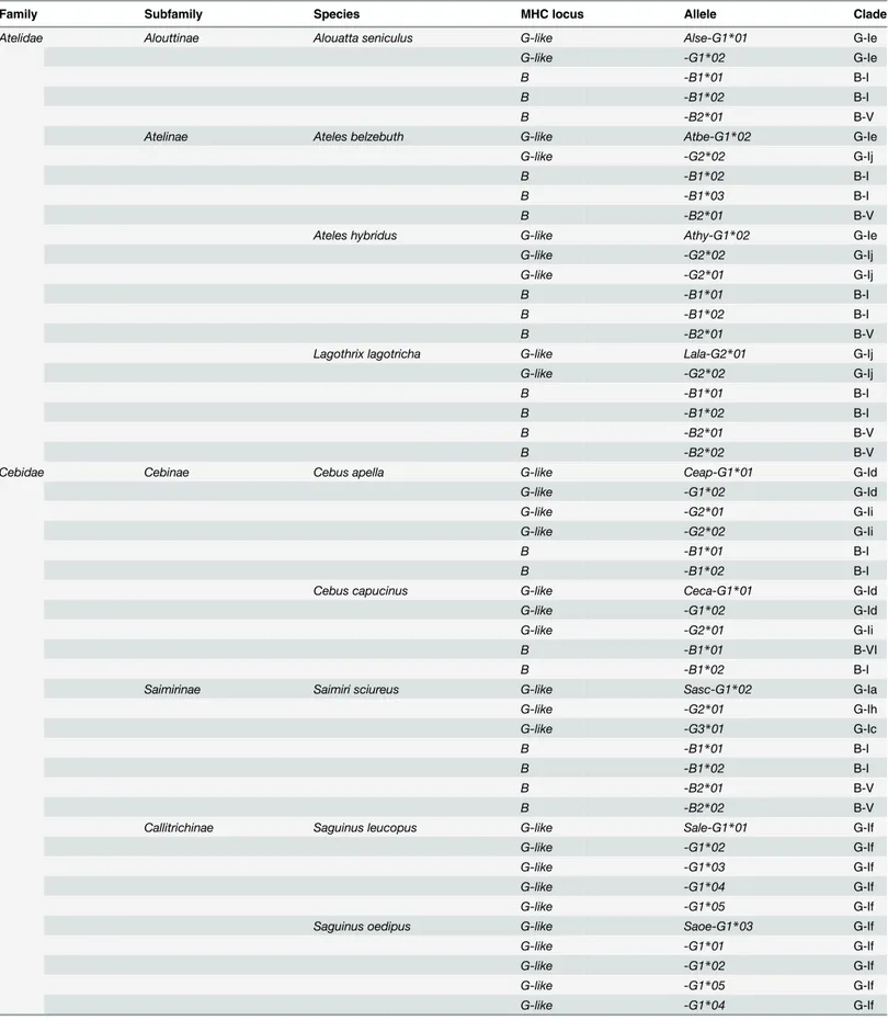
Table 1. MHC-G-likeand–BcDNAs identified in representative species of the Platyrrhini familiesAtelidaeandCebidae.
Family Subfamily Species MHC locus Allele Clade
Atelidae Alouttinae Alouatta seniculus G-like Alse-G1*01 G-Ie
G-like -G1*02 G-Ie
B -B1*01 B-I
B -B1*02 B-I
B -B2*01 B-V
Atelinae Ateles belzebuth G-like Atbe-G1*02 G-Ie
G-like -G2*02 G-Ij
B -B1*02 B-I
B -B1*03 B-I
B -B2*01 B-V
Ateles hybridus G-like Athy-G1*02 G-Ie
G-like -G2*02 G-Ij
G-like -G2*01 G-Ij
B -B1*01 B-I
B -B1*02 B-I
B -B2*01 B-V
Lagothrix lagotricha G-like Lala-G2*01 G-Ij
G-like -G2*02 G-Ij
B -B1*01 B-I
B -B1*02 B-I
B -B2*01 B-V
B -B2*02 B-V
Cebidae Cebinae Cebus apella G-like Ceap-G1*01 G-Id
G-like -G1*02 G-Id
G-like -G2*01 G-Ii
G-like -G2*02 G-Ii
B -B1*01 B-I
B -B1*02 B-I
Cebus capucinus G-like Ceca-G1*01 G-Id
G-like -G1*02 G-Id
G-like -G2*01 G-Ii
B -B1*01 B-VI
B -B1*02 B-I
Saimirinae Saimiri sciureus G-like Sasc-G1*02 G-Ia
G-like -G2*01 G-Ih
G-like -G3*01 G-Ic
B -B1*01 B-I
B -B1*02 B-I
B -B2*01 B-V
B -B2*02 B-V
Callitrichinae Saguinus leucopus G-like Sale-G1*01 G-If
G-like -G1*02 G-If
G-like -G1*03 G-If
G-like -G1*04 G-If
G-like -G1*05 G-If
Saguinus oedipus G-like Saoe-G1*03 G-If
G-like -G1*01 G-If
G-like -G1*02 G-If
G-like -G1*05 G-If
G-like -G1*04 G-If
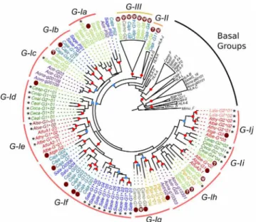
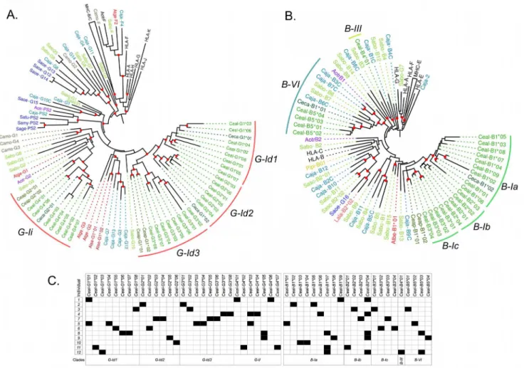
and—Ih,Callithrixsequences in clusters G-Ib and—If, andSaguinussequences in cluster If (Fig 3). Finally,Pitheciidae MHC-G-likesequences (Pithecia pithecia and C.moloch) grouped in cluster G-Ig and -Ih. Thus,MHC-G-likecDNAs had a general tendency to cluster by families or genera, indicating that this locus has diversified relatively recently after the divergence of such taxa.
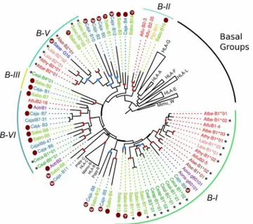
A gene tree of primateMHC-Bsequences showed that the Platyrrhini cDNAs clustered in five clades (I–VI) out the nine defined with the genomic sequences (Fig 4). These clades, with the exception of B-III that had a single cDNA, contained sequences from two different Platyr-rhini families, eitherAtelidaeandCebidaeorAtelidaeandPitheciidae. Cluster B-I contained 57% of the analyzed cDNAs (21 out of 37) and included sequences from the familiesAtelidae (generaAteles,Lagothrix,Alouatta) andCebidae(generaCebus,SaimiriandAotus). Clade B-II contained sequences from the familiesAtelidae(genusAteles) andPitheciidae(genusPithecia), clade B-III had a single cDNA from theCebidaefamily (Cebus), clade B-V included seven cDNAs from the familiesAtelidae(Ateles,Lagothrix,Alouatta) andCebidae(Saimiri), and clade B-VI had cDNA sequences from theAtelidae(Ateles) andCebidae(CallithrixandCebus) families (Fig 4). Hence, the clustering pattern ofMHC-BandMHC-G-likecDNAs differs in that the former shows five major clades that include sequences from different Platyrrhini fami-lies, whereas the latter displays only one major clade forming groups composed by cDNAs from a single family or even from a single genus. This indicates thatMHC-G-likeloci have diversified more recently thanMHC-Bloci, and that this rapid diversification has often coin-cided with the adaptive radiation of the Platyrrhini genera [13].
Fig 3. Bayesian phylogenetic tree based on exons 4–8 of PlatyrrhiniMHC-G-likesequences. Sequences marked with an asterisk (*) are cDNAs from this work, whereas those marked with a dark red dot are genomic sequences used to assign cDNAs to a given clade. Red dots on the branching points represent a support with a posterior probability>0.9, and light blue dots represent a support with a posterior probability
between 0.7 and 0.9. Lineages and clusters are indicated at the periphery of the tree. Caja,Callithrix jacchus; Sabo,Saimiri boliviensis; Sasc,Saimiri sciureus; Camo,Callicebus moloch; Pipi,Pithecia pithecia; Atge,
Ateles geoffroyi; Atfu,Ateles fusciceps; Athy,Ateles hybridus; Atbe,Ateles belzebuth; Alse,Alouatta seniculus; Lala,Lagothrix lagotricha; Saoe,Saguinus oedipus; Sage,Saguinus geoffroyi; Sala,Saguinus labiatus; Safu,Saguinus fuscicollis; Sale,Saguinus leucopus; Aotr,Aotus trivirgatus; Aona,Aotus nancymae; Aovo,Aotus vociferans; Ceap,Cebus apella; Ceal,Cebus albifrons; Ceca,Cebus capucinus. Accession numbers for published Platyrrhini MHC class I sequences used in the analysis are listed inS3 Table.
doi:10.1371/journal.pone.0131343.g003
Polymorphism of
MHC-G-like
and
MHC-B
loci in the capuchin monkey
Cebus albifrons
MHC class I polymorphism in the capuchin monkey (C.albifrons)was assessed by cloning and sequencing cDNAs from 12 unrelated individuals. The MHC class I cDNAs derived from PCR amplifications using generic primers designed to amplify bothMHC-G-likeand-Bloci. The resulting sequences were 1,041 bp long, included exon 1 to 8, and had no alterations that might prevent their proper translation. In total, 29 uniqueMHC-G-likecDNAs were identified in this population, with one to five sequences per individual, and a mean sequence distance of d= 9 ± 0.6% (GenBank accession numbers KM219732—KM219782). Twenty-six out of the 29 sequences were not shared between individuals whereas the other three cDNAs where shared by two individuals each (Fig 5). A phylogenetic tree ofC.albifrons MHC-G-likecDNAs showed that they grouped in the two clusters exclusive for the genus Cebus, G-Id and G-Ii, having a mean inter-cluster distance ofd= 11.5 ± 1% (Fig 5A). The G-Id clade was constituted by three groups withC.albifronssequences, G-Id1, G-Id2, and G-Id3, with an average within-group distance of 8.1 ± 0.6%, 5.9 ± 0.6%, and 5.8 ± 0.5%, respectively. Only one group was distin-guished in clade G-Ii, having an average within-group distance ofd= 5.8 ± 0.4% (Fig 5A). These four sequence groups might represent the transcribedMHC-G-likeloci in this popula-tion, which is supported not only by the clustering pattern in the phylogenetic tree, but also by the fact that there is no individual with more than two sequences in each group (Fig 5C). How-ever, no individual had representative sequences in all four groups/loci, suggesting that there exist haplotypic variability in gene number among the individuals of this population. Fig 4. Bayesian phylogenetic tree based on exons 4–8 of PlatyrrhiniMHC-Bsequences.Sequences marked with an asterisk (*) are cDNAs from this work, whereas those marked with a dark red dot are genomic sequences used to assign cDNAs to a given lineage. Red dots on the branching points represent a support with a posterior probability>0.9, and light blue dots represent a support with a posterior probability between 0.7 and 0.9. Lineages and clusters are indicated at the periphery of the tree. Caja,Callithrix jacchus; Sabo,
Saimiri boliviensis; Sasc,Saimiri sciureus; Pipi,Pithecia pithecia; Athy,Ateles hybridus; Atbe,Ateles belzebuth; Atfu,Ateles fusciceps; Alse,Alouatta seniculus; Lala,Lagothrix lagotricha; Ceap,Cebus apella; Ceal,Cebus albifrons; Ceca,Cebus capucinus; Aotr,Aotus trivirgatus; Aona,Aotus nancymae; Aovo,Aotus vociferans; Saoe,Saguinus oedipus. Accession numbers for published Platyrrhini MHC class I sequences used in the analysis are listed inS3 Table.
Twenty-two uniqueMHC-BcDNAs were identified in theC.albifronspopulation with two to four sequences per individual and with an average sequence distance ofd= 10.7 ± 0.7%. The majority of sequences (19) were unique of a single individual, whereas three cDNAs were pres-ent in two, three, and five individuals, respectively (Fig 5C).C.albifronscDNAs clustered in three of the clades identified in the phylogenetic analysis of genomic sequences, B-I, B-III, and B-VI, having a mean distance between clades B-I and B-III ofd= 17.0 ± 1.4%, between clades B-I and B-VI ofd= 18.8 ± 1.4%, and between clades B-III and B-VI ofd= 20.3 ± 1.7. Clade B-I contained 17 of the 22 cDNAs (77.3%) having a mean sequence distance ofd= 5.8 ± 0.5%. Sequences from clade B-I were in turn organized into three groups, B-Ia, B-Ib, and B-Ic (Fig 5B), having an average within-group distance of 3.7 ± 0.3%, 5.1 ± 0.5%, and 4.8 ± 0.5%, respec-tively. Clade B-III contained a single cDNA (Ceal-B401) and clade B-VI contained four cDNAs clustered together and having an average distance of 4.1 ± 0.5%. As discussed for MHC-G-likesequences, it is possible that the five groups segregating in the phylogenetic tree represent different loci, as there are no individuals with more than two cDNAs per group. Also, there are no individuals withMHC-BcDNAs in each of the five groups, suggesting that there are different haplotypes in the population varying in gene content.
Fig 5. Diversification ofMHC-G-likeandMHC-Bloci in a population of 12 unrelated white-fronted capuchin monkeys (Cebus albifrons).Bayesian phylogenetic trees ofMHC-G-like(A) andMHC-B(B) cDNAs based on exons 4–8, showing at the periphery of the tree the lineages and clusters as defined
in the text. Red dots on branching points indicate a support with a posterior probability>0.7. C, Assignment of putative alleles to each individual of the population, where black squares indicate presence and white squares indicate absence.
doi:10.1371/journal.pone.0131343.g005
Natural Selection has promoted the diversification of MHC-G-like and -B
loci in
C.
albifrons
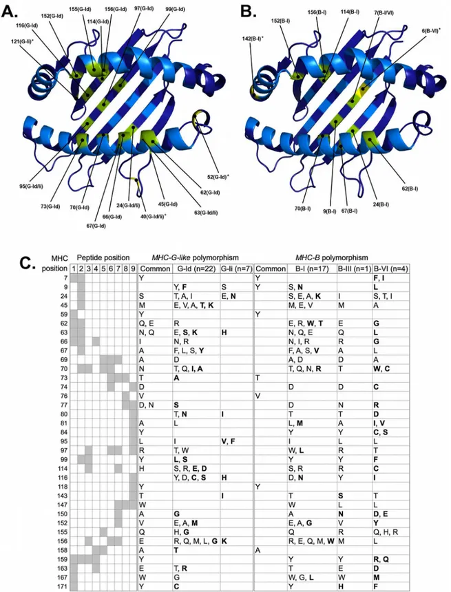
Natural selection analysis ofMHC-G-likecDNAs fromC.albifronsresulted in the identifica-tion of 19 positively selected posiidentifica-tions from the peptide binding region (PBR), 16 of which have been predicted to interact with the peptide within 4.0 Å [35] (Fig 6). Three of the posi-tively selected positions contacting the peptide were identified in both G-Id and G-Ii lineages (positions 24, 63, and 95), whereas 13 of such positions (45, 62, 66, 67, 70, 73, 97, 99, 114, 116, 152, 155, and 156) were unique of the G-Id lineage but none for of the G-Ii lineage (Fig 6A). MHC-G-likecDNAs encoded two or more alternative amino acids in 27 of the 35 peptide-con-tacting positions, although the majority of variable positions occurred in the G-Id lineage (26) in comparison to the G-Ii lineage (7) (Fig 6C). In turn, MHC-B predicted proteins had 11 posi-tively selected residues in the PBR and nine of them contacting the peptide within 4.0 Å (Fig 6B). One of the latter positions (position 7) was shared by lineages B-I and B-VI, eight posi-tions (9, 24, 62, 67, 70, 114, 152, and 156) were detected only in lineage B-I, and none was unique for the B-VI lineage. Furthermore, seven positively selected positions contacting the peptide (24, 62, 67, 70, 114, 152, 156) were shared between MHC-G-like and MHC-B predicted proteins (Fig 6A and 6B).MHC-B cDNAs encoded 30 variable peptide-contacting positions, 16 from lineage B-I and nine from lineage B-VI (Fig 6C). These MHC-B variable residues tended to be unique of a given clade, in contrast to MHC-G-like predicted proteins where clades shared most of the amino acids in variable positions. Indeed, in 24 out of the 27 MHC-G-like variable peptide-contacting positions, clades G-Id and G-Ii had at least one amino acid in common, whereas in only three of the 30 MHC-B variable positions the three clades had at least one amino acid in common (Fig 6C). However, the number of variable posi-tions sharing amino acids was different between clades, as there were 17 posiposi-tions with shared amino acids between clades B-I and B-III, six between B-I and B-VI, and six between B-III and B-VI. This suggests that the degree overlapping between peptides presented by MHC-G-like is greater than that of MHC-B molecules, and that the overlapping of peptides presented by pro-teins of the B-I and B-III clades is greater than that of propro-teins from clade B-VII with any of the other two MHC-B clades. The most variable peptide-contacting position for both MHC-G-like and—B was position 156, located at the PBR alpha-2 helix and contacting peptide posi-tions 3, 6, and 7, with seven different amino acids for each locus class (Fig 6C). Yet, most other variable positions (e.g., positions 24, 45, 63, 81, 116) were predicted to interact with the peptide anchor positions 2 and 9, suggesting that both MHC-G-like and—B can present a wide spec-trum of processed peptides (Fig 6C).
Fig 6. Natural selection has promoted the diversification ofC.albifronsMHC class I loci at PBR positions predicted to interact with the processed peptide.PBR residues evolving under positive selection in Platyrrhini MHC-G-like (A) and MHC-B (B) lineages are indicated on the structural model with their corresponding positions. In parenthesis next to the position number is indicated the particular lineage where positive selection was detected. Residues colored in green are positively selected positions predicted to contact the peptide, whereas residues colored in yellow and marked with an asterisk are positively selected positions that do not contact the peptide. C, variability of MHC-G-like and MHC-B positions that interact with the processed peptide within
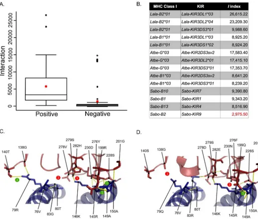
In order to identify potential MHC:KIR interacting pairs in Platyrrhini, an interaction index I = kh / ln dGwas proposed. This index takes into account the average energy of an H-bond (k), the number of H-bonds in the pair (h), and the desolvation energy (dG). The index was first applied to 116 human, chimpanzee and Rhesus monkey MHC:KIR pairs that had been experimentally evaluated. The meanI-value for interacting pairs was 5,250 (n = 45) and that for non-interacting pairs was 1,376 (n = 71) (Fig 7A). Some positive interactions had lowI -val-ues and some negative interactions had highI-values (i.e., false positives and false negatives), representing 15% of the 116 interactions. Then, MHC:KIR pairs were evaluated for Platyrrhini species with available sequence information for both molecules [36], revealing some potential interacting pairs, including cases of an MHC class I molecule interacting with more than one KIR (Fig 7B). Structural modeling of MHC:KIR pairs with a high and a lowIindex (Lala-B201:Lala-KIR3DL103 and Sabo-B2:Sabo-KIR9), revealed a much more stable interaction in theL.lagotrichapair having aI= 26,615 (Fig 7C) than in itsS.boliviensiscounterpart with a I= 2,975 (Fig 7D). This indicates that the interaction index proposed here has the power to predict MHC:KIR interactions based on protein sequences.
Discussion
For many years, the study of the MHC class I system in Platyrrhini species focused mostly on characterizing the diversity ofMHC-G-likeloci. Although few studies indicated the presence of MHC-Bloci in Platyrrhini, the impressive complexity of the region in this taxon was only real-ized until the sequencing of the MHC class I beta block in the common marmoset (C.jacchus) [16]. Moreover, unexpected complexity was also found in the common marmoset MHC class I alpha block, with more than a dozenMHC-G-likeand fiveMHC-Fgenes and pseudogenes [15]. This great expansion of theMHC-G-likeandMHC-Bloci is here reported for another Cebidae species, the Bolivian squirrel monkey (S.boliviensis), which contained at least 10 MHC-G-likesequences (five putative genes and five pseudogenes) and 15MHC-Bsequences (13 putative genes and two pseudogenes). This high number of MHC class I sequences identi-fied in the squirrel monkey’s draft genome is unlikely to be an assembly artifact. Assembly of high depth Illumina-based sequenced genomes typically results in underrepresentation of recent, rapidly evolving, lineage-specific genomic regions, due to the collapse of segmental duplications and recent repeat regions [37]. Therefore, assembly artifacts would involve miss-ing segmental duplications instead of increasmiss-ing the number of duplicates. The expansion of the MHC class I subgenomic regions, however, does not appear to be a common trait to all Pla-tyrrhini species. Indeed, theMHC-G-likeregion of the spider monkey (A.geoffroyi), which was completely covered by two BAC clones, contained only threeMHC-G-likeand twoMHC-F genes. Differential expansion of the MHC class I genes between related species has been previ-ously observed. Humans and apes contain singleHLA-Aand–Bgenes whereas Rhesus mon-keys contain multiple copies of bothMHC-AandMHC-Bloci [9]. Similarly, in Galliform birds, the gene number and organization of MHC class I genes varies between closely related species [38]. Thus, the MHC class I alpha and beta blocks in Platyrrhini have evolved under a birth-and-death model where some taxa have undergone a rapid process of gene or regional duplications, whereas others have maintained a low copy number either as an ancestral 4.0Å. In the left-hand panel, the rows give the MHC numbering positions, the columns give the peptide positions, and the intersections between them (in gray
background) mark the predicted interactions between MHC positions and each of the 9 residues of the peptide, according to [35]. The center and right-hand panels show the different amino acids at each position of MHC-G-like and MHC-B predicted proteins, respectively. Residues shared by lineages and those unique for a lineage within either MHC-G-like or—B are indicated separately. Residues in bold type are unique for a lineage and are not shared between
MHC-G-like and MHC-B loci.
condition or as a consequence of a taxon-specific gene deletions. The process of regional expansion evidenced inCebidaespecies appears to be more accelerated in the alpha block than in the beta block as can be inferred by the limited number of orthologousMHC-G-likegenes in comparison with the more frequent instances of orthology betweenMHC-Bloci. The high rate of diversification of the MHC class I alpha and beta blocks contrasts with the highly conserved kappa block containing theMHC-Elocus, which has remained virtually unchanged since the divergence of Catarrhini and Platyrrhini (S1 Fig).
In most of the species analyzed in this work there was evidence for various transcriptionally activeMHC-G-likeand–Bloci, having a number of unique cDNAs per individual that raged from two to five for both classes of loci. In the twoCallitrichinaespecies analyzed (S.oedipus andS.leucopus), however, noMHC-BcDNAs were found, although fiveMHC-G-likecDNAs were identified in each species. A similar case has been reported in the common marmoset, also a Callitrichinae, that, despite having severalMHC-Bgenes, no evidence forMHC-B tran-scripts was found [17]. Yet, with more sequencing effortMHC-Bhas been detected in this spe-cies [16]. Hence, it is possible that theMHC-Bloci inCallitrichinaespecies have limited Fig 7. Interaction (I) index proposed to predict stable interaction between MHC class I and KIR molecules, based on the number of hydrogen bonds and the desolvation energy.A, Box plot ofIindex values from experimentally validated human, chimpanzee and Rhesus monkey MHC:KIR pairs with functional interactions (positive, n = 45) and lack of interaction (negative, n = 71). The red star indicates the averageIindex value for positive and negative interactions. B,Iindex values derived from models of Platyrrhini MHC:KIR interacting pairs. Structural model of interacting pairs Lala-B2*01:Lala-KIR3DL1*03 withI= 26,615 (C) and Sabo-B2:Sabo-KIR9I= 2,975 (D). Number of hydrogen bond gained (numbers in green circles) and lost (numbers in red circles) with respect to the human HLA-B*5701:KIR3DL1 (PDB: 3VH8) interaction are indicated along with the MHC class I and KIR residues critical for the interaction [29]. Accession numbers for the Platyrrhini KIR sequences used in this analysis are in theS3 Table.
doi:10.1371/journal.pone.0131343.g007
expression levels in peripheral blood cells and/or their expression is not universal but restricted to selected tissues.
The number of MHC class I cDNAs per species identified in this work may appear low in light of the large number of genes exhibited by some Platyrrhini species. AlthoughS.boliviensis andA.geoffroyiwere not sampled, their sister species (S.sciureusandA.belzebuth/A. hybri-dus) showed two or threeMHC-G-likecDNAs and three or fourMHC-BcDNAs. Assuming that the complexity of the MHC class I region ofS.boliviensisandA.geoffroyiis similar to that of their sister species, it can be hypothesized that the number of MHC class I loci with high transcriptional activity in Platyrrhini species is small and relatively fixed (two to four), regard-less of the MHC class I genomic complexity of the species. This pattern has been observed in the highly expanded MHC class I region of the Rhesus monkey, where there are fewMHC-A and–Bgenes with high expression levels with the remaining genes displaying low expression levels [39]. However, it is also possible that the generic primers used to amplify cDNAs were not efficient enough to amplify the entire set ofMHC-G-likeand–Bsequences from the sam-pled species. Solving this issue would require access to genomic sequences to design locus-spe-cific primers and a quantitative methodology to evaluate differential expression of genes.
The analysis of MHC class I diversity in 12 unrelatedC.albifronsindividuals showed a max-imum fiveMHC-G-likeand fourMHC-BcDNAs per individual. The vast majority of cDNAs were unique for a given individual, although there were sequences shared by up to five individ-uals, indicating that most MHC class I loci are highly polymorphic. In gene trees, these cDNAs clustered into fourMHC-G-likeand fiveMHC-Bgroups, likely corresponding to the highly transcribed loci. Not all individuals had representative cDNAs in each group, suggesting that there is haplotypic diversity in both families MHC class I genes. Haplotypic diversity of MHC class I genes has been described in several species, including the Rhesus monkey [39], cattle [40], seals [41], and the zebra fish [42]. The variability in the MHC class I region in Platyrrhini has likely been promoted by natural selection for ligand binding. InC.albifrons, positions of the PBR predicted to interact with the processed peptide and the Ig domains of KIR molecules were generally highly variable and with signatures of positive selection within the major sequence clades. This indicates that most of theC.albifrons MHC-G-likeand–Bloci act as clas-sical loci presenting peptides to TcRs and modulating the activity of NK cells.
The MHC class I system in Platyrrhini has been traditionally seen as a product of a relatively rapid diversification of an ancestral locus related toMHC-GorMHC-A, which occurred after the divergence of Platyrrhini and Catarrhini. The genomic analysis of the common marmoset MHC class I region [15] and the present work unveiled an unsuspected complexity of this gene family in Platyrrhini characterized by a differential expansion and contraction ofMHC-G-like andMHC-Bgenes between species. This process, that appears to be more rapid in the MHC-G-likeregion than in theMHC-Bregion, generates loci with different transcriptional activities, which might be correlated with different functional roles. Thus, the variability of the MHC class I system is generated at different levels, from genetic polymorphism to gene number vari-ation and differential expression of loci.
Supporting Information
S1 Fig. Draft genomic map of the MHC class I kappa block (MHC-E) ofSaimiri boliviensis compared to its syntenic region in humans and the common marmoset.
(TIF)
(MHC-B/C) ofSaimiri boliviensis. (DOCX)
S2 Table. Variability of MHC-G-like and—B positions predicted to interact with KIR domains D1 and D2 inC.albifrons.
(DOCX)
S3 Table. Additional Platyrrhini MHC class I sequences used for phylogenetics analyses. (DOCX)
Acknowledgments
We thank Dr. Claudia Brieva and the staff from URRAS—Universidad Nacional de Colombia for providing blood samples. We thank Michael Graham, Julie Karl, and David O'Connor for their insightful comments on this work.
Author Contributions
Conceived and designed the experiments: JSL LFC. Performed the experiments: JSL. Analyzed the data: JSL LFC. Contributed reagents/materials/analysis tools: JSL LFC. Wrote the paper: JSL LFC.
References
1. Parham P, Moffett A. Variable NK cell receptors and their MHC class I ligands in immunity, reproduction and human evolution. Nat Rev Immunol. 2013; 13(2):133–44. Epub 2013/01/22. nri3370 [pii] doi:10. 1038/nri3370PMID:23334245.
2. Nikolich-Zugich J, Fremont DH, Miley MJ, Messaoudi I. The role of mhc polymorphism in anti-microbial resistance. Microbes Infect. 2004; 6(5):501–12. Epub 2004/04/28. doi:10.1016/j.micinf.2004.01.006
S1286457904000553 [pii]. PMID:15109966.
3. Horton R, Wilming L, Rand V, Lovering RC, Bruford EA, Khodiyar VK, et al. Gene map of the extended human MHC. Nat Rev Genet. 2004; 5(12):889–99. Epub 2004/12/02. nrg1489 [pii] doi:10.1038/ nrg1489PMID:15573121.
4. Le Bouteiller P. HLA class I chromosomal region, genes, and products: facts and questions. Crit Rev Immunol. 1994; 14(2):89–129. Epub 1994/01/01. PMID:7702749.
5. Gaudieri S, Kulski JK, Dawkins RL, Gojobori T. Different evolutionary histories in two subgenomic regions of the major histocompatibility complex. Genome Res. 1999; 9(6):541–9. Epub 1999/07/13.
PMID:10400921.
6. Fukami-Kobayashi K, Shiina T, Anzai T, Sano K, Yamazaki M, Inoko H, et al. Genomic evolution of MHC class I region in primates. Proc Natl Acad Sci U S A. 2005; 102(26):9230–4. Epub 2005/06/22.
0500770102 [pii] doi:10.1073/pnas.0500770102PMID:15967992; PubMed Central PMCID: PMC1153716.
7. Adams EJ, Parham P. Species-specific evolution of MHC class I genes in the higher primates. Immunol Rev. 2001; 183:41–64. Epub 2002/01/10. 1830104 [pii]. PMID:11782246.
8. Abi-Rached L, Kuhl H, Roos C, ten Hallers B, Zhu B, Carbone L, et al. A small, variable, and irregular killer cell Ig-like receptor locus accompanies the absence of MHC-C and MHC-G in gibbons. J Immunol. 2010; 184(3):1379–91. Epub 2009/12/23. jimmunol.0903016 [pii] doi:10.4049/jimmunol.0903016
PMID:20026738.
9. Daza-Vamenta R, Glusman G, Rowen L, Guthrie B, Geraghty DE. Genetic divergence of the rhesus macaque major histocompatibility complex. Genome Res. 2004; 14(8):1501–15. Epub 2004/08/04.:
doi:10.1101/gr.2134504 14/8/1501[pii]. PMID:15289473; PubMed Central PMCID: PMC509259. 10. Schrago CG. On the time scale of New World primate diversification. Am J Phys Anthropol. 2007; 132
(3):344–54. Epub 2006/11/30.: doi:10.1002/ajpa.20459PMID:17133436.
11. Perelman P, Johnson WE, Roos C, Seuanez HN, Horvath JE, Moreira MA, et al. A molecular phylogeny of living primates. PLoS Genet. 2011; 7(3):e1001342. Epub 2011/03/26.: doi:10.1371/journal.pgen. 1001342PMID:21436896; PubMed Central PMCID: PMC3060065.
12. Watkins DI, Chen ZW, Hughes AL, Evans MG, Tedder TF, Letvin NL. Evolution of MHC class I genes of a New World primate from ancestral homologues of human non-classical genes. Nature. 1990; 346:60–3. PMID:2114550
13. Cadavid LF, Shufflebotham C, Ruiz FJ, Yeager M, Hughes AL, Watkins DI. Evolutionary instability of the major histocompatibility complex class I loci in New World primates. Proc Natl Acad Sci U S A. 1997; 94(26):14536–41. Epub 1998/02/07. PMID:9405648; PubMed Central PMCID: PMC25046.
14. Knapp LA, Cadavid LF, Watkins DI. The MHC-E locus is the most well conserved of all known primate class I histocompatibility genes. J Immunol. 1998; 160(1):189–96. Epub 1998/04/29. PMID:9551971.
15. Kono A, Brameier M, Roos C, Suzuki S, Shigenari A, Kametani Y, et al. Genomic sequence analysis of the MHC class I G/F segment in common marmoset (Callithrix jacchus). J Immunol. 2014; 192(7):3239–
46. Epub 2014/03/07. jimmunol.1302745 [pii] doi:10.4049/jimmunol.1302745PMID:24600031. 16. Shiina T, Kono A, Westphal N, Suzuki S, Hosomichi K, Kita YF, et al. Comparative genome analysis of
the major histocompatibility complex (MHC) class I B/C segments in primates elucidated by genomic sequencing in common marmoset (Callithrix jacchus). Immunogenetics. 2011; 63(8):485–99. Epub
2011/04/21. doi:10.1007/s00251-011-0526-8PMID:21505866.
17. van der Wiel MK, Otting N, de Groot NG, Doxiadis GG, Bontrop RE. The repertoire of MHC class I genes in the common marmoset: evidence for functional plasticity. Immunogenetics. 2013; 65 (12):841–9. Epub 2013/09/11. doi:10.1007/s00251-013-0732-7PMID:24018468.
18. Sawai H, Kawamoto Y, Takahata N, Satta Y. Evolutionary relationships of major histocompatibility com-plex class I genes in simian primates. Genetics. 2004; 166(4):1897–907. Epub 2004/05/06. 166/4/1897
[pii]. PMID:15126407; PubMed Central PMCID: PMC1470823.
19. Darling AE, Mau B, Perna NT. progressiveMauve: multiple genome alignment with gene gain, loss and rearrangement. PLoS One. 2010; 5(6):e11147. Epub 2010/07/02. doi:10.1371/journal.pone.0011147
PMID:20593022; PubMed Central PMCID: PMC2892488.
20. Edgar RC. MUSCLE: a multiple sequence alignment method with reduced time and space complexity. BMC Bioinformatics. 2004; 5:113. Epub 2004/08/21. doi:10.1186/1471-2105-5-113 1471-2105-5-113
[pii]. PMID:15318951; PubMed Central PMCID: PMC517706.
21. Tamura K, Peterson D, Peterson N, Stecher G, Nei M, Kumar S. MEGA5: molecular evolutionary genet-ics analysis using maximum likelihood, evolutionary distance, and maximum parsimony methods. Mol Biol Evol. 2011; 28(10):2731–9. Epub 2011/05/07. msr121 [pii] doi:10.1093/molbev/msr121PMID: 21546353; PubMed Central PMCID: PMC3203626.
22. Tamura K, Nei M, Kumar S. Prospects for inferring very large phylogenies by using the neighbor-joining method. Proc Natl Acad Sci U S A. 2004; 101(30):11030–5. Epub 2004/07/20. doi:10.1073/pnas. 0404206101[pii]. PMID:15258291; PubMed Central PMCID: PMC491989.
23. Posada D, Crandall KA. MODELTEST: testing the model of DNA substitution. Bioinformatics. 1998; 14 (9):817–8. Epub 1999/01/27. btb117 [pii]. PMID:9918953.
24. Huelsenbeck JP, Ronquist F. MRBAYES: Bayesian inference of phylogenetic trees. Bioinformatics. 2001; 17(8):754–5. Epub 2001/08/29. PMID:11524383.
25. Ronquist F, Huelsenbeck JP. MrBayes 3: Bayesian phylogenetic inference under mixed models. Bioin-formatics. 2003; 19(12):1572–4. Epub 2003/08/13. PMID:12912839.
26. Yang Z. PAML 4: phylogenetic analysis by maximum likelihood. Mol Biol Evol. 2007; 24(8):1586–91.
Epub 2007/05/08. msm088 [pii] doi:10.1093/molbev/msm088PMID:17483113.
27. Yang Z, Wong WS, Nielsen R. Bayes empirical bayes inference of amino acid sites under positive selection. Mol Biol Evol. 2005; 22(4):1107–18. doi:10.1093/molbev/msi097PMID:15689528.
28. Arnold K, Bordoli L, Kopp J, Schwede T. The SWISS-MODEL workspace: a web-based environment for protein structure homology modelling. Bioinformatics. 2006; 22(2):195–201. Epub 2005/11/23.
bti770 [pii] doi:10.1093/bioinformatics/bti770PMID:16301204.
29. Vivian JP, Duncan RC, Berry R, O'Connor GM, Reid HH, Beddoe T, et al. Killer cell immunoglobulin-like receptor 3DL1-mediated recognition of human leukocyte antigen B. Nature. 2011; 479(7373):401–
5. Epub 2011/10/25. nature10517 [pii] doi:10.1038/nature10517PMID:22020283.
30. Champ PC, Camacho CJ. FastContact: a free energy scoring tool for protein-protein complex struc-tures. Nucleic Acids Res. 2007; 35(Web Server issue):W556–60. Epub 2007/06/01. gkm326 [pii] doi: 10.1093/nar/gkm326PMID:17537824; PubMed Central PMCID: PMC1933237.
31. Moesta AK, Abi-Rached L, Norman PJ, Parham P. Chimpanzees use more varied receptors and ligands than humans for inhibitory killer cell Ig-like receptor recognition of the MHC-C1 and MHC-C2 epitopes. J Immunol. 2009; 182(6):3628–37. Epub 2009/03/07. 182/6/3628 [pii] doi:10.4049/jimmunol. 0803401PMID:19265141; PubMed Central PMCID: PMC2854666.
specificity for MHC class I. Immunogenetics. 2011; 63(8):543–7. Epub 2011/05/05. doi:10.1007/ s00251-011-0527-7PMID:21541786; PubMed Central PMCID: PMC3160831.
33. Older Aguilar AM, Guethlein LA, Adams EJ, Abi-Rached L, Moesta AK, Parham P. Coevolution of killer cell Ig-like receptors with HLA-C to become the major variable regulators of human NK cells. J Immunol. 2010; 185(7):4238–51. Epub 2010/09/02. jimmunol.1001494 [pii] doi:10.4049/jimmunol.1001494
PMID:20805421; PubMed Central PMCID: PMC3124317.
34. Rosner C, Kruse PH, Hermes M, Otto N, Walter L. Rhesus macaque inhibitory and activating KIR3D interact with Mamu-A-encoded ligands. J Immunol. 2011; 186(4):2156–63. Epub 2011/01/25.
jimmu-nol.1002634 [pii] doi:10.4049/jimmunol.1002634PMID:21257962.
35. Nielsen M, Lundegaard C, Blicher T, Lamberth K, Harndahl M, Justesen S, et al. NetMHCpan, a method for quantitative predictions of peptide binding to any HLA-A and -B locus protein of known sequence. PLoS One. 2007; 2(8):e796. Epub 2007/08/30. doi:10.1371/journal.pone.0000796PMID:
17726526; PubMed Central PMCID: PMC1949492.
36. Cadavid LF, Palacios C, Lugo JS. Bimodal evolution of the killer cell Ig-like receptor (KIR) family in New World primates. Immunogenetics. 2013; 65(10):725–36. doi:10.1007/s00251-013-0719-4PMID: 23846852.
37. Baker M. De novo genome assembly: what every biologist should know. Nat Meth. 2012; 9(4):333–7.
doi:10.1038/nmeth.1935
38. Eimes JA, Reed KM, Mendoza KM, Bollmer JL, Whittingham LA, Bateson ZW, et al. Greater prairie chickens have a compact MHC-B with a single class IA locus. Immunogenetics. 2012; 65(2):133–44.
Epub 2012/11/28. doi:10.1007/s00251-012-0664-7PMID:23179555.
39. Otting N, Heijmans CM, Noort RC, de Groot NG, Doxiadis GG, van Rood JJ, et al. Unparalleled com-plexity of the MHC class I region in rhesus macaques. Proc Natl Acad Sci U S A. 2005; 102(5):1626–
31. Epub 2005/01/25. 0409084102 [pii] doi:10.1073/pnas.0409084102PMID:15665097; PubMed Central PMCID: PMC545086.
40. Ellis S. The cattle major histocompatibility complex: is it unique? Vet Immunol Immunopathol. 2004; 102(1–2):1–8. Epub 2004/09/29. doi:10.1016/j.vetimm.2004.06.007S0165-2427(04)00181-3 [pii].
PMID:15451610.
41. Hammond JA, Guethlein LA, Norman PJ, Parham P. Natural selection on marine carnivores elaborated a diverse family of classical MHC class I genes exhibiting haplotypic gene content variation and allelic polymorphism. Immunogenetics. 2012; 64(12):915–33. Epub 2012/09/25. doi: 10.1007/s00251-012-0651-zPMID:23001684; PubMed Central PMCID: PMC3518486.
42. McConnell SC, Restaino AC, de Jong JL. Multiple divergent haplotypes express completely distinct sets of class I MHC genes in zebrafish. Immunogenetics. 2014; 66(3):199–213. Epub 2013/12/03. doi: 10.1007/s00251-013-0749-yPMID:24291825; PubMed Central PMCID: PMC3965299.
![Fig 1. Comparison of the MHC class I region of S. boliviensis, A. geoffroyi, and C. moloch using as reference that of humans [3] and C](https://thumb-eu.123doks.com/thumbv2/123dok_br/16307131.186560/6.918.64.840.122.704/comparison-class-region-boliviensis-geoffroyi-moloch-reference-humans.webp)