Universidade de Lisboa
Faculdade de Ciências
Departamento de Biologia Vegetal
Carbon monoxide and brain sequestration of
Plasmodium berghei
ANKA in experimental
cerebral malaria
Ana Catarina Dias Pena
MESTRADO EM BIOLOGIA CELULAR E
BIOTECNOLOGIA
Universidade de Lisboa
Faculdade de Ciências
Departamento de Biologia Vegetal
Carbon monoxide and brain sequestration of
Plasmodium berghei
ANKA in experimental
cerebral malaria
Ana Catarina Dias Pena
Dissertação de mestrado orientada por:
Doutora Ana Pamplona
Instituto de Medicina Molecular
Faculdade de Medicina da Universidade de Lisboa
Professora Doutora Rita Zilhão
Faculdade de Ciências da Universidade de Lisboa
MESTRADO EM BIOLOGIA CELULAR E
BIOTECNOLOGIA
TABLE OF CONTENTS
ACKNOWLEDGEMENTS... I
ABBREVIATIONS ... II
1. RESUMO ... 1
2. ABSTRACT ... 6
3. INTRODUCTION... 8
3.1. Malaria: a worldwide burden ... 8
3.2. Plasmodium life cycle in the mammalian host ... 8
3.2.1. Liver stage - The exo-erythrocytic cycle
... 9
3.2.2. Blood stage - The intra-erythrocytic cycle
... 10
3.2.3. Sexual stage
... 11
3.3. Cerebral malaria (CM) ... 11
3.4. Experimental cerebral malaria (ECM): the benefit of rodent models .... 12
3.5. Pathogenesis of CM: sequestration, inflammation and hemostasis
dysfunction ... 13
3.5.1. Sequestration of iRBCs
... 13
3.5.2. Sequestration of iRBCs in the murine model
... 14
3.5.3. Inflammation
... 15
3.5.3.1. Pro-inflammatory cytokines: tumor necrosis factor-α (TNF-α),
lymphotoxin-α (LT-α) and interferon-γ (IFN-γ)
... 16
3.5.3.2. Leukocyte accumulation in the brain: the importance of CD8
+T cells
... 17
3.5.4. Hemostasis dysregulation
... 19
3.5.5. The integration of events: blood-brain barrier (BBB) damage
... 20
3.6. Heme-oxygenase-1 (HO-1) and carbon monoxide (CO) ... 21
3.6.1. Pivotal role of HO-1 and CO in the protection against ECM
... 22
3.7. CO-releasing molecules (CO-RMs) ... 25
3.7.1. CO-releasing molecule-2 (CORM-2): vasorelaxing,
anti-proliferative, anti-ischaemic, anti-oxidant and anti-inflammatory
properties
... 26
4. AIMS OF THE PROJECT ...
ERRO! MARCADOR NÃO DEFINIDO.
5. RESULTS ... 31
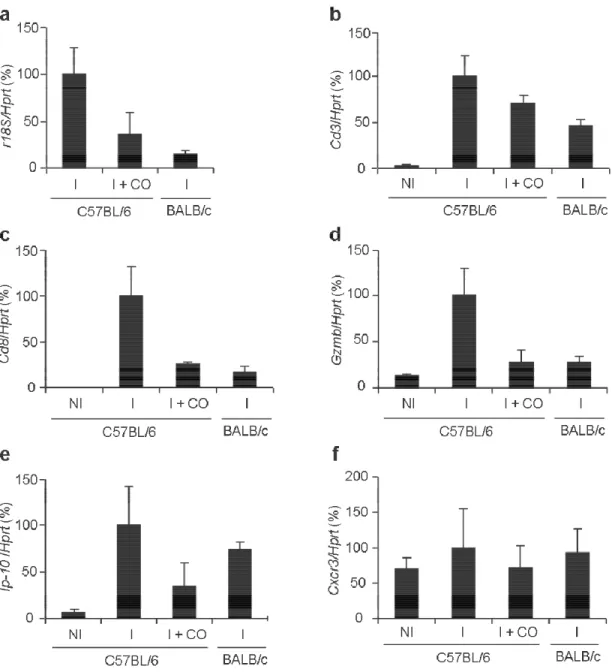
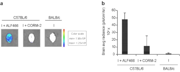
5.1. CO inhalation prevents parasite accumulation/sequestration in the
brain and neuroinflammation ... 31
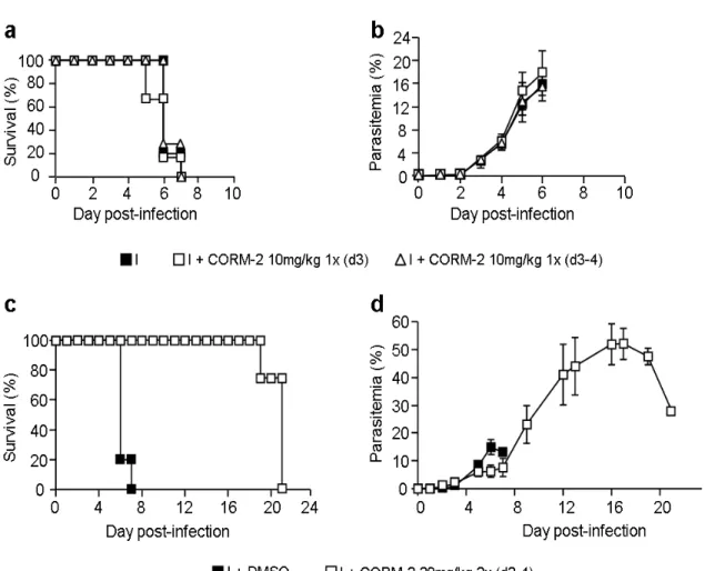
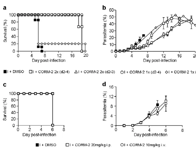
5.2. CORM-2 protects against ECM development... 36
5.3. CORM-2 protective effects is dependent on CO release but does not
involve COHb formation ... 39
5.4. CORM-2 prevents parasite accumulation/sequestration in the brain and
neuroinflammation - CORM-2 mimics CO inhalation ... 41
5.5. CORM-2 changes the profile of intra-erythrocytic stages of P.berghei
ANKA ... 44
5.6. CORM-2 does not alter reticulocytosis significantly... 45
5.7. CORM-2 treated mice show a reduced hematocrit and platelet volume
... 47
5.8. CORM-2 treatment increases the number of phagocytic cells in
circulation ... 49
5.9. Spleens from CORM-2 treated mice have increased weights ... 50
5.10. CORM-2 prevents loss of RBC deformability... 51
6. DISCUSSION... 53
7. CONCLUDING REMARKS AND FUTURE WORK ... 63
8. MATERIALS AND METHODS ... 65
8.1. Mice ... 65
8.2. Parasites, infection and disease assessment... 65
8.3. CO exposure ... 65
8.4. Visualization and quantification of bioluminescence in organs ex vivo
... 66
8.5. Quantitative Real-Time PCR ... 66
8.6. CO-releasing molecules administration ... 67
8.7. Quantification of O
2Hb, and MetHb and COHb ... 67
8.8. BBB permeability ... 68
8.9. Flow cytometry analysis ... 68
8.10. Hematocrit and Hemogram... 69
8.11. RBC deformability ... 70
8.12. Statistical analysis ... 70
A
cknowledgements
First of all, and even though this may seem odd, I would like to thank to myself.
I must be grateful for being able to overcome difficulties during the past two years, and most of all, for the willing to learn the so many things I learned and that would not be possible without this work.
Secondly, I want to express my gratitude to my supervisor, Ana Pamplona, for all the support, teaching and friendship. Above all, besides the scientific skills, with you I learned that “bosses” can be important friends! And what a good surprise that was! I must thank Maria Mota for receiving me in her lab, for all the support and knowledge and for being the enthusiastic scientist she is.
I would also like to thank to my supervisor, Professor Rita Zilhão, for the guidance and comprehension and the admirable ability to make her students believe in their capacities!
To Bruno Silva-Santos for welcoming me in his lab, and for bringing chocolates every time he travels.
To all the crew from UMA, UNIMOL, UNICEL, and the most recent groups at IMM, “UNIBESE” and “UNILAMPAS”, thank you a lot for the companionship, the precious advices, and the joy! How could I have done this thesis without your dear help and the smile you have put on my face?
I must also thank to Dr. Carlota Saldanha and Dr. Teresa Freitas from Microvascular Biology and Inflammation Unit at IMM, for contributing with their expertise to the present work.
I acknowledge Pedro Alves, because he also acknowledges me in his thesis! Obviously, for much more than that…for being a shoulder, for his pacience, tenderness and cheerfulness.
To nature, dance and art, particularly music, for nourish me in such a unique way.
To all my friends, you know who you are, and that strong embrace that only you can give me.
Abbreviations
BBB blood-brain barrier cAMP cyclic guanosine
monophosphate CCL5 CC ligand-5 CCR5 chemokine receptor-5 CM cerebral malaria COHb carboxyhemoglobin CXCR3 CXC chemokine receptor-3 DMSO dimethyl sulfoxide
EC endothelial cell ECM experimental cerebral
malaria
GFP green fluorescent protein
Hb hemoglobin
HO-1 heme oxygenase-1 i.p. intraperitoneally i.v. intravenously
ICAM-1 intercellular adhesion molecule-1
IFN-γ interferon-γ
iNOS inducible nitric oxide IP-10 IFN-γ-inducible protein-10 iRBC infected red blood cells LPS lypopolisaccharide
LT-α lymphotoxin-α
MAPK mitogen-activated protein kinase
MetHb methemoglobin MIG monokine induced by
IFN-γ
NADPH nicotinamide adenine dinucleotide phosphate NF-κb nuclear factor
κ-light-chain-enhancer in B cells NO nitric oxide
O2Hb oxyhemoglobin
p.i. post-infection PbA Plasmodium berghei
ANKA
PBS phosphate-buffered saline
RBC red blood cells
ROS reactive oxygen species sGC soluble guanylate cyclise TNF-α tumour necrosis factor-α VCAM-1 vascular adhesion
1. Resumo
A malária é uma das doenças infecciosas mais importantes em todo o mundo, afectando cerca de 200-300 milhões de pessoas todos os anos. A infecção por malária é causada por um protozoário do filo Apicomplexa do género Plasmodium. Estes parasitas possuem um ciclo de vida complexo que requer um mosquito Anopheles como vector e um hospedeiro vertebrado. Em mamíferos, o ciclo de vida do parasita inclui uma fase hepática, assintomática, seguida de uma fase eritrocítica, onde surgem os sintomas de malária e a patologia. Na fase eritrocítica, o parasita no estádio de merozoíto, oriundo da fase hepática, entra na circulação sanguínea e invade um glóbulo vermelho (GV) onde se replica assexuadamente. O desenvolvimento intra-eritrocítico do parasita é constituído por vários estádios que incluem um forma inicial em anel, seguida de uma fase de trofozoíto maduro que por sua vez se desenvolve em esquizonte. Durante a maturação do esquizonte ocorre um processo de reprodução assexuada designado esquizogenia através do qual são originados vários merozoítos, que levam à ruptura do glóbulo vermelho. Após a ruptura, os merozoítos libertados re-iniciam o ciclo intra-eritrocítico, invadindo novos glóbulos vermelhos. Este ciclo de ruptura e re-invasão é responsável pela hemólise e consequente anemia que se verifica durante uma infecção por malária.
Apesar da infecção por malária geralmente não originar um perfil clínico grave, alguns casos resultam em patologia severa, responsável pela morte de aproximadamente 1 milhão de pessoas anualmente. Em humanos, a malária severa ocorre sobretudo devido a infecções por Plasmodium falciparum. A malária cerebral (MC) é uma das formas mais letais de malária severa ainda sem tratamento e afectando sobretudo crianças com idades inferiores a 5 anos. A patogénese da MC é muito complexa, e os mecanismos que levam ao desenvolvimento da doença ainda são pouco conhecidos. No entanto, a patogénese da MC tem sido explicada por três processos principais: sequestração, inflamação e disfunção hemostática.
Uma melhor compreensão da patogénese da MC é fundamental para o desenvolvimento de novas estratégias terapêuticas contra este síndroma. O modelo de malária cerebral experimental (MCE) em murganhos C57BL/6 infectados com P.
modelo apresenta várias caraterísticas histopatológicas e imunológicas semelhantes à neuropatologia humana. No entanto, uma das principais diferenças é o tipo de células envolvido no processo de sequestração. Nos humanos, sabe-se que a sequestração de glóbulos vermelhos infectados (GVis) na microvasculatura do cérebro é um processo crucial para o desenvolvimento da patologia. Contudo, em murganhos, os leucócitos são as células que maioritariamente sequestram no cérebro. Esta observação tem lançado dúvidas sobre a importância da sequestração de GVis no cérebro no modelo de murganho, e contribuído para a controvérsia existente relativamente ao uso deste modelo como um modelo adequado ao estudo da MC. Desta forma, é importante compreender o papel da sequestração de glóbulos vermelhos infectados no cérebro e o seu papel no desenvolvimento da MCE em murganhos C57BL/6 infectados com PbA. Para além do processo de sequestração, também a inflamação e a disfunção do sistema hemostático têm sido envolvidos na patogénese da MC. Pensa-se que uma resposta imune exacerbada contra o parasita poderá desencadear eventos patogénicos que se sobrepõem à função protectora do sistema imunitário, entre os quais a neuroinflamação será um processo crítico. Neste contexto, várias citocinas pró-inflamatórias têm sido implicadas na MC, como o factor de necrose tumoral-α (TNF-α), o interferão-γ (IFN-γ) e a linfotoxina-α (LT-α). A expressão no endotélio vascular cerebral de moléculas de adesão celular, como a molécula de adesão intercelular-1 (ICAM-1), parece também ser um processo importante para a patologia, mediando a sequestração de leucócitos e GVis no cérebro. Para além disto, o recrutamento de linfócitos T, em particular de linfócitos T CD8+, para a microvasculatura cerebraldesempenha um papel
crucial na patogénese da MCE. Nesse sentido, a expressão no cérebro de quimiocinas, i.e. moléculas quimiotácticas que dirigem a migração de leucócitos, tais como a proteína induzida pelo IFN-γ-10 (IP-10), foi demonstrada contribuir para o estabelecimento da patologia em murganhos. Por outro lado, pensa-se que o desequilíbrio hemostático também desempenhe um papel importante na patogénese da MC. A existência de um estado pro-coagulante e o potencial papel das plaquetas na sequestração de GVis e na congestão vascular parecem ser intervenientes importantes neste processo.
A orquestração dos processos de sequestração, inflamação e desequilíbrio hemostático parecem culminar na lesão da barreira hemato-encefálica, um processo que parece decisivo na manifestação clínica da MC e cuja extensão poderá ditar sequelas neurológicas permanentes ou mesmo a morte.
O conhecimento mais aprofundado dos factores e mecanismos implicados na MC tem mostrado que várias moléculas do hospedeiro participam no desenvolvimento da doença, quer com uma acção patogénica, quer com uma função essencialmente protectora. Foi demonstrado recentemente que a expressão da heme oxigenase-1 (HO-1) do hospedeiro tem um papel preponderante em determinar a susceptibilidade à MC. De facto, foi provada uma importante acção protectora da enzima contra o desenvolvimento da MCE, que parece ser mediada sobretudo pela produção de monóxido de carbono (CO), um produto final da actividade da HO-1. Surpreendentemente, verificou-se que a administração de CO por inalação a murganhos C57BL/6 infectados com PbA, impedia o desenvolvimento de MCE em 100% dos casos. O efeito protector do CO manifestava-se na inibição da congestão microvascular cerebral, da neuroinflamação e do aumento da permeabilidade da barreira hemato-encefálica. A inibição da neuroinflamação reflectia-se na redução da expressão no cérebro de TNF-α, IFN-α, LT-α e ICAM-1 e inibição do recrutamento de células T CD8+ para o cérebro. O efeito terapêutico exercido pelo CO foi sugerido
depender da supressão da oxidação da hemoglobina (Hb) em circulação, através da ligação do CO à Hb, formando carboxihemoglobina. Isto impediria a conversão da Hb em metahemoglobina e a libertação de heme, uma molécula com efeitos inflamatórios e oxidantes nocivos.
Neste trabalho pretendeu-se aprofundar o mecanismo protector do CO contra a MCE, em particular no que respeita ao seu efeito na sequestração de glóbulos vermelhos infectados no cérebro. Utilizou-se um método desenvolvido recentemente que permite revelar em tempo real a presença do parasita, por detecção de bioluminescência. Os murganhos foram infectados com um parasita PbA transgénico que expressa luciferase, o que permitiu a sua detecção nos órgãos por emissão de bioluminescência. Verificou-se que apesar do cérebro Verificou-ser o órgão com menor acumulação de GVis em murganhos C57BL/6 infectados com PbA, a presença de parasita neste órgão é nitidamente detectável por este sistema. De especial importância é o facto de se verificar que a acção protectora do CO parece estar associada com uma diminuição evidente da sequestração de GVis no cérebro. Estes resultados, para além de indicarem que o mecanismo protector do CO inclui a redução da sequestração de parasita no cérebro, reforçam que o processo de sequestração cerebral de GVis é fundamental para o
desenvolvimento da MCE, reforçando a relevância do modelo de murganho C57BL/6 infectado com PbA como um modelo muito semelhante à patologia humana e, por isso, adequado ao seu estudo. Adicionalmente, confirmou-se que o CO tem uma actividade anti-inflamatória e uma acção anti-quimiotáctica, uma vez que inibe a expressão de IP-10 no cérebro. Isto parece contribuir para uma menor migração de células T CD8+ para
o cérebro, processo crucial para a patologia no murganho.
O segundo objectivo deste trabalho foi estudar a bio-actividade e o potencial terapêutico das moléculas libertadoras de CO (CO-RMs) no modelo experimental de malária cerebral. De facto, tem sido demonstrado que estas moléculas reproduzem os efeitos anti-oxidantes e anti-inflamatórios do CO em vários modelos de patologia, afigurando-se como potenciais novos fármacos. Uma das vantagens destas moléculas é a possibilidade de exercerem os efeitos benéficos do CO sem o perigo de toxicidade associado ao CO por inalação.
Surpreendentemente, observámos que a administração de CORM-2 com uma concentração de 20 mg/kg de peso corporal, por via intravenosa, duas vezes por dia, do dia 2 ao 3 após infecção, confere uma protecção de 100% contra o desenvolvimento de MCE. Adicionalmente, verificou-se que o CORM-2 não produz carboxihemoglobina, indicando que não induz a mesma toxicidade que o CO inalado, o que tem especial relevância para futuras aplicações terapêuticas. Tal como para o CO administrado por inalação, verificou-se uma diminuição nos níveis de metahemoglobina em circulação em murganhos tratados com CORM-2, reforçando o papel benéfico do CO no controlo do stress oxidativo causado pela infecção. Os resultados mostram também que o CORM-2 diminui a sequestração de Gvis no cérebro e a neuroinflamação em murganhos infectados com PbA, tal como anteriormente demonstrado para o tratamento com CO inalado. Adicionalmente, a administração de CORM-2 inibe a agregação plaquetária e a perda de deformabilidade eritrocitária durante a infecção. Estas observações são importantes porque durante a infecção o desequilíbrio do sistema de coagulação parece contribuir para a patogénese da MC e, por outro lado, a diminuição da deformabilidade eritrocitária representa um forte biomarcador de severidade em malária humana. O CORM-2 parece contribuir assim para o restabelecimento do equilíbrio hemostático no modelo C57BL/6-PbA de CM, e, deste modo, para o não desenvolvimento da MCE. Contrariamente ao que se observa com o CO inalado, o tratamento com CORM-2
mantém constante, durante os dias 5 e 8 após infecção, a percentagem de glóbulos vermelhos infectados. No entanto, a relevância desta observação para a protecção conferida pelo CORM-2 no desenvolvimento da MCE deverá ser melhor estudada em experiências futuras.
No seu conjunto, estes resultados indicam que as CO-RMs podem representar uma nova classe de compostos com potencial terapêutico na protecção do hospedeiro contra o desenvolvimento da malária cerebral.
Palavras-chave:
Malária Cerebral, CO, CO-RMs, Plasmodium berghei ANKA, Sequestração de glóbulos vermelhos infectados
2. Abstract
Malaria is a major infectious disease worldwide, causing ~1 million deaths each year due to severe complications, one of the most lethal being cerebral malaria (CM). Thereby, understanding CM pathogenesis is of vital importance for developing effective therapies against it.
The experimental cerebral malaria (ECM) model of C57BL/6 mice infected with P.
berghei ANKA (PbA) shares many similarities with human CM. However, whereas in
humans a critical event is sequestration of Plasmodium-infected red blood cells (iRBCs) in the brain microvasculature, in rodents is mostly leukocyte sequestration that occurs. Thereby, the pathologic significance of iRBCs brain sequestration during ECM is controversial and remains to be clarified.
Recently, it was shown that heme-oxygenase-1 (HO-1) plays a crucial role in protection against ECM, which appears to be mediated by carbon monoxide (CO) production, an end-product of its enzymatic activity. In fact, administration of CO by inhalation rescues all C57BL/6 PbA-infected mice from developing ECM.
The present study shows that CO protection comprises the reduction of iRBC brain sequestration in infected mice, supporting the importance of this process in ECM pathogenesis and the relevance of C57BL/5 PbA-infected mouse model to study CM. Moreover, our results indicate that CO has a therapeutic potential as a molecule with anti-inflammatory and anti-chemotaxis effects.
We also demonstrate that CORM-2, a CO-relasing molecule (CORM-2), mimics CO protection against ECM, suppressing neuroinflammation and parasite sequestration in the brain. Importantly, CORM-2 does not induce formation of carboxyhemoblogin, circumventing CO inhalation toxicity. Moreover, CORM-2 inhibits platelet aggregation and loss of RBC deformability, which likely contributes to prevent disease development. Additionally, CORM-2 also leads to an arrest in parasite load, which origin and relevance is not clear.
Altogether, these results indicate that CO-RMs seem to represent a novel class of drugs with therapeutic potential to protect the host from cerebral malaria.
Key-words:
Cerebral malaria, CO, CO-RMs, Plasmodium berghei ANKA, Sequestration of infected red blood cells
3. Introduction
3.1. Malaria: a worldwide burden
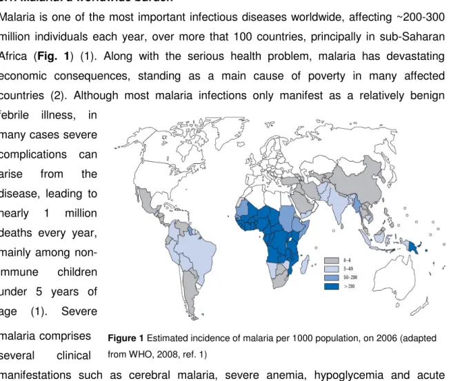
Malaria is one of the most important infectious diseases worldwide, affecting ~200-300 million individuals each year, over more that 100 countries, principally in sub-Saharan Africa (Fig. 1) (1). Along with the serious health problem, malaria has devastating economic consequences, standing as a main cause of poverty in many affected countries (2). Although most malaria infections only manifest as a relatively benign febrile illness, in
many cases severe complications can arise from the disease, leading to nearly 1 million deaths every year, mainly among non-immune children under 5 years of age (1). Severe malaria comprises several clinical
manifestations such as cerebral malaria, severe anemia, hypoglycemia and acute respiratory distress, often occurring in combination within the same patient (3).
3.2. Plasmodium life cycle in the mammalian host
Malaria infection is caused by apicomplexan protozoa of the genus Plasmodium. The parasites have a complex life cycle that requires two hosts: a mosquito vector and a vertebrate host, which can be a mammalian, a bird or a lizard, depending on the
Plasmodium species. There are five parasite species reported to infect humans, namely P. falciparum, P. vivax, P. malariae, P. ovale and, most recently, P. knowlesi (4). P. falciparum is by far the most widespread and dangerous of all parasites, standing as the major responsible for severe illness and mortality associated with malaria (1, 3). The entire life cycle as well as the morphology of the different developmental stages is
Figure 1 Estimated incidence of malaria per 1000 population, on 2006 (adapted from WHO, 2008, ref. 1)
conserved between mammalian parasites, including those infecting rodents, namely P.
berghei, P. chabaudi, P. yoelli and P. vinckei, which have been extensively used in experimental malaria models.
3.2.1. Liver stage - The exo-erythrocytic cycle
Malaria parasites are transmitted to a mammalian host trough the bite of a female
Anopheles spp. mosquito during a blood meal, inoculating the first infective form of the
parasite, the sporozoite, into the host dermis. Further on, sporozoites move from the skin and enter the blood circulation, travelling readily to the liver where they will initiate asexual replication. In the liver, the sporozoites transverse several hepatocytes before invading and developing in a final hepatocyte. Between 2 to 6 days later, according to the Plasmodium species (for human Plasmodium species from 6-16 days later), the infected hepatocytes rupture and thousands of merozoites per invading sporozoite are released into the bloodstream. This stage is the so-called liver stage of the parasite and is totally asymptomatic (Fig. 2) (reviewed in ref. 5).
Figure 2 Plasmodium life cycle in the human host (adapted from an image from Malaria Unit at Instituto de Medicina Molecular)
3.2.2. Blood stage - The intra-erythrocytic cycle
In contrast to the clinically silent liver stage, the blood stage of infection is when the malaria-associated pathology occurs. Each merozoite released from the liver invades a red blood cell (RBC) where asexually replicates during 24-72h, depending on the
Plasmodium species (human Plasmodium species have a replication cycle of 48-72h), to
produce merozoites. The replication cycle ends with the burst of infected RBCs (iRBCs) and the release of merozoites, which in turn will infect other RBCs and initiate a new cycle of asexual replication (Fig. 2) (reviewed in ref. 6). The typical waves of fever and chills observed during a malaria infection correspond to the cycles of erythrocyte rupture and re-infection by the parasites. Some Plasmodium species show a preference for invading reticulocytes, i.e. immature RBCs, whether others invade both mature RBCs and reticulocytes. In humans, P.
vivax and P. ovale predominantly infect reticulocytes, as well as P.
berghei and P. yoelli in rodents.
During the asexual intra-erythrocytic cycle, parasites mature through several developmental stages. Inside the RBC, the parasite first establishes as a ring-stage, surrounded by a vacuolar membrane
(parasitophorous vacuolar
membrane) created from the invagination of the RBC plasma membrane that occurs during the invasion step. The ring-stage is characterized by a large vacuole where hemoglobin starts to be digested to form hemozoin or the so-called “malarial pigment”, the characteristic brown pigment of Plasmodium. Parasites feed on host RBC hemoglobin to obtain free amino acids and iron. However, digestion generates a noxious by-product, the free heme. To overcome heme toxicity the parasite converts it into hemozoin, which is an inert crystal (7). The young ring parasite further grows into a trophozoite, which displays a different shape and a larger cell size, and where DNA replication begins. Next, the parasite enters the schizont stage where nuclear division occurs. During schizogony the parasite replicates its DNA and divides its nuclei several times, forming a syncytial cell. Finally, as the schizont matures, cytoplasm divides, the Figure 3 Asexual blood stages of Plasmodium falciparum
red blood cell rupture and 16 to 32 merozoites egress to invade fresh erythrocytes and perpetuate the cycle (Fig. 3) (reviewed in ref. 8, 9).
3.2.3. Sexual stage
The sexual life cycle of Plasmodium proceeds when some merozoites invade RBCs and undergo sexual differentiation (in response to stress or other stimuli) into gametocytes (Fig. 4), which can be taken up by mosquitoes during a blood meal. In the mosquito midgut, where fertilization occurs, the gametocytes fuse to produce a zygote denominated ookinete that develops into an oocyst in which thousands of sporozoites are formed. Finally, the sporozoites migrate into the mosquito’s salivary glands where they remain ready to be transmitted (Fig. 2) (reviewed in ref. 6).
3.3. Cerebral malaria (CM)
Cerebral malaria is the most severe neurological complication and one of the most life-threatening occurring from Plasmodium falciparum infection. CM affects mainly children and manifests clinically as motor and behaviour abnormalities, fever, consciousness impairment, convulsions and coma, ultimately leading to neurological sequelae or death (3). The usual treatment with anti-malaria drugs, like quinine and artemisin derivatives, is not sufficient to rescue CM patients from death or cognitive deficits. Along with anti-parasitic therapy, the patients of cerebral malaria must receive supportive treatment, such as exchange transfusion, ventilation support, treatment of renal failure, correction of acid-base imbalance and administration of anti-convulsants.These treatments are not always easily available in the sub-developed countries where malaria is endemic. Other adjunctive treatments have been suggested however none of them have shown proved benefits and some are reported as deleterious (reviewed in ref. 11, 12). Regardless of the efforts to improve CM treatment, there is still no successful therapy and the disease continues to exhibit high mortality and morbidity rates. The threat of this health problem is underlined by the observation that in endemic areas ~ 20% of children suffering from CM still succumb to the disease (13). A clear understanding of the mechanisms underlying CM pathogenesis is hampered by the complexity and multi-factorial Figure 4 Sexual blood stages of
Plasmodium falciparum
(adapted from Coatney, G.R. et
dependence of the pathology. Besides the parasite transmission, virulence and drug resistance characteristics, many nutritional, genetic and immunological host factors have been implicated in the disease (14). In this way, despite the important insights already provided, many aspects underlying CM etiology remain unclear and subject of debate. Novel studies are crucial to shed light on the pathogenic mechanisms behind it and improve our knowledge of the disease in order to develop new therapeutic strategies that effectively combat it.
3.4. Experimental cerebral malaria (ECM): the benefit of rodent models
Studying CM pathology in humans is obviously not simple. Many of the studies rely on
post-mortem analyses which do not provide insights into the full pathogenic events
leading to the disease and hinder a direct correlation with clinical symptoms. Besides, the presence of other severe malaria complications or non-malaria pathologies often brings difficulties to the correct diagnosis of CM and the establishment of CM-specific associations. Therefore, experimental animal and in vitro models have been invaluable tools to study this pathology (reviewed in ref. 15). The most widely used and best characterized animal model of experimental cerebral malaria (ECM) is Plasmodium
berghei ANKA (PbA) infection in C57BL/6 mice. In this ECM model 80-100% of C57BL/6
mice develop neurological signs identical to human CM (mono-, hemi-, para- ou tetraplegia, tendency to roll over on stimulation, ataxia, convulsions and coma) and die between days 6 and 12 after inoculation with P. berghei ANKA iRBCs. Parasitemia, the percentage of iRBCs in circulation, is generally low at the time of death. ECM resistant strains of mice such as BALB/c mice are also used for comparative purposes. Only 0 to 20% of BALB/c mice infected with the same dose of P .berghei ANKA iRBCs develop ECM while the remaining BALB/c mice die 2 to 3 weeks after infection with severe anemia and hyperparasitemia but without neurological signs (16, 17).
The ECM model of C57BL/6 mice infected with P. berghei ANKA shares many similarities with the human pathology, including the referred neurological symptoms, development of brain petechial hemorrhages, edema, disruption of blood-brain barrier (BBB), systemic increase of pro-inflammatory cytokines, up-regulation of adhesion molecules in the brain endothelium, changes in the brain metabolism and establishment of a pro-coagulant state (18-20). The main distinction argued between the mouse and human CM is that whereas in humans are mainly iRBCs that sequester in the brain microvasculature and that this is a key event for the human disease, in rodents is mostly
leukocyte sequestration that takes place. Hence, the pathologic importance of iRBCs sequestration in the brain vasculature during ECM has long been a matter of controversy and remains to be clarified. This issue will be discussed further on.
3.5. Pathogenesis of CM: sequestration, inflammation and hemostasis dysfunction Until know there are three main hypothesis to explain CM pathogenesis: the sequestration (or mechanical), the inflammation and the hemostasis hypothesis. Currently, it is accepted that these three main processes do not act independently in the development of CM but there is an intricate interplay between them, responsible for the complexity of the disease (21).
3.5.1. Sequestration of iRBCs
Since the first studies about severe malaria that the presence of infected and uninfected sequestered RBCs in the microvasculature of organs such as brain, heart, lungs, liver, kidney, spleen, intestines, adipose tissue and placenta has been a persistent histological finding in patients suffering from severe pathology (22-24). Given the constancy of this observation, erythrocyte sequestration is generally accepted as a hallmark and crucial event for the pathogenesis of severe malaria. The distribution of parasite sequestration in the organs varies and tends to reflect the clinical outcome of disease. Indeed, it has been reported that patients with CM show an increased RBC sequestration in the brain microvasculature, when compared with other organs (25). Sequestration of both non-infected and non-infected RBCs at later erythrocytic developmental stages, namely mature trophozoites and schizonts (22), occurs possibly via different possible mechanisms. The sequestration hypothesis, proposed as early as 1894 by Marchiafava and Bignami (26), establishes sequestration as the primary cause of severe malaria. According to it, the cytoadherence of iRBCs to endothelial cells (ECs) of capillaries and post-capillary venules results in blood flow obstruction that leads to petechial hemorrhages, metabolic dysfunction, hypoxia and deficient removal of waste products, such as lactate, giving rise to disease (reviewed in 27). In one hand, the iRBCs might adhere to the ECs via P.
falciparum proteins expressed at the erythrocyte surface, principally through P. falciparum erythrocyte membrane protein-1 (PfEMP-1) (28). Antigenic variability of
PfEMP-1 molecules is a major mechanism used by the parasite to evade host immune responses (reviewed in 29). PfEMP-1 binds to receptors up-regulated during infection in the host vascular endothelium, namely intercellular adhesion molecule-1 (ICAM-1),
vascular adhesion molecule-1 (VCAM-1), CD36, E-selectin, P-selectin, thrombospondin (TSP) or chondroitin sulphate A (CSA) carbohydrates, the last one particularly associated with placental malaria (reviewed in ref. 30, 31). On the other hand, sequestration can be amplified through the adhesion of iRBCs to other iRBCs (autoagglutination) or to non-infected RBCs (rosetting), or through platelets bound to iRBCs (platelet-mediated clumping) (32, 33). In addition to the iRBCs cytoadherence capacity, during a P. falciparum infection both infected and uninfected RBCs become less deformable (34) and consequently exhibit a higher tendency to plug vessels, further contributing to microcirculation obstruction (35). Cytoadherence of iRBCs is a process thought to confer survival advantage to parasite by providing a microaerophilic venous environment that is better suited for its maturation and by escaping clearance by the spleen, which recognizes iRBCs loss of deformability (34, 36) and opsonisation with antibodies and/or complement components (37).
3.5.2. Sequestration of iRBCs in the murine model
As stated before it is still a mater of debate whether the murine model closely resembles the pathogenesis of human CM, particularly in what concerns iRBCs sequestration in the brain microvasculature, a hallmark of CM. The sequestration phenomena appear to be a common denominator in human and murine CM, although the main cell type sequestered in the host organs differs between human and murine pathology. It has been shown that parasitized RBCs accumulate in the brains of different CM-susceptible mouse strains infected with P.berghei ANKA at the time of neurological symptoms (38-41). Nevertheless, leukocytes are the principal cell type consistently found to sequester in the mouse (38, 39, 42, 43) rather than iRBCs, as seen in humans (22). Given this fact, the pathological relevance of iRBC sequestration in the murine model has been discussed. Recently, Frank-Fayard and colleagues have investigated iRBCs sequestration in the C57BL/6 murine model making use of a real-time in vivo imaging system and a transgenic luciferase-expressing P. berghei ANKA parasite. The work indicates that iRBC sequestration in mice is largely mediated by CD36, and occurs mainly in the lungs but also in the adipose tissue, however is negligible in the brain. The study also shows that parasite sequestration does not appear to correlate with pathology since CD36-/- mice develop ECM (44). On the contrary, other studies using the same
mouse model and imaging system show an association between cerebral pathology and increased iRBCs accumulation in the brain (45, 46). Also, quantitative PCR data
suggests that significantly higher numbers of P. berghei ANKA parasites sequester in the brain and other organs of ECM-susceptible mice, when compared with the organs of resistant mice (15). Moreover, a very recent work by Baptista et al. strongly sustains that the concomitant presence of iRBCs and CD8+ T lymphocytes are a prerequisite for ECM
onset in P. berghei ANKA-infected mice (47, submitted).CD8+ T cells arekey mediators
of ECM that will be considered in more detail further on. Nevertheless there is still no consensus, in the malaria research field, about the relevance of PbA iRBCs sequestration in ECM. Other parasite species and mouse strains have been suggested as alternative models to study iRBCs sequestration, although none of them shares as many similarities with human CM as the PbA infection in C57BL/6 mice model. One example is the lethal (17XL) strain of P. yoelli which induces ECM in Swiss mice. In this model, a prominent intravascular sequestration of iRBCs occurs but with few or any leukocyte accumulation in the brain, resembling human CM histopathology. However, this strain causes a virulent infection that gives rise to very high levels of parasitemia (48). Also, P. chabaudi AS iRBCs exhibit a significant cytoadherent capacity, sequestrating mainly in the liver and to a lesser extent in the spleen and brain of CBA mice. Nonetheless, the P. chaubadi AS infection self-resolves and do not develop into severe disease (49).
In fact, although iRBCs sequestration is widely accepted as a key feature of human CM, it has become evident that alone can not explain the pathogenesis of the disease. The sequestration process during P. falciparum is a common event in patients with severe illness or asymptomatic malaria. For this reason parasite sequestration in the tissues is not sufficient to induce a specific pathology (13). Indeed, some studies defend that brain sequestration does not occur in all patients who have succumbed from clinically diagnosed CM (50, 51) and, similarly to what is observed in mouse models, brain intravascular infiltrates with leukocytes and platelets have also been associated with human cerebral pathology (52-55). These findings approach mouse and human pathology and suggest other mechanisms underlying CM pathology apart from RBC sequestration.
3.5.3. Inflammation
Now, it is generally accepted that host immune responses also play an essential role in the malaria-associated pathology. According to the inflammation theory, the inflammatory processes triggered by infection are important for parasite control and
clearance; however, the immune system might set up an exacerbated inflammatory response that leads to multi-organ failure and death (reviewed in ref. 56). The inflammatory cascade is complex, probably including different players of the innate and adaptive immune system and the sequence of events is still far from being completely elucidated. Studies have been focused on identifying the crucial regulators of the inflammatory state responsible for CM pathology. These include pro-inflammatory cytokines such as tumour necrosis factor-α (TNF-α), lymphotoxin-α (LT-α) and interferon-γ (IFN-γ), cellular adhesion molecules, like ICAM-1, and T lymphocytes, particularly CD8+ T lymphocytes, which have been closely associated with the
pathogenesis of the disease and used as markers of murine cerebral pathology (reviewed in ref. 57).
3.5.3.1. Pro-inflammatory cytokines: tumor necrosis factor-α (TNF-α), lymphotoxin-α (LT-α) and interferon-γ (IFN-γ)
TNF-α is generally considered an essential element in the immunopathogenesis of CM. Increased levels of the cytokine in circulation and brain expression have been commonly associated with human and murine pathology (58-61). In the CBA murine model of CM, depletion of the cytokine prevents development of the disease (42). However, the abrogation of ECM development in TNF-α-LT-α double gene-knockout mice not only supports a role for TNF-α but also for LT-α, a molecule related to TNF-α family, in the pathogenesis of CM (62). In fact, LT-α -/- mice infected with PbA were shown to be
completely protected against ECM whereas TNF-α -/- showed the same incidence of
ECM as control C57BL/6 wild-type mice, suggesting that LT-α, and not TNF-α, is a principal cytokine mediator of pathology in this murine model (63). TNF receptor 2 (TNFR2) also appears crucial for the pathogenesis of the disease since TNFR2 -/- mice
are significantly resistant to ECM development. The central role of TNFR2 would probably lie on the shared usage of this receptor by TNF-α and LT-α (64). Recently, also LT-α signalling mediated by lymphotoxin-β receptor (LTβR) has been shown to be an essential pathway in ECM (65, 66).
Other pro-inflammatory cytokine, IFN-γ, has been considered a key mediator in CM pathogenesis. Elevated concentrations of IFN-γ are also observed in circulation during human acute malaria (67, 68) and a recent study reports that heterozygotes for an IFN-γ receptor (IFN-γR) polymorphism had lower incidence and mortality from CM (69). The central role of IFN-γ in the cerebral pathology is strongly corroborated in the murine
model in which depletion or deficiency for IFN-γ or its receptor totally prevents disease onset (70, 71).
3.5.3.2. Leukocyte accumulation in the brain: the importance of CD8+ T cells As aforementioned, brain accumulation of leukocytes occurs in both murine and human CM although leukocytes accumulate more than RBCs in the murine model. During ECM, brain intravascular leukocytes are mainly constituted of monocytes/macrophages, neutrophils and T lymphocytes (38, 39, 41, 72). Several studies using neutralising antibodies and T-cell deficient mice, demonstrated the requirement of CD4+ and CD8+ T
cells for the cerebral pathology of P. berghei ANKA-infected mice, among which CD8+ T
cells were proved to play a central role (71-77). There is a selective increase in brain CD8+ T cells in mice that developed ECM compared with mice that do not develop ECM.
Moreover antibody depletion of CD8+ T cells on the day before the development of
neurological symptoms totally abrogates ECM (72, 74). The crucial involvement of CD8+
T cells as direct effectors in ECM pathology is further supported by the observation that their recruitment to the brain just precedes the onset of neurological signs, demonstrating their pivotal role on the late stages of the disease (78). These results were corroborated by adoptive transfer experiments where spleen CD8+ T cells, since
the spleen is the main organ of CD8+ T cell activation, of mice with ECM, adoptively
transferred to T-cell deficient mice (RAG-2 deficient mice) were found to migrate to the brain and lead to ECM (76). CD8+ T cells are essential effector cells of the adaptive
immune system directed for killing of cells invaded by pathogens or malignant cells. Still, these cells have to be tightly regulated in a way to prevent immunopathogenesis. Thus, CD8+ T cell-mediated apoptosis of neurovascular ECs and consequent BBB damage is
thought to underlie the role of these cells during ECM (78). Granzyme B and perforin are molecules produced by activated CD8+ T cells that mediate their cytotoxic action by
inducing apoptosis through caspase activation. The pathogenic significance of EC killing by activated CD8+ T cells is strongly supported by the observation that these molecules
are highly up-regulated in the brain during ECM and that perforin-deficient mice do not develop the pathology (76, 78, 80). Of special importance is a recent work showing that, albeit the importance of CD8+ T cells in ECM pathology, these cells are not sufficient per
se to induce ECM unless iRBCs are also present in the brain microvasculature. Thereby,
the simultaneous recruitment of both cells to the brain is required for the murine cerebral pathology to occur (47, submitted). As mentioned before, also CD4+ T cells have been
shown to participate in ECM pathogenesis of P. berghei ANKA-infected mice. However, these cells seem to be involved at an induction phase of the disease rather than exerting a direct action as CD8+ T cells (72, 73, 75). Besides T lymphocytes, also
monocytes/macrophages and neutrophils accumulate in the brain microvasculature during ECM. These phagocytic cells are part of the innate immune response and seem more implicated in the early establishment and amplification of the inflammatory state, both systemically and locally in the brain, by producing high levels of pro-inflammatory cytokines, such as IFN-γ and TNF-α, and secreting chemokines, i.e. chemotactic molecules that trigger leukocyte migration, like CC ligand-5 (CCL5), IFN-γ-inducible protein-10 (IP-10) and monokine induced by IFN-γ (MIG). Indeed, depletion of macrophages or neutrophils early in infection prevents the development of ECM, down-regulating the expression of pro-inflammatory cytokines in the brain and markedly decreasing the sequestration of leukocytes (81, 82).
In fact, due to their chemotaxis function of leukocytes, chemokines have been investigated as possible mediators in CM pathogenesis. Mice deficient in the CC chemokine receptor-5 (CCR5), the receptor of CCL5, have been reported to be significantly resistant against ECM development or exhibit a delayed onset of the pathology, probably due to impaired CD8+ T cell trafficking to the brain (76, 83). Recent
studies have drawn more attention over CXC chemokine receptor-3 (CXCR3) and its ligands IP-10 and MIG. These chemokines are up-regulated in response to PbA infection and mice knock-out for CXC3, IP-10 or MIG genes are significantly protected against disease, clearly implicating these molecules on ECM pathogenesis Moreover, splenic T cells from CM susceptible mice but not resistant mice, induce the expression of CXCR3, indicating a correlation between CXCR3 expression and disease. In fact, the pathogenic contribute of CXCR3 during ECM was correlated with lymphocyte recruitment since CXCR3 deficient mice show a marked impairment of CD8+ T cell migration to the brain
(46, 84-86). Importantly, IP-10 was found to be a biomarker of CM-associated mortality in P. falciparum malaria (87, 88). These studies suggest that chemotactic factors such as IP-10, and their respective receptors, play an important role in cerebral pathogenesis, at least in part by mediating CD8+ T cell trafficking to the brain.
3.5.3.3. Adhesion molecules: intercellular adhesion molecule-1 (ICAM-1)
Other key event that appears crucial to CM pathogenesis is the stimulation of a pro-adhesive phenotype in the vascular endothelium, through up-regulation of adhesion
molecules. Pro-inflammatory cytokines such as TNF-α, LT-α and IFN-γ, induced during infection, can activate the vascular endothelium inducing the expression of adhesion molecules such as ICAM-1, VCAM-1 and P-selectin This in turn promotes sequestration of parasitized erythrocytes and leukocytes to brain microvasculature, which likely contributes to neurovascular endothelial damage and BBB derangement (65, 71, 89, 90). ICAM-1 has been an adhesion molecule strongly associated with CM pathogenesis. An high up-regulation of the molecule in the brain correlates with cerebral pathology in humans and rodents (91-93) and ICAM-1-/- mice were shown to be fully protected
against the pathology (94).
3.5.4. Hemostasis dysregulation
Besides iRBC sequestration and inflammation, dysfunction of hemostasis system is another process argued to significantly contribute to CM pathogenesis. Supporting this hypothesis is the persistent observation that patients suffering from P. falciparum malaria exhibit dysregulation of the coagulation system including prolonged bleeding times and prothrombin times, decreased levels of anti-coagulants, platelet hyperaggregability, thrombocytopenia (low levels of circulating platelets), generation of pro-coagulant microparticles and occurrence of microthrombi and haemorrhages in brain vessels (95, 96). Several evidences during murine and human CM indicate an important role for platelets, major players of the hemostasis system, in the microvascular injury occurring during pathology (reviewed in ref. 97). Platelets can cooperate with iRBC to amplify sequestration by mediating iRBCs clumping, i.e. aggregation, and iRBCs adhesion to brain endothelium (33, 98). As a consequence, vascular obstruction and damage to brain ECs would be enhanced. In fact, the thrombocytopenia observed during severe malaria and particularly in CM is hypothesized to be a host protective mechanism against platelet-mediated clumping of iRBCs (99). On the other hand, platelets are also immune effector cells, that could augment the pro-inflammatory environment associated with CM. A recent study supporting such an immuno-modulatory role for platelets during pathology, demonstrates that platelet activation by iRBCs induces the production of platelet factor 4 (PF4), a platelet-derived chemokine, which acts as an important immune activator and mediator of T cell recruitment to the brain during ECM (100). In addition, microparticles, other elements of the hemostasis system, have been suggested as relevant effectors in the pathogenesis of the disease. Microparticles are submicrometer elements resulting from the shedding of the plasma membrane from various cell types,
which circulate in the peripheral blood and have been implicated in thrombosis, inflammation and vascular activation (20, 101).
3.5.5. The integration of events: blood-brain barrier (BBB) damage
The integrated action of the pathogenic mechanisms of sequestration, inflammation and hemostasis deregulation is generally accepted to culminate in the dysfunction and, ultimately the breakdown of BBB, during both murine and human CM (39, 102, 103). BBB is a highly specialised interface between the intravascular space and the central nervous system that function as a selective diffusion barrier which tightly controls molecular and cellular trafficking into the brain, maintaining homeostasis. The structural and functional integrity of BBB depends on endothelial cells (ECs) with characteristic tight and adherens cell junctions, astrocytes, pericytes, perivascular macrophages, neurons and microglia i.e. resident macrophages in the brain (104). During cerebral pathology, activation of microglia, damage and apoptosis of astrocytes appear as crucial events in BBB disruption (105-109). Moreover, injury and apoptosis of microvasculature ECs were also implicated in the BBB damage, and associated with CD8+ T cell
perforin-mediated cytotoxicity, as already mentioned (76, 79, 80, 110). Additionally, CD8+ T cells
can also impair BBB tight junctions without inducing apoptosis, though in a perforin-dependent manner (111). Furthermore, neuronal apoptosis, which is observed during ECM, is likely to play a role in human CM and mediate the irreversible neurological sequelae that can occur in non-lethal CM cases (112, 113).
In summary, although the precise sequence of events leading to CM is unknown, we know now many events involved in the pathology that allow us a more extended view of the mechanisms that lead to CM. The comprehensive knowledge of these processes is fundamental to discover new drug targets and develop novel treatment strategies against the pathology. Scientific work has made evident the involvement of different host factors in the progression of CM and keeps extending the range of cells and host molecules implicated either in pathogenesis or protection against the disease. In this way, recently, heme-oxygenase-1 (HO-1) and carbon monoxide (CO), one of its products, were identified as molecules playing a critical role in the host protection against CM in mice (114).
3.6. Heme-oxygenase-1 (HO-1) and carbon monoxide (CO)
Heme-oxygenase (HO) is the rate limiting enzyme in the catabolism of free heme (iron protophorphyrin IX). HO breaks down the porphyrin ring of heme rendering equimolar amounts of biliverdin, free iron (Fe2+) and carbon monoxide (CO) (115). In mammals,
biliverdin is rapidly converted by biliverdin reductase into bilirubin (116). Heme is a prosthetic group of many proteins, for this reason called hemoproteins, such as hemoglobin (Hb), cytochromes, peroxidases (117), as well as cyclooxygenase-2 (COX-2) (118), inducible nitric oxide synthase (iNOS) (119) and indoleamine 2,3-dioxygenase (IDO) (120), which are likely implicated in host protective response against severe malaria. Besides, heme is also directly involved in many cellular processes such as gene modulation, cell differentiation, cell proliferation and immune stimulation. However, when excessive concentrations of heme are present in the free state, this leads to oxidative damage of cells and tissues (reviewed in 117). Three isoforms of HO enzyme have been identified, respectively HO-1, HO-2 and HO-3, codified by different genes with different expression patterns. While HO-3 expression profile is still not characterized, HO-2 expresses constitutively and HO-1 expression is inducible (121, 122). HO-1 expression is up-regulated as part of the host protective response against stress conditions, and can be triggered by a wide range of agents particularly those that increase oxidative stress such as heavy metals, bacterial lipopolysaccharides (LPS), hypoxia, hyperoxia, heat shock, ischemia, UV radiation, H2O2, cytokines, nitric oxide (NO), and its substrate
heme. HO-1 not only plays a role in important biological functions such as vasodilatation and neurotransmission, but also exerts a remarkable cytoprotective action in different pathological conditions due to its anti-inflammatory, anti-apoptotic, anti-oxidative and anti-proliferative effects. Such activities result both from degradation of excessive free heme by the enzyme and from the biological actions of the three products generated (reviewed in ref. 123). From these products, CO is the best studied and the one that more closely mimics HO-1 effects when administered exogenously, sustaining the notion that HO-1 protective actions are principally mediated by CO production (117). The biological significance of the molecule is reinforced by the observation that CO functions as a signalling mediator in important physiological processes such as vasorelaxation and neurotransmission (124). Similarly to HO-1, CO apoptotic, proliferative, anti-oxidative and anti-inflammatory properties and capacity to inhibit platelet aggregation have been associated with its ability to ameliorate cardiac, lung, liver and vascular injuries, including protection against experimental allo- and xenotransplantation
rejection, oxidative damage, ischemic injury, thrombosis and sepsis (125-134). Although the direct molecular targets of CO responsible for its bioactivity remain to be indentified, its effects are known to be mediated by at least two major pathways: the soluble guanylate cyclase (sGC)/cyclic guanosine monophosphate (cGMP) system and mitogen-activated protein kinases (MAPKs) (123). Other molecules seem to be involved in CO signalling like stress-responsive transcription factors including nuclear factor κ-light-chain-enhancer of activated B cells (NF-κB), peroxisome proliferator-activated receptor-γ (PPAR-γ) and hypoxia-inducible factor-1α (HIF-1α). Also, the affinity of CO for metal atoms supports that metalloproteins could act as CO biological sensors, in particular heme-containing proteins such as hemoglobin, myoglobin, sGC, COX-2, cytochromes, iNOS and nicotinamide adenine dinucleotide phosphate (NADPH) oxidase (123, 124). Recently, also CO-induced production of reactive oxygen species (ROS) has been defended to mediate CO signalling (135). The CO pleiotropic effects thus rely on modulation of different pathways, depending on tissue and cell types and the biological effect exerted.
3.6.1. Pivotal role of HO-1 and CO in the protection against ECM
Reinforcing the function of HO-1/CO system as a host defence against pathological conditions, a recent work by Pamplona and colleagues (114), revealed that HO-1 plays a crucial role in the control of CM progression in P.berghei ANKA-infected mice. Upon infection, the enzyme is up-regulated at lower levels in the ECM-susceptible strain of C57BL/6 mice, which develop ECM, compared with the ECM-resistant strain of BALB/c mice that do not develop the pathology. The involvement of HO-1 in the susceptibility to ECM was further corroborated by showing that BALB/c resistance to disease was lost by the majority of mice due to inhibition of HO-1 activity or HO-1 deficiency. HO-1 protective effect appears to be mediated by CO production since exogenous administration of CO, by inhalation, rescues 100% of C57BL/6 infected mice from developing ECM, whereas biliverdin treatment failed to do so. The protection against ECM development conferred by HO-1 induction and CO exposure was not related with parasitemia alterations, but rather with abolishment of brain microvasculature congestion with leukocytes and RBCs, brain hemorrhages and prevention of BBB disruption. Importantly, the CO protective effect correlated with inhibition of neuroinflammation in the C57BL/6-infected mice, verified by reduction of brain expression of TNF-γ, LT-α, ICAM-1 and VCAM-1 and prevention of activated CD8+ T cell recruitment to the brain (114). The HO-1/CO
protective roles during ECM were shown to rely, at least in part, on reducing free heme in circulation. During a malaria infection extensive hemolysis occurs, due to rupture of iRBCs as a consequence of parasite replication, and to decreased life-span of uninfected RBCs (136). This originates the release of considerable amounts of hemoglobin (Hb) into circulation. In the presence of ROS, hemoglobin, HbFe2+, is readily
oxidized to methemoglobin (MetHb), HbFe3+, which is highly unstable and promptly
releases free heme. Thereby, the homeostasis of these molecules must be tightly regulated by the organism in order to protect cells and tissues from heme-induced inflammation and oxidative damage. Mammals possess several defense mechanisms to maintain Hb/MetHb/heme homeostasis including the RBC MetHb reductase system, HOs and the scavengers haptoglobin, hemopexin and albumin (137, 138). However, in situations of hemolysis, such as observed during a malaria infection, the physiological mechanisms of defence may become exhausted and insufficient to avoid excessive free heme release. In line with this, a significant increase in free heme in the plasma was shown to correlate with ECM onset in C57BL/5 PbA-infected mice, whereas BALB/c PbA-infected mice or C57BL/6 P.berghei NK65-infected mice, which do not developed ECM, had significantly lower levels of free heme in circulation (114). Moreover, CO protection against ECM was shown to be mediated by inhibition of free heme generation since C57BL/6 P. berghei ANKA- infected mice protected by CO exposure have reduced free heme in the plasma and, reversely, CO protection against ECM can be eliminated by heme administration in these mice. Importantly, it was also shown that MetHb plasm levels were significantly increased in ECM-developing C57BL/6 infected mice, while CO protected mice had MetHb levels similar to those observed in uninfected mice. Altogether, the results point towards an essential role of free heme in the pathogenesis of ECM. Moreover, the protective action of HO-1/CO system against pathology does not target the parasite but is hypothesized to result, at least partially, by preventing Hb oxidation to MetHb and subsequent heme release. This is probably attained through the CO binding to Hb, producing carboxyhemoglobin (COHb) (114, 138). Noteworthy, CO is also able to inhibit CD8+ T cell recruitment to the brain, preventing neuroinflammation
and BBB disruption. The interdependence of these events is not established but together, they seem to underlie the protective action of CO against CM in the murine model (114). These results give an important contribution for the better understanding of CM pathogenesis, which could lead to the development of novel therapeutic strategies, possibly by targeting the Hb/MetHb/heme system. Moreover, an important implication
from these observations is the hypothesis that measurement of free heme or MetHb in the blood could serve as a prognostic tool of CM (138). Additionally, an interesting link between CO and NO, another gaseous endogenous messenger, could be drawn, supporting the involvement of cell-free Hb in CM pathogenesis. NO and CO share many similarities including vasodilation, neurotransmission, anti-inflammatory properties (139). Evidences suggesting a protective function of NO in CM further close the two molecules. Actually, despite the fact that NO is produced as part of the inflammatory response against the parasite, low, rather than high NO bioavailability is associated with human severe malaria and with ECM onset in the C57BL/6 PbA-infected mice (140, 141). Strikingly similar to CO, exogenous NO administration markedly protected mice against disease development, without changes in parasitemia, by decreasing pro-inflammatory markers in the blood, BBB permeability and brain hemorrhages (141). NO low bioavailability was presumed to result from hypoargininemia (deficiency in arginine, the substrate for NO synthesis by nitric oxide synthase, NOS), by low blood and RBC nitrite production and, importantly, by the increase of free Hb in the blood, which scavanges NO (141). What is more, a significant correlation between cell-free Hb, low NO bioavailability, endothelial dysfunction and inflammation was also established for severe malaria in humans (142). It is possible that CO, by binding to Hb, prevents NO scavenging and this in turn would lead to increased NO bioavailability, and thus contribute to suppression of ECM. On the other side, NO is a potent inducer of HO-1 and thereby its protective action against disease might be mediated by stimulating CO production (114).
Besides prevention against ECM, HO-1/CO protective role was also demonstrated by our lab in another model of severe malaria. The P. berghei-ANKA-infected DBA/2 mice were shown to constitute a good model of malaria-associated acute respiratory distress syndrome (MARDS). During MARDS, lung injury is a central event, sharing histopathological similarities with BBB disruption during ECM, including increased permeability, hemorrhages and edema. Remarkably, HO-1 induction and CO exposure totally prevented MARDS incidence. Moreover, CO treatment blocked the increase in vascular endothelial growth factor (VEGF) in circulation, which appears crucial to the establishment of the respiratory syndrome (143, submitted).
Interestingly, although HO-1 and its product CO, have a pivotal role in preventing malaria pathology during the blood stage of infection, a work by our lab revealed that the anti-inflammatory activity of HO-1 and CO promotes the liver stage of infection. In fact,
Plasmodium sporozoite infection on the liver induces HO-1 expression, which in turn
down-regulates the inflammatory response, protecting the infected hepatocytes, and leading to increased parasite loads in the liver (144). This suggests that HO-1 expression can play different regulatory roles during a malaria infection, depending on the tissue and parasite stage.
Discovery of CO signalling properties surprised the scientific community and attracted attention for a molecule formerly regarded as physiological insignificant and an air pollutant. In fact, CO was best known for its strong toxicity when present in high concentrations, by virtue of CO ability to tightly bind to the heme-dependent proteins haemoglobin (forming COHb), myoglobin and cytochromes, blocking oxygen delivery and leading to tissue hypoxia, ultimately causing death (145). However, it should be underlined that CO beneficial effects have been experimentally obtained in mice at concentrations (250 p.p.m, parts per million) far below its toxic levels (123).
In this way, while more studies to disclose the CO protective mechanisms are imperative, namely in the context of protection against severe malaria, a potential application of CO as a therapeutic agent, at low doses, has being increasingly predicted. 3.7. CO-releasing molecules (CO-RMs)
Indeed, the discovery that CO plays a significant role in life processes together with the observation that exogenous administration of CO gas could mimic the cytoprotective effects induced by HO-1 stimulation, prompted scientists to search for strategies aiming at CO application as a pharmacological agent. Major problems emerging from the potential use of CO gas therapy are the dangerous effects associated with its toxicity besides the fact that CO cellular targets responsible for its cytoprotective effects are still poorly known. Recently, to overcome these problems, the majority of research has been dedicated to the so-called CO-releasing molecules (CO-RMs), i.e. molecules that could carry and deliver CO in a more specific and controllable manner to cells and tissues, exerting the same protective effects of gaseous CO but overcoming CO toxicity. On the other hand, due to their more specific CO release, for instance through ligand substitution, these compounds would serve as important tools to identify the intracellular targets of CO and clarify the mechanism responsible for its bioactivity (148).