The endogenous cytokine profile and nerve fibre density in mouse ear
Leishmania major-
induced lesions related to nociceptive thresholds
Silvia D. Cangussú
a,b, Carolina C. Souza
a, Maria Salete A. Castro
c, Leda Q. Vieira
d,
Fernando Q. Cunha
e, Luís Carlos C. Afonso
f, Rosa Maria E. Arantes
a,⇑aDepartamento de Patologia Geral, Instituto de Ciências Biológicas, Universidade Federal de Minas Gerais, Belo Horizonte, Brazil bDepartamento de Ciências Biológicas, Instituto de Ciências Exatas e Biológicas, Universidade Federal de Ouro Preto, Ouro Preto, Brazil cDepartamento de Farmacologia, Instituto de Ciências Biológicas, Universidade Federal de Minas Gerais, Belo Horizonte, Brazil
dDepartamento de Bioquímica e Imunologia, Instituto de Ciências Biológicas, Universidade Federal de Minas Gerais, Belo Horizonte, Brazil eDepartamento de Farmacologia, Faculdade de Medicina de Ribeirão Preto, Universidade de São Paulo, Ribeirão Preto, Brazil
fDepartamento de Ciências Biológicas, Instituto de Ciências Exatas e Biológicas – NUPEB, Universidade Federal de Ouro Preto, Ouro Preto, Brazil
h i g h l i g h t s
"Fibres density decrease with the hyperalgesia inLeishmania major infected C57BL/6 mice.
"The pro-inflammatory cytokines upregulate during hyperalgesia in C57BL/6.
"The anti-inflammatory cytokines downregulate during hypoalgesia in BALB/c.
"Cytokines, innervation and tissue damage are correlated in leishmaniasis.
g r a p h i c a l
a b s t r a c t
a r t i c l e
i n f o
Article history: Received 10 July 2012
Received in revised form 6 November 2012 Accepted 7 November 2012
Available online 30 November 2012
Keywords:
Cutaneous leishmaniasis Cytokines
Hyperalgesia Hypoalgesia
Intradermal ear infection
a b s t r a c t
Several reports have shown that cutaneous leishmaniasis lesions are painless, suggesting thatLeishmania infection interferes with pain perception. Comparisons of inflammation-induced hyperalgesia between BALB/c and C57BL/6 mice have been little explored in the literature, and comparative data regarding nociception in leishmaniasis are non-existent. In susceptible BALB/c mice and resistant C57BL/6 mice that were intradermally inoculated with a low dose ofLeishmania majorin the ear, we investigated the vari-ation in nociception over a 12-wk period post-infection and this varivari-ation’s associvari-ation with the structure of nerve fibres and the presence of endogenous cytokines that are classically considered hyper- or hypo-nociceptive. Infected BALB/c mice presented susceptibility and severe lesions. Infected C57BL/6 mice exhibited resistance and healing lesions. The immune response involved pro- and anti-inflammatory cytokine secretion, respectively. The infection-induced hypoalgesia in BALB/c mice after wks 9 was accompanied by decreased levels of IL-6 and IL-10 in ear tissue with intact nerves. C57BL/6 mice showed short-lived hyperalgesia in wks 2, which was related to increased local levels of IL-6, KC/CXCL-1, TNF-a and IL-10 and a decrease in nerve density. The increase in pro-inflammatory cytokine IL-6, KC/CXCL-1 and TNF-alevels during hyperalgesia suggested a role for these mediators in afferent nerve sensitisation, which was secondary to the inflammatory damage of nerve fibres stained by PGP 9.5.
In contrast, the mechanisms of hypoalgesia may include the downregulation of cytokines, the preservation of the structure of nerve endings, and as yet uninvestigated unidentified differences in
0014-4894Ó2012 Elsevier Inc.
http://dx.doi.org/10.1016/j.exppara.2012.11.015
⇑ Corresponding author. Address: Departamento de Patologia Geral, Instituto de Ciências Biológicas, Universidade Federal de Minas Gerais, Av. Antônio Carlos, 6627,
Pampulha, CEP 31270-901 Belo Horizonte, MG, Brazil. Fax: +55 31 34092879. E-mail address:[email protected](R.M.E. Arantes).
Contents lists available atSciVerse ScienceDirect
Experimental Parasitology
j o u r n a l h o m e p a g e : w w w . e l s e v i e r . c o m / l o c a t e / y e x p r
neurotransmitter release or a direct role of the parasites in the context of the progressive and permissive inflammatory response of BALB/c mice.
Ó2012 Elsevier Inc.
1. Introduction
Leishmaniasis lesions in humans generally progress into pain-less ulcerations (Sotiropoulos and Wilbur, 2001; Morris-Jones and Weber, 2004a, 2004b; Daboul, 2008); however, hyperalgesia has also been reported in humans (Kubba et al., 1987) and BALB/ c mice (Kanaan et al., 2000). Endogenous cytokine production has been associated with hyperalgesia in patients with inflammatory diseases (Seitz et al., 1994) and animal models (Kanaan et al., 2000; Cunha et al., 2005).
The immunopathology of cutaneous leishmaniasis (CL) has been studied, but few authors have addressed the correlation be-tween neural involvement, cytokine profile and nociception. Cur-rent paradigms of the subset of T helpers present in the lesions of infected mice lead to the progressive increase in lesion size in BALB/c mice, while C57BL/6 mice present lesion resolution in both the classical model (high doses, footpad, subcutaneous) (Heinzel et al., 1989) and low-dose model (ear, intradermic) (Belkaid et al., 1998; Cangussú et al., 2009).
The intraplantar injection of cytokines that are generally re-garded as pro-inflammatory (IL-1b, IL-6, IL-8 and TNF-
a
) has been implicated in hyperalgesia, and anti-inflammatory cytokines (IL-4, IL-10 and TGF-b) have been described to have hyponociceptiveef-fects (Cunha et al., 1999, 2000; Vale et al., 2003). However, there is a lack of information about the profile of endogenous cytokines in analgesia and hypoalgesia.
The nociceptive phenomenon in human CL induced by
Leish-mania major has been associated with inflammatory peripheral
neural involvement (Kubba et al., 1987; Satti et al., 1989). More-over, among the studies dealing with CL only, hyperalgesia has been investigated using classical models and BALB/c mice (Ahmed et al., 1998; Kanaan et al., 2000; Karam et al., 2006; Amaral et al., 2008).
The role of cytokines in the hyperalgesia detected in the clas-sical BALB/c model ofL. majoris controversial. Some studies re-ported increased levels of IL-6 and IL-1bin the infected footpad
(Kanaan et al., 2000; Karam et al., 2006), while others found that IL-6 and, more importantly, IL-1bdid not play a direct role inL.
major-induced hyperalgesia (Karam et al., 2011). Moreover, TNF
was not increased in this model (Kanaan et al., 2000; Karam et al., 2006), although it has been shown to produce hyperalgesia through a cascade of events (Ferreira et al., 1993; Cunha et al., 2005).
Because the endogenous cytokine profile has largely been stud-ied in experimental CL lesions, we used the ear model ofL. major
infection to investigate the susceptible and resistance responses in nociception. To clarify the role of peripheral innervation and the relevance of the cytokine profile in the ear model of infec-tion-induced disturbed nociception, we conducted this long-term study. Throughout 12 weeks (wks) ofL. majorinfection in BALB/c mice, we observed permissive intracellular growth of the patho-gen, which was associated with non-healing lesions. In contrast, C57BL/6 mice presented severe tissue damage and clearance of the parasite in healing lesions (Cangussú et al., 2009). The relation-ships between changes in nociceptive thresholds, the endogenous production of IL-1b, IL-6, KC/CXCL-1 (chemokine (C-X-C motif)
li-gand 1), IL-10 and TNF-
a
cytokines, and the structural changes of nerve fibres were investigated.2. Methods
2.1. Mice
Female BALB/ and C57BL/6 mice that were 8–9 wks old were obtained from CEBIO, Instituto de Ciências Biológicas, UFMG (Belo Horizonte, MG, Brazil). Animals were handled in accordance with international guidelines for the use and handling of experimental animals, and the protocol was approved by the Animal Care Com-mittee of the Universidade Federal de Minas Gerais (CETEA/UFMG protocol 128/05). Pain tests were carried out with strict adherence to the ethical guidelines for the study of experimental pain in con-scious animals (Zimmermann, 1983).
2.2. Parasite and intradermal infection
C57BL/6 and BALB/c mice were inoculated intradermally in the right ear using a 26-gauge needle in a volume of 10
l
l PBS, as described previously (Belkaid et al., 1998). We inoculated 1103metacyclicL. majorclone V1 promastigotes (WHO/MHOM/80/Friedlin), which were isolated from stationary cultures (5–6 days old) using a gradient of FicollÒ PM 400 (Amersham
Biosciences – Uppsala, Switzerland) (Spath and Beverley, 2001). The left ear dermis received an inoculation of 10
l
l PBS only and was used as a control. Contralateral sham-injected ears and uninfected animal ears did not present statistically significant differences in the local cytokine profile or nociceptive profile.2.3. Behavioural tests
A thermoalgesimeter test (thermal test) was used as a variation of the hot plate (HP) and tail flick (TF) tests used in other studies (Kanaan et al., 2000; Karam et al., 2006) and was adapted by our group to detect heat-evoked nociception-related behaviour in mouse ears. A device (Fig. 1) was built to assess the variations of heat-evoked nociceptive thresholds in mouse ears by the adapta-tion of a five-phase transmission pyrography machine (Palante, Model CM-10, São Paulo, Brazil). The factory conditions of the apparatus were adapted with a solid metal point that reached a maximum temperature of 45 degrees Celsius (°C), which was
con-trolled by an thermostat (electrical resistance 0.41X, voltage
alter-nating current 0.88 and electric current 2.1 Å) when operating in phase one. The apparatus had overload protection and double insu-lation to ensure safety.
To confirm the development of the thermal response, the ani-mals were subjected to pain tests in both ears once per wk starting 1 wk before and for 12 wks after the intradermal inoculation of 1103metacyclicL. majorpromastigotes into the right ear
der-mis. The thermoalgesimetric evaluations were performed as adapted fromSavernini et al. (2012). The experiments were always conducted between 12:00 and 17:00. The heat test was performed on the dorsal side of both ears with a single stimulus each time, starting with the infected ear. The stimuli on each ear in the same animal were separated by an interval of at least 30 min. Between measuring the response in the right and left ears, the device was placed in the test area without thermal stimulation to confirm the specificity of the thermal response. The test was performed at the border of the lesion regardless of its size or aspect. All the 194 S.D. Cangussú et al. / Experimental Parasitology 133 (2013) 193–200
mice were subjected to the same experimental conditions. The re-sults represent data from two different experiments.
There was no variation in the pattern of nociceptive behaviour in the uninfected ear. Uninfected animals were subjected to pain tests over the 12 wks post-infection period, and their nociceptive thresholds were compared to the results obtained from the contra-lateral ears (uninfected) of infected animals. There was no signifi-cant variation between the groups in nociceptive thresholds (p> 0.05) or cytokine measurements (p> 0.05; data not shown).
In the control group (left ear dermis), only vehicle (PBS) was in-jected. In a quiet room that was used exclusively for the tests, the mice were gently restrained, and after a few seconds (s) during which they stopped struggling, the ear of each animal was placed on the heated metal extremity with the temperature maintained at 45°C. The time until a withdrawal reaction was measured as
the latency of the test and manually recorded with a stop watch. The end point was characterised by the removal of the ear or the head from the heated metal by the animal. Measurements were made for each ear in each of the 18 animals per group. The exper-iment was repeated twice. The effects ofL. majorinfection were as-sessed by comparing the latency of the ipsilateral side and the contralateral side. A maximum time of 20 s was established to pre-vent tissue damage.
2.4. Cytokine measurements
Before infection and at 2 and 12 wks after infection, C57BL/6 and BALB/c mice were euthanised (n= 10–15 each), and the in-fected and control ears were removed and stored at 80°C before
processing. Briefly, ear samples were homogenised in 100
l
l of the appropriate buffer (PBS containing 0.4 M NaCl, 0.05% Tween 20, 0.5% bovine serum albumin (BSA), 0.1 mM phenyl-methyl-sulphonyl fluoride, 0.1 mM benzethonium chloride, 10 mM EDTA and 0.001% aprotinin) according to Safieh-Garabedian et al. (1995)andCunha et al. (2008). After homogenisation, the samples were centrifuged (3000 rpm/10 min). The cytokine/chemokine levels were measured in the supernatant. IL-6, KC/CXCL-1, IL-10,TNF-
a
and IL-1b levels were determined by an enzyme-linkedimmunosorbent assay (ELISA) according to the manufacturer’s instructions. The results were expressed as picograms of each cyto-kine per millilitre (pg/ml), and the control concentrations of these cytokines were determined in ears injected with PBS.
2.5. Immunohistochemical expression of PGP 9.5 in ears of BALB/c and C57BL/6 mice
The mice were euthanised at 2 and 12 wks post infection (p.i.). Infected and control ears were removed and fixed in picric acid/ formaldehyde as previously described (Stefanini et al., 1967). After fixation, the ears were sectioned longitudinally through the inoculum site (previously demarcated) or, when noticeable, through the induration area. The two halves were dehydrated, cleared, embedded in paraffin and cut into sections 4–5
l
m thick. For immunohistochemistry, endogenous peroxidase activity was abolished with 3.5% H2O2for 30 min, followed by incubation with 1:40 normal goat serum (NGS) for 30 min at room temperature. The labelled streptavidin–biotin (LSAB) method was applied in at least 3 consecutive sections of each ear (infected and contralat-eral). Consecutive paraffin sections that were 4–5l
m thick from ears sampled at 2 and 12 wks p.i. were stained using a primary rabbit polyclonal antibody against the neural marker protein gene product 9.5 (PGP 9.5) (1:250; Ultraclone, UK) for 2 h at room temperature. The secondary biotinylated antibodies used were goat anti-rabbit antibodies, followed by streptavidin-peroxidase complexes (DAKO-LSABR 2 system; DAKO, Carpinteria, CA). The reaction was visualised by incubating the section with 3,3-diaminobenzidine tetrahydrochloride (Sigma, St. Louis, MO) and counterstaining with haematoxylin. Negative controls were prepared by incubating sections with preadsorbed antiserum and by replacing the primary antiserum with immunoglobulins of the same class and concentration. The images were captured and segmented to produce binary images with a computer-assisted image analysis system (Kontron Elektronic/Carl Zeiss, Germany). Automated morphometrical quantification of the PGP 9.5-positivefibre density/total area was obtained with a 40X objective (20 fields).
2.6. Data analysis
The results were expressed as the average and standard devia-tion (SD) of two independent experiments. The degree of signifi-cance was calculated by a one-way ANOVA followed by Tukey’s post hoc test if the data were normally distributed or Kruskall Wal-lis and Mann–Whitney (Wilcoxon) tests for non-parametric data. Probability values (p) of 0.05 or less were considered significant.
3. Results
3.1. Nociceptive thresholds
The ears of the infected animals injected with PBS (control ears) showed stable latency when they were subjected to the thermal test (Fig. 2). These results were similar to the levels of latency of the thermal test in the non-injected ears of uninfected animals (data not shown). Differences in nociception were observed follow-ing the i.d. inoculation of 1103L. majormetacyclic
promastig-otes in both strains of mice (Fig. 2).
In BALB/c mice, the i.d. injection ofL. majorproduced hypoalge-sia, with a significant increase in the thermal latency at wks 9 (p< 0,001), 10 (p< 0.001), 11 (p= 0.04) and 12 (p= 0.03) p.i. com-pared with the control ear (Fig. 2A).
In C57BL/6 mice, differences in nociception were only observed at wks 2 p.i (p< 0.001), when the latency of the thermal test de-creased significantly (hyperalgesia) compared with the control ear (Fig. 2B).
3.2. Morphological analysis of nerve fibres in ears of BALB/c and C57BL/6 mice
In our study, nervous fillets stained by PGP 9.5 were visible in both lineages in contralateral (control) and infected ears (Fig. 3A–D). In uninfected contralateral ears, the PGP 9.5 density did not vary among strains of mice, but the density decreased in C57BL/6 infected ears simultaneously with the hyperalgesia de-tected in wks 2 (Fig. 3E).
However, in BALB/c mice, nerves were often intact despite the degree of surrounding tissue inflammation at both wks 2 and 12 p.i. (Fig. 3A and C). In contrast, in C57BL/6 mice, there were signs of axonal degeneration associated with inflammatory cells visible inside the limits of the perineurum, especially at wks 2 (Fig. 3B and D). Our morphological analysis indicated a variation in the density of peripheral nerve fibres using the PGP marker, and the staining was clearly less intense in C57BL/6 mice at wks 2 com-pared to the other groups, indicating structural damage. The mor-phometric quantification of PGP 9.5-positive fibre density is shown inFig. 3E.
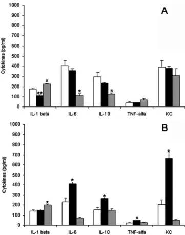
3.3. Variations in cytokine levels
The levels of IL-6, KC/CXCL-1, IL-10, TNF-
a
and IL-1bweremea-sured in the infected and controls ears of BALB/c and C57BL/6 mice (n= 10–15 each). Mice were euthanised at wks 2 and 12 following the i.d. injection ofL. major(Fig. 4).
In BALB/c mice, the levels of IL-6 and IL-10 did not change at wks 2 p.i but decreased significantly at wk 12 (p< 0.001 for all). In these mice, the baseline TNF-
a
and KC/CXCL-1 levels did not change between 2 and 12 wks p.i. (Fig. 4A).In C57BL/6 mice, the levels of IL-6, IL-10, TNF-
a
and KC/CXCL-1 increased significantly (p= 0.007,p< 0.001,p< 0.001 andp< 0.001, respectively) at wks 2 p.i and decreased at wks 12 p.i. (p= 0.007,p< 0.001,p< 0.001 andp< 0.001, respectively) (Fig. 4B). These lev-els returned to the control levlev-els at wks 12 p.i. (p= 0.174).Fig. 4A shows the discrete variation in the level of IL-1bin both lineages.
The levels of IL-1bin the BALB/c mice decreased from an average
of 175 ± 5 pg/ml in the control ears to 111 ± 9 pg/ml in wks 2 p.i and increased to 223 ± 6 pg/ml by the end of the 12 wks post-injection period. In C57BL/6 mice, IL-1blevels did not differ from
the baseline level at 2 wks p.i. (Fig. 4B). These levels were 141 ± 11 pg/ml in the control and 147 ± 11 pg/ml in the infected ears at wks 2 p.i. and increased to 201 ± 6 pg/ml at wks 12 p.i.
4. Discussion
We investigated the nociceptive effect of low doses ofL. major
promastigotes that were inoculated intradermally in the ears of BALB/c (susceptible) and C57BL/6 (resistant) mice and its relation-ship with nerve injury and the level of pro- and anti-inflammatory cytokines that are considered hyper- or hypo-nociceptive (Verri et al., 2006). In BALB/c mice,L. majorproduced prolonged hypoal-gesia from wks 9. In C57BL/6 mice, the latency of the thermal test decreased significantly (hyperalgesia) compared with the control ear only at wks 2 p.i.
Our results reveal two important aspects of cutaneous leish-maniasis: first, the nociceptive response and the cytokine profile vary for each mouse lineage; second, in agreement with the hypothesis that nociceptive phenomena are directly associated
Fig. 2.Time course of the effects of the intradermal injection ofL. majoron the thermal nociception of BALB/c (A) and C57BL/6 (B) mice, as assessed by the thermoalgesimeter test. The latencies observed in infected ears (N) were compared
to the latencies in control ears (j). Data are presented as the means ± standard error for each group (n= 18, two repeated experiments per mouse strain). (⁄) Significant difference between infected and controls ears (p< 0.05).
with the sensitisation of nerve fibres, we detected morphological and morphometrical differences in nerve fibre lesions and density between the mouse lineages at wks 2 p.i but not at wks 12.
Neural involvement in cutaneous leishmaniasis induced by L.
majorhas been considered in mouse and human lesions, in which
inflammatory infiltrate is observed around (perineuritis) and with-in cutaneous nerves (neuritis) with-in addition to the presence of the parasite in the perineural space, as confirmed by our previous mor-phological results (Cangussú et al., 2009) and results from other researchers (Kubba et al., 1987; Satti et al., 1989; Kanaan et al., 2000). Through the systematic histological examination of ear tis-sues, we excluded the massive destruction of nerves, even in the presence of severe architectural changes detected in ear lesions. We detected a slight but significant decrease in fibre density that was correlated with inflammatory changes in resistant mice at the peak of hyperalgesia. The presence of oedema related to the
acute inflammatory phenomena, which are more intense during the initial phase of the disease, may explain why both strains showed a decrease in relative PGP staining compared to the other time points. The quantification of intradermal nerve fibres is not an easy task, especially in small animals. There are few references concerning the evaluation of nerve fibre density in rodents, and in leishmaniasis, they are restricted to a few authors (Kanaan et al. 2000). We did not observe intra-epidermal fibres in mouse ears, and we believe that only a few small nerve fibres associated with the dermal vessels and glands were properly quantified by our method despite the limitations we faced, such as the small thickness. To compensate for this limitation of the model, we ana-lysed at least 3 consecutive sections of each ear (infected and con-tralateral). We systematically measured the stained area, which included both the easily visible and the smaller nerve profiles, by automatic morphometry. The results are presented as the PGP
Fig. 3.PGP 9.5 expression in contralateral and infected ears of BALB/c and C57BL/6 mice inoculated with 103L. majormetacyclic promastigotes. Stained nerves in the dermis
of contralateral ears of BALB/c and C57BL/6 mice (upper insets, A and B, respectively). In lesions in BALB/c mice at 2 wks (A, arrows), the nerves were often intact, but C57BL/6 mice showed unstained nerves (B, arrows and lower inset) with signs of axonal degeneration and visible inflammatory cells (arrows) inside the limits of the perineurum (neuritis). At 12 wks (C, D), despite the severe inflammation in the surrounding tissue in BALB/c mice (C, arrowheads), the nerves remained intact in both lineages (arrows). (E) PGP 9.5 nerve density decreased similar to hyperalgesia in C57BL/6 mice at 2 wks p.i. BALB/c mice did not present quantitative or qualitative nerve damage.⁄Represents a
statistically significant difference in C57BL/6 mice between control ears and infected ears 2 wks p.i. (p= 0.01) and⁄⁄represents statistically significant differences in C57BL/6
9.5 area/total area and compared to the controls. We found peri-neuritis at wks 2–4.5, especially in C57BL/6 mice, in which the tis-sue damage and debris were more pronounced (Cangussú et al., 2009). At this time, we also detected hyperalgesia in C57BL/6 mice. We did not observe a significant difference in nerve density at any time point for BALB/c mice. The more intense inflammatory re-sponse and tissue damage in C57BL/6 mice at initial times and their rapid resolution may be responsible for the acute hyperalge-sia and nerve sensitisation and damage (loss of staining) in this group compared to the different lesion pattern observed in BALB/ c mice.
In the intradermal low-dose ear model, our results provide a clear demonstration of an early period of hyperalgesia that lasts for approximately 15 days in C57BL/6 mice, after which sensitivity to the thermal test returns to normal levels. In contrast, infected BALB/c mice showed a sustained increase in the thermal latency (hypoalgesia) after wks 9, which remained until 12 wks p.i. At that time, we did not detect axonal damage with PGP 9.5 staining.
Previous studies have demonstrated the presence of a sustained hyperalgesic state in BALB/c mice inoculated with a high dose ofL.
major(3105and 2.5106promastigotes/hind footpad,
respec-tively), as shown by a decrease in their pain thresholds (Kanaan et al., 2000; Karam et al., 2006), and short-lived (10 days) hyperal-gesia in BALB/c mice inoculated with 4103 promastigotes per
hind paw (Karam et al., 2006). These studies differ from ours by the higher injected dose and the location of the injection (footpads instead of ears), conditions that alter the inflammatory response and could potentially alter the nociceptive response.
We cannot associate the macroscopic evolution or size of the le-sion with the algesic patterns. In both animals, the lele-sions were
discrete and not ulcerated at 2 wks p.i., although only C57BL/6 mice presented thermal sensitivity compared to the uninfected ear. Moreover, BALB/c lesions presented progressive ulceration beginning at 5 wks, but hypoalgesia began at wks 9, and these ani-mals never presented hyperalgesia, despite intense and persistent ulceration until euthanasia. Between 5 and 7 wks, only a few C57BL/6 mice presented discrete areas of superficial ulceration, which were healed by 12 wks. However, these animals exhibited a thermal response between wks 4 and wks 12, which was similar to the controls.
We believe that the differences in nociception that were detected between the strains might be related to variations in the degree of nerve damage in C57BL/6 mice, which was directly inflicted by inflammatory cells or mediated by cytokines after infection-induced tissue injury. This damage was paradoxically less severe in the large BALB/c mouse lesions. The nociceptive phenomena studied here are also associated with variations in the tissue levels of several cytokines considered hyper- or hypo-nociceptive, as previously described byCunha et al. (1991, 1992, 1999), Inoue et al. (1999), and Poole et al. (1995). The capacity to evoke hyperalgesia can be mediated by a prostaglan-din-dependent mechanism and/or a prostaglandin-independent mechanism. KC/CXCL-1 results in hyperalgesia involving the sympathetic nervous system. Because KC/CXCL-1 is released by activated macrophages and endothelial cells, it may be a humoral link between tissue injury and sympathetic hyperalgesia (Cunha et al. 1991). IL-6 activates the IL-1/prostaglandin hyperalgesic pathway, whereas TNF
a
activates both pathways (Cunha et al., 1992). According to Inoue et al. (1999), interleukin-1b inducessubstance Prelease from primary afferent neurons through the cyclooxygenase-2 system, and this phenomenon may contribute to inflammation-induced hyperalgesia in the intact primary afferent neuro-spinal cord pathway.Poole et al. (1995)suggested that IL-10 limits the inflammatory hyperalgesia evoked by carra-geenan and bradykinin through two mechanisms: the inhibition of cytokine production and the inhibition of IL-lb-evoked PGE2
production. These authors suggest that the latter effect is not mediated via IL-10-induced IL-Ira and may result from the suppression of prostaglandin H synthase-2 (COX-2) by IL-10.
In C57BL/6 mice, it is noticeable that the control latency level measured at wks 12 was approximately half of the level measured at the beginning of the experiment (and was also approximately half of the levels found in control BALB/c mice). It would therefore be interesting to study the subjacent mechanism involved in the host-parasite response. Previous studies also detected differences in the pain behaviour between mouse strains (Larsson et al., 2006), including a higher visceromotor response (VMR) to colorec-tal distension before and after dextran sodium sulphate treatment in C57BL/6 mice compared to BALB/c mice, and suggested that there are strain- and/or model-related differences in visceral pain responses to inflammation.
The levels of the cytokine IL-6 were significantly upregulated during hyperalgesia in C57BL/6 mice and downregulated during hypoalgesia in BALB/c mice. These results are in line with previ-ous reports that indicated that IL-6 plays a role in nociception in the rat model of mechanical hyperalgesia (Cunha et al., 1992; Sommer and Kress, 2004). Other studies have also indicated an increase in IL-6 levels in L. major-injected footpads of BALB/c mice, which is associated with a decrease in the latency (Kanaan et al., 2000; Karam et al., 2006). There is evidence indicating that IL-6 exerts its role in the development and maintenance of pain through resident cell activation, polymorphonuclear cell infiltra-tion, cytokine producinfiltra-tion, prostanoid and sympathomimetic amine release and the activation of intracellular pathways, espe-cially mitogen-activated protein kinase (MAPK) (Manjavachi et al., 2010).
Fig. 4.The effect ofL. majorinfection on the levels of cytokines measured by ELISA in BALB/c (A) and C57BL/6 (B) mice at wks 2 and 12 following ear dermis inoculation with 1103L. majormetacyclic promastigotes. Levels of cytokines in the control ears (open bars), at 2 wks (solid bars) and 12 wks p.i. (grey bars). The bars represent the mean of 10–15 animals ± SD. (⁄)And (⁄⁄)represent statistically
significant differences compared with the other values for each cytokine (p< 0.05).
In our study, the levels of TNF-
a
and KC/CXCL-1 were signifi-cantly upregulated during hyperalgesia in C57BL/6 mice. As men-tioned previously, TNF-a
and KC/CXCL-1 produce hyperalgesia by both exogenous administration (Cunha et al., 1991, 1992; Sommer and Kress, 2004; Cunha et al., 2005) and endogenous production (Cunha et al., 2003, 2005) in several models other than leishmani-asis. However, in BALB/c mice, the levels of TNF-a
and KC/CXCL-1 did not vary significantly throughout the period of observation, in agreement with the results ofKanaan et al. (2000), who did not de-tect changes in the levels of TNF-a
in the ‘classical model’ of infec-tion by L. major using the same mouse strain. The KC/CXCL-1 nociceptive profile has not been studied before in experimental leishmaniasis.IL-1bhas pro-inflammatory activity and induces hyperalgesia
after subcutaneous injection into the rat paw (Ferreira et al., 1988; Cunha et al., 1992) and after intraperitoneal administration in mice (Watkins et al., 1994). Our results showed a discrete in-crease in the levels of IL-1bin both strains after 12 wks of infection,
but this increase was accompanied by hypoalgesia only in BALB/c mice. The IL-1blevels in C57BL/6 mice were also upregulated at
12 wks compared to the controls, despite having a nociception pro-file similar to the baseline of the control ear, in which the IL-1b
levels were not increased. These variations are discrete and may not have biological relevance. Therefore, in our model, no direct relationship between the levels of IL-1band variations in
nocicep-tion was observed. Our results are in agreement with those of
Karam et al. (2011), who did not detect a direct role for IL-1bin
L. major-induced hyperalgesia in the classical model of infection
in BALB/c mice.
IL-10 is a potent anti-inflammatory cytokine (Poole et al., 1995; Vale et al., 2003; Karam et al., 2007; Amaral et al., 2008), and its administration (exogenous IL-10) limits the inflammatory hyperal-gesia evoked by carrageenan and bradykinin in rats ( Safieh-Garab-edian et al., 1995) and mice (Vale et al., 2003; Karam et al., 2007) through two mechanisms: the inhibition of cytokine production and the inhibition of IL-1b-evoked PGE2production by suppressing prostaglandin H synthase-2 (COX-2). However, none of these mechanisms were unequivocally demonstrated to operate in asso-ciation with the endogenous cytokine profile in leishmaniasis and should be further investigated.
Unexpectedly, in the present study, the levels of the cytokine IL-10 were significantly upregulated during hyperalgesia in C57BL/6 mice and downregulated during hypoalgesia in BALB/c mice. How-ever, in support of our observation, recent studies have indicated increased levels of IL-10 in the seminal plasma of patients with chronic pelvic pain syndrome, which was strictly correlated with patient pain severity (Miller et al., 2002). Moreover, Tu et al. (2003) found that IL-10 / mice and IL-10+/+ mice treated with an antibody against IL-10 had an increased latency time for the paw licking response compared to untreated IL-10+/+mice. In their studies, the authors proposed that endogenous IL-10 may be in-volved in the induction of hyperalgesia. The increase in IL-10 in C57BL/6 mice at 2 wks is likely a secondary response due to the presence of pro-inflammatory cytokines and may not play a direct role in hyperalgesia. These aspects should be further investigated by blocking candidate cytokines, including IL-13.
There are qualitative similarities and differences between mice and rats in terms of the cytokines released by inflammatory stimuli and the hypernociceptive mechanisms of action of cytokines.
TNF-a
, IL-1b, IL-6, and KC/CXCL-1 are released in rats and micechal-lenged with nociceptive stimuli (Karam et al., 2006, 2007; Cunha et al., 2005). The different results reported in the literature on the profile of cytokines in mouse inflammatory nociception reflect the complexity of the mechanisms involved in the variations of the pain threshold. Moreover, differences in methodology can explain the variations in proposed mechanisms. For example, there are
dif-ferences in the stimulus applied before measuring the threshold pain (thermal, mechanical), the location of stimulation (ear, foot-pad, tail), the different strains of rats and mice used and the vehicle administered (inflammatory stimulus and/or hypernociceptive mediator).
The data on cytokine levels discussed here should be interpreted as one element in the complex scenario of inflammatory nocicep-tive phenomena, and the specificity of the evolution ofL. major -induced lesions in resistant and susceptible strains of mice should be considered. For example, a direct association of IL-6, which was decreased with hypoalgesia in BALB/c mice (12 wks) and increased with hyperalgesia in C57BL/6 mice (2 wks), should be interpreted in the context of basal differences in cytokine levels between the mouse strains. The production of IL-6 at 2 and 12 wks in BALB/c mice and C57BL/6 mice were similar (p= 0.056 andp= 0.06; respectively); however, a significant decrease in IL-6 levels compared to uninfected levels at 12 wks (p< 0.001) and 2 wks p.i (p< 0.001) was only detected in BALB/C mice. In contrast, C57BL/6 mice presented increased levels compared to control mice (p= 0.007) at wks 2 and returned to the basal level at 12 wks p.i. It is interesting to observe that basal levels of IL-6 in BALB/c mice are twice the values in C57BL/6 mice (p= 0.033), adding value to the variation necessary to provoke changes in the algesic limiar more than to the absolutes values measured. Our findings on the association between IL-6 and hypoalgesia in BALB/c mice are in accordance with the reduction in pro-inflammatory cytokines, primarily IL-6, which has been associated with hypoalgesia by other authors (Cunha et al., 1992).
Our results also highlight the importance of considering the impact of inflammatory changes in L. majorlesions that lead to nerve damage, which in turn modulate nerve susceptibility to pain mediators as an additional complicating factor in the nociceptive panorama. Changes include local blood flow and vascular perme-ability changes, the activation and migration of immune cells and changes in the release of growth and trophic factors from sur-rounding tissues. The data on the cytokine profile discussed here should be interpreted as one element in the complex scenario of inflammatory nociceptive phenomena, and the specificity of the evolution ofL. major-induced lesions in resistant and susceptible strains of mice should be considered.
In summary, we presented a model to study inflammatory nociception in mice that reinforces a role for inflammatory cytokines and nerve damage in afferent nerve sensitisation and increases our knowledge of the histopathologic component in models of hyper- or hypoalgesia. Further investigations are ongoing and will address aspects of pain perception in human cutaneous leishmaniasis.
5. Conclusion
Our work uses a multidisciplinary approach that not only as-sesses the complexity of pain in the context of a long-term infec-tious condition that is extremely important to the public health field, but also indicates that specific mechanisms related to the parasite-induced immune response should be further explored to understand the correlation between inflammatory and anti-inflammatory cytokines, nerve sensitisation and tissue damage. Our data may also explain why the advanced destructive lesions observed in some clinical forms of this disease are favoured by the higher limiar of pain perception in patients who only receive a clinical diagnosis and treatment late in the course of this disease.
Acknowledgments
571093/2008-6), FAPEMIG (PPM 2009) and PRPq/UFMG. S.D. Cangussú received a fellowship from Coordenação de Aperfeiçoa-mento de Pessoal do Ensino Superior (CAPES). L.Q. Vieira, L.C.C. Afonso and R.M.E. Arantes are fellows supported by Conselho Nac-ional de Desenvolvimento Científico e Tecnológico (CNPq).
We are indebted to Mirna Maciel d´Auriol-Souza and Vânia Aparecida da Silva for preparing the tissue samples, Lucélia Pinhe-iro and Maria Marta Figueiredo for animal care, Juan Pereira de Macedo for parasites, and Átila Savernini for developing the ther-moalgesimeter and helping to manipulate it.
References
Ahmed, A.A.A., Theodorsson, M.E., Nordlind, K., 1998. Neuropeptide Y concentrations in experimental Leishmania major cutaneous leishmaniasis. Neuroreport 9, 3271–3277.
Amaral, F.A., Sachs, D., Costa, V.V., Fagundes, C.T., Cisalpino, D., Cunha, T.M., Ferreira, S.H., Cunha, F.Q., Silva, T.A., Nicoli, J.R., Vieira, L.Q., Souza, D.G., Teixeira, M.M., 2008. Commensal microbiota is fundamental for the development of inflammatory pain. PNAS 105, 2193–2197.
Belkaid, Y., Kamhawi, S., Modi, G., Valenzuela, J., Noben-Trauth, N., Rowton, E., Ribeiro, J., Sacks, D.L., 1998. Development of a natural model of cutaneous leishmaniasis: powerful effects of vector saliva and saliva preexposure on the long-term outcome ofLeishmania majorinfection in the mouse ear dermis. J. Exp. Med. 188, 1941–1953.
Cangussú, S.D., Souza, C.C., Campos, C.F., Vieira, L.Q., Afonso, L.C.C., Arantes, R.M.E., 2009. Histopathology of Leishmania major infection: revisiting L. major histopathology in the ear dermis infection model. Mem. Inst. Oswaldo Cruz. 104, 918–922.
Cunha, F.Q., Lorenzetti, B.B., Poole, S., Ferreira, S.H., 1991. Interleukin-8 as a mediator of sympathetic pain. Br. J. Pharmacol. 104, 765–767.
Cunha, F.Q., Poole, S., Lorenzetti, B.B., Ferreira, S.H., 1992. The pivotal role of tumour necrosis factor alpha in the development of inflammatory hyperalgesia. Br. J. Pharmacol. 107, 660–664.
Cunha, F.Q., Poole, S., Lorenzetti, B.B., Veiga, F.H., Ferreira, S.H., 1999. Cytokine-mediated inflammatory hyperalgesia limited by interleukin-4. Br. J. Pharmacol. 126, 45–50.
Cunha, J.M., Cunha, F.Q., Poole, S., Ferreira, S.H., 2000. Cytokine-mediated inflammatory hyperalgesia limited by interleukin-1 receptor antagonist. Br. J. Pharmacol. 130, 1418–1424.
Cunha, J.M., Sachs, D., Canetti, C.A., Poole, S., Ferreira, S.H., Cunha, F.Q., 2003. The critical role of leukotriene B4 in antigen-induced mechanical hyperalgesia in immunised rats. Br. J. Pharmacol. 139, 1135–1145.
Cunha, T.M., Verri, W.A., Silva Jr, J.S., Poole, S., Cunha, F.Q., Ferreira, S.H., 2005. A cascade of cytokines mediates mechanical inflammatory hypernociception in mice. Proc. Natl. Acad. Sci. USA 102, 1755–1760.
Cunha, T.M., Verri, W.A., Schivo, L.R., Napimoga, M.H., Parada, C.A., Poole, S., Teixeira, M.M., Ferreira, S.H., Cunha, F.Q., 2008. Crucial role of neutrophils in the development of mechanical inflammatory hypernociception. J. Leukoc. Biol. 83, 824–832.
Daboul, M.W., 2008. Is the Amastigote form of Leishmania the only form found in humans infected with Cutaneous Leishmaniasis? Science 39, 38–41. Ferreira, S.H., Lorenzetti, B.B., Bristow, A.F., Poole, S., 1988. Interleukin-1 beta as a
potent hyperalgesic agent antagonized by a tripeptide analogue. Nature 334, 698–700.
Ferreira, S.H., Lorenzetti, B.B., Poole, S., 1993. Bradykinin initiates cytokine-mediated inflammatory hyperalgesia. Br. J. Pharmacol. 110, 1227–1231. Heinzel, F.P., Sadick, M.D., Holaday, B.J., Coffman, R.L., Locksley, R.M., 1989.
Reciprocal expression of interferon gamma or interleukin four during the resolution or progression of murine leishmaniasis. Evidence for expansion of distinct helper T cell subsets. J. Exp. Med. 169, 59–72.
Inoue, A., Ikoma, K., Morioka, N., Kumagai, K., Hashimoto, T., Hide, I., Nakata, Y., 1999. Interleukin-1b induces substance P release from primary afferent neurons through the cyclooxygenase-2 system. J. Neurochem. 73, 2206–2213. Kanaan, S.A., Saade, N.E., Karam, M., Khansa, H., Jabbur, S.J., Jurjus, A.R., 2000.
Hyperalgesia and upregulation of cytokines and nerve growth factor by cutaneous leishmaniasis in mice. Pain 85, 477–482.
Karam, M.C., Al-Kouba, J.E., Bazzi, S.I., Smith, C.B., Leung, L., 2011. Interleukin-13 reduces hyperalgesia and the level of interleukin-1bin BALB/c mice infected withLeishmania majorwith an up-regulation of interleukin-6. J. Neuroimmunol. 234, 49–54.
Karam, M.C., Hamdan, H.G., Abi Chedid, N.A., Bodman-Smith, K.B., Baroody, G.M., 2007. Interleukin-10 reduces hyperalgesia and the level of Interleukin-1beta in BALB/c mice infected withLeishmania majorwith no major effect on the level of Interleukin-6. J. Neuroimmunol. 183, 43–49.
Karam, M.C., Hamdan, H.G., Abi Chedid, N.A., Bodman-Smith, K.B., Eales-Reynolds, L.J., Baroody, G.M., 2006.Leishmania major: low infection dose causes short-lived hyperalgesia and cytokines upregulation in mice. Exp. Parasitol. 113, 168– 173.
Kubba, R., el-Hassan, A.M., Al-Gindan, Y., Omer, A.H., Bushra, M., Kutty, M.K., 1987. Peripheral nerve involvement in cutaneous leishmaniasis (Old World). Int. J. Dermatol. 26, 527–531.
Larsson, M.H., Rapp, L., Lindström, E., 2006. Effect of DSS-induced colitis on visceral sensitivity to colorectal distension in mice. Neurogastroenterol. Motil. 18, 144– 152.
Manjavachi, M.N., Motta, E.M., Marotta, D.M., Leite, D.F., Calixto, J.B., 2010. Mechanisms involved in IL-6-induced muscular mechanical hyperalgesia in mice. Pain 151, 345–355.
Miller, L.J., Fischer, K.A., Goralnick, S.J., Litt, M., Burleson, J.A., Albertsen, P., Kreutzer, D.L., 2002. Interleukin-10 levels in seminal plasma: implications for chronic prostatitis–chronic pelvic pain syndrome. J. Urol. 167, 753–756.
Morris-Jones, S., Weber, M., 2004a. A medical mystery – painless ulcers. N. Engl. J. Med. 350, 1442.
Morris-Jones, S., Weber, M., 2004b. Medical mystery: painless ulcers – the answer. N. Engl. J. Med. 350, 2313–2314.
Poole, S., Cunha, F.Q., Selkirk, S., Lorenzetti, B.B., Ferreira, S.H., 1995. Cytokine-mediated inflammatory hyperalgesia limited by interleukin-10. Br. J. Pharmacol. 115, 684–688.
Safieh-Garabedian, B., Poole, S., Allchorne, A., Winter, J., Woolf, C.J., 1995. Contribution of interleukin-1 beta to the inflammation-induced increase in nerve growth factor levels and inflammatory hyperalgesia. Br. J. Pharmacol. 115, 1265–1275.
Satti, M.B., el-Hassan, A.M., al-Gindan, Y., Osman, M.A., al-Sohaibani, M.O., 1989. Peripheral neural involvement in cutaneous leishmaniasis. A pathologic study of human and experimental animal lesions. Int. J. Dermatol. 28, 243–247. Savernini, A., Savernini, N., Amaral, F.A., Romero, T.R.L., Duarte, I.D.G., Castro, M.S.A.,
2012. Assay of therapeutic ultrasound induced-antinociception in experimental trigeminal neuropathic pain. J. Neurosci. Res. 90, 1639–1645.
Seitz, M., Loetscher, P., Dewald, B., Towbin, H., Ceska, M., Baggiolini, M., 1994. Production of interleukin-1 receptor antagonist, inflammatory chemotactic proteins, and prostaglandin E by rheumatoid and osteoarthritic synoviocytes-regulation by IFN-gamma and IL-4. J. Immunol. 152, 2060–2065.
Sommer, C., Kress, M., 2004. Recent findings on how proinflammatory cytokines cause pain: peripheral mechanisms in inflammatory and neuropathic hyperalgesia. Neurosci. Lett. 361, 184–187.
Sotiropoulos, G., Wilbur, B., 2001. Clinical communications: two cases of cutaneous leishmaniasis presenting to the emergency department as chronic ulcers. J. Emerg. Med. 20, 353–356.
Spath, G.F., Beverley, S.M., 2001. A Lipophosphoglycan-independent method for isolation of infective Leishmania metacyclic promastigotes by density gradient centrifugation. Exp. Parasitol. 99, 97–103.
Stefanini, M., De Martino, C., Zamboni, L., 1967. Fixation of ejaculated spermatozoa for electron microscopy. Nature 216, 173–174.
Tu, H., Juelich, T., Smith, E.M., Tyring, S.K., Rady, P.L., Hughes Jr., T.K., 2003. Evidence for endogenous interleukin-10 during nociception. J. Neuroimmunol. 139, 145– 149.
Vale, M.L., Marques, J.B., Moreira, C.A., Rocha, F.A.C., Ferreira, S.H., Poole, S., Cunha, F.Q., Ribeiro, R.A., 2003. Antinociceptive effects of interleukin-4, -10, and -13 on the writhing response in mice and zymosan-induced knee joint incapacitation in rats. J. Pharmacol. Exp. Therap. 304, 102–108.
Verri, W.A., Cunha, T.M., Parada, C.A., Poole, S., Cunha, F.Q., Ferreira, S.H., 2006. Hypernociceptive role of cytokines and chemokines: targets for analgesic drug development? Pharmacol. Ther. 112, 116–138.
Watkins, L.R., Wiertelak, E.P., Goehler, L.E., Smith, K.P., Martin, D., Maier, S.F., 1994. Characterization of cytokine-induced hiperalgesia. Brain. Res. 654, 15–16. Zimmermann, M., 1983. Ethical guidelines for investigations of experimental pain