2017
UNIVERSIDADE DE LISBOA
FACULDADE DE CIÊNCIAS
DEPARTAMENTO DE BIOLOGIA VEGETAL
Characterization and expression of subtilases involved in
grapevine resistance to Plasmopara viticola
Clemente da Silva
Mestrado em Biologia Molecular e Genética
Dissertação orientada por:
Doutora Marta Sousa Silva Doutora Andreia Figueiredo
i
Acknowledgements
Quando Deus nos dá a vida, temos a nosso dispor diferentes caminhos, a minha escolha é tentar seguir o caminho por Ele escrito, claro com as minhas limitações e dificuldades pelo simples facto de ser humano. Agradeço a Deus pelo dom da vida, por me guiar com sabedoria que só Ele detém, por saber quando e onde colocar as pessoas certas para cruzar o meu caminho. Tudo que aconteceu comigo para chegar a esta etapa da minha formação e terminá-la com êxito não tem nenhuma explicação humana se não a providência daquele que me fortalece, Deus.
Quando falava de saber quando e onde colocar as pessoas certas, me referia a essas duas mulheres, as minhas orientadoras nomeadamente, Doutora Andreia Figueiredo e Professora Marta Sousa Silva. Por me terem transformado num jovem com capacidade crítica no que concerne a ciência e terem ajudado a melhorar significativamente o meu domínio nas técnicas laboratoriais, sem me esquecer da paciência que tiveram para comigo nos primeiros meses de adaptação. Aos meus colegas Joana, Marisa, Daniela, Rui e Gonçalo o meu obrigado por facilitar a minha adaptação no grupo e pela amizade. Mas, tudo começou quando conheci a Professora Rita Zilhão, que em 15 dias que esteve na Universidade Eduardo Mondlane-Moçambique, foram suficientes para acreditar no meu potencial e convidar para vir fazer o mestrado. E nessa vinda para cá não só cresci na vida académica como também como pessoa, porque ela entregou-me nas mãos de uma das pessoas que tenho uma admiração profunda e amor imensurável, o Padre Mário Rui Pedras, que cuidou de mim como um filho. Saí de Moçambique deixei meu pai e minha mãe cheguei a Lisboa ganhei um pai (Padre Mário) e uma mãe (Rita).
A família Nhangumele (Teofilo, Luisa, Gladys, Rosy, Teolisa), que me apoiou quando tudo parecia estar perdido, no que concerne a minha vinda para Lisboa para efetuar um estágio de 6 meses no Laboratório de Doutor Prudêncio a montante do mestrado, o meu sincero kanimambo.
Agradeço à família Matos que desde a Licenciatura tem me acompanhado e apoiado incondicionalmente.
Agradeço ao Doutor Prudêncio e ao Bruno Cardoso por me ter ajudado a conhecer as minhas limitações no laboratório, o que facilitou a minha integração no grupo no qual fiz esta tese de mestrado.
A Dra. Mariamo, Dra. Iris, ao Dr. Hermógenes e ao Dr. Sumbana que fizeram despertar em mim esse gosto pela investigação, o meu obrigado.
Agradeço ao Doutor Fernando Vaz, ao Professor Carlos Cordeiro, pela disponibilidade em ajudar sempre que necessitasse.
A todos os meus amigos em especial Arsénia, Nélio, Salomão, Sérgio, Bernardo, Nahida, Inocência, Timóteo, Luísa, Arivalter e Graciela que direta e indiretamente contribuíram para realização deste trabalho.
A toda família São Nicolau, o acompanhamento que todos padres deram e os restantes residentes a amizade que ofereceram, tenham a certeza que ajudaram a moldar esse jovem numa pessoa melhor, Obrigado.
ii
Aos meus pais Moisés F. da Silva e Luísa Tembe da Silva, agradeço pelo apoio incondicional que vem deste o dia do meu nascimento. A toda família Silva o meu kanimambo. Sem me esquecer de uma miúda que não faz parte da família Silva, mas quando apareceu na minha preencheu uma boa parte do meu coração, a minha namorada Nilza Uamusse.
iii
Abstract
One of the most important fruit plant cultivated worldwide is grapevine (Vitis vinifera L.), mainly due to its economic importance in the wine industry. In 2016 the world area under vines was 7.5 million hectares representing a production over 75.8 million tones, being the European Union the leading producer of wine (OIV, 2017). The domesticated grapevine is highly susceptible to downy mildew caused by the obligatory oomycete
Plasmopara viticola. This pathogen affects the leaves and shoots, being downy mildew
disease characterized by the presence of oil spots on the surface of leaves and white down that can be seen on the underside of the leaves, canes and bunches in periods of high humidity. This disease results in great losses in entire vineyards when control measures are not implemented. The current disease control strategies include the massive use of fungicides, which are very prejudicial to human health. A deeper understanding of the resistance mechanisms is crucial to define alternative control methods.
Subtilisin-like proteases (subtilases) belong to a large group of serine proteases present among all organisms such as archaea, bacteria, eukarya, fungi and yeast. My research group has previously characterized the grapevine subtilase gene family, highlighting the involvement of some subtilases in P. viticola resistance. The action mechanisms of subtilases involved in plant defense against pathogens are still unknown; however recent studies have identified prosystemin as the subtilase SBT3 substrate and highlighted the role of the processed systemin in the octadecanoid pathway for jasmonic acid (JA) biosynthesis.
In the present work, we have selected 5 subtilase genes namely, VviSBT3.19 Isoform X2, VviSBT5.3a, VviSBT4.19 Isoform X1, VviSBT3.20, VviSBT3.21 Isoform X1, and analysed their expression in two Vitis genotypes (resistant and susceptible to downy mildew), after inoculation with P. viticola and elicitation with either JA or salicylic acid (SA). Our results showed that the expression of VviSBT5.3a and VviSBT4.19 increase after both P. viticola inoculation and JA elicitation in resistant grapevine genotype in the first hours after inoculation and elicitation. These results suggest subtilases’ involvement in the grapevine immunity.
The grapevine subtilase VviSBT4.19 was selected for further functional characterization aiming to unravel its structure and function The VviSBT4.19 coding sequence was isolated and cloned into both propagation vector (pJET1.2/blunt) and expression vector (pET28a(+)). Two bacteria strains, E. coli BL21codon plus and Turner, were tested for recombinant protein production.
VviSBT4.19 expression was tested by induction with IPTG. Although the SDS-PAGE gel did not show a band corresponding to the VviSBT4.19 protein size, a dot blot analysis was performed. Anti-poly-His-tag antibodies were used and a positive result was obtained for IPTG induction. The determination of this subtilase structure will be crucial to understand its importance and function in grapevine resistance against P.
viticola.
Keywords: Vitis vinifera L., Plasmopara vitícola, VviSBT4.19, recombinant protein expression, elicitation, inoculation, Jasmonic acid
iv
Resumo Alargado
A videira (Vitis vinifera L.) é a planta de fruto mais cultivada em todo o mundo devido à sua importância económica na indústria vinícola. A sua área de cultivo mundial atinge os 7,5 milhões de hectares com uma produção de 75,8 milhões de toneladas em 2016, sendo a União Europeia líder na produção mundial de vinho (OIV, 2017). Atualmente, uma das grandes ameaças para a indústria vinícola é o míldio da videira, doença causada pelo oomycete obrigatório Plasmopara viticola que afeta todas as castas de
Vitis vinifera frequentemente usadas na produção de vinho. No seu ciclo de vida, o P. viticola hiberna sob a forma de oósporos, estas estruturas são altamente resistentes às
condições climáticas adversas, podendo conservar a sua vitalidade durante 2 anos. Sob condições adequadas (temperatura entre os 22-25ºC e humidade elevada) os oósporos germinam, dando origem a zoosporângios que libertam zoósporos que germinam e penetram através dos estomas. Na página superior da folha surgem manchas translúcidas e oleosas coincidentes com o aparecimento de um enfeltrado de micélio branco na página inferior levando ao aparecimento de infeções secundárias. Se não forem utilizadas medidas de controlo apropriadas esta doença pode levar a perdas avultadas durante a época de cultivo. Atualmente, as estratégias de controlo da doença assentam exclusivamente na aplicação preventiva de fitoquímicos durante a época de cultivo, acarretando problemas ambientais graves. O conhecimento profundo dos mecanismos de resistência das plantas resistentes a infeção por Plasmopara vitícola é importante para definir métodos alternativos de controlo.
Em estudos anteriores a interação entre a videira e o P. viticola foi caracterizada por uma abordagem de biologia de sistemas. Uma análise detalhada das diferenças entre a resposta de genótipos suscetíveis (interação compatível) e resistentes (interação incompatível) a este patogénio, ao nível da transcritómica, metabolómica e proteómica, permitiu a identificação de mecanismos e de candidatos associados ao estabelecimento da interação incompatível. Um dos candidatos é uma subtilisin-like protein, também denominada subtilase. As subtilases são proteases serínicas que exercem funções altamente específicas no desenvolvimento das plantas e em cascatas de sinalização. Na década passada, vários estudos realçaram o papel das subtilases na resposta de defesa a agentes patogénicos nomeadamente no reconhecimeno dos efetores dos patogénios, sinalização e ativação de uma resposta imunitária da planta. Apesar do mecanismo de acção das subtilases associadas à resposta de defensa contra patogénios não estar ainda elucidado, estudos recentes em plantas modelo como a Arabidopsis e o tomate identificaram a prosistemina (o precursor da sistemina) como o substrato das subtilases durante a ataque por herbívoros e/ou por patógenos. A sistemina é um polipeptídeo com 18 resíduos de aminoácidos ativo presente em concentração muito baixas, que actua na via sinalizadora octadecanóide responsável pela biossíntese do ácido jasmónico (JA). O JA é uma fitohormona vegetal associada a mecanismos de resposta a stress biótico, nomeadamente por patógenios necrotróficos ou herbivoria. Muito recentemente, estudos do nosso grupo de investigação demonstraram o envolvimento do JA na resposta da videira ao patogénio biotrófico Plasmopara viticola. A sistemina, como substrato das subtilases foi inicialmente isolada das folhas de tomate Lycopersicum esculentum Mill.,
v
sendo demonstrado que quando uma folha é ferida por herbivoria ou mecanicamente, os genes codificadores de sistemina são expressos, transcritos e rapidamente transportados via floema para outras partes da planta ainda intactas. A sistemina atua sobre cinases proteicas activadas por mitogénio, levando à ativação de fosfolipases e consequente libertação do ácido linoléico das membranas plastidiais. O ácido linoléico é utilizado na síntese do JA que regula, via feedback positivo, a expressão génica da prosistemina. O ácido jasmónico produzido move-se através do sistema vascular onde alcança folhas intactas, desencadeando o processo de defesa. Em estudos anteriores, a família de subtilases de videira foi caracterizada e candidatos putativamente associados à resposta de defesa da videira à infeção com o Plasmopara viticola foram identificados. Com o presente trabalho pretende-se validar o envolvimento desses candidatos na resposta de defesa da videira ao P.viticola, avaliar a sua relação com a elicitação por fitohormonas (JA e ácido salicílico (SA)), e selecionar um candidato para validação funcional.
A expressão de 5 genes que codificam as subtilases VviSBT3.19 Isoforma X2, VviSBT5.3a, VviSBT4.19 Isoforma X1, VviSBT3.20, VviSBT3.21 Isoforma X1, previamente identificadas como candidatos associados à resistência da videira ao P.
viticola foi avaliada por Reação de Polimerização em cadeia em tempo real (qPCR). A
sua expressão foi avaliada em dois genótipos de videira, suscetível e resistente ao P.
viticola, após inoculação com o patogénio e elicitação com JA e SA. A expressão das
subtilases VviSBT5.3a e VviSBT4.19 é aumentada nas primeiras horas (6h), tanto após inoculação como também após elicitação por JA no genótipo resistente, sugerindo o seu envolvimento na resposta imunitária da videira ligada à sinalização por JA.
O gene VviSBT4.19 foi selecionado para uma caracterização molecular e funcional de forma a elucidar a sua estrutura e função na resistência da videira ao P. vitícola. A região codificante do gene VviSBT4.19 foi amplificada usando oligonucleótidos específicos para a mesmo. Posteriormente foi efectuada a clonagem da região codificante desta subtilase num vetor de propagação (pJET1.2/blunt) e subsequentemente no vetor de expressão (pET28a(+)). A clonagem e a expressão foram feitas em bactérias adequadas para cada etapa nomeadamente, E. coli TOP10 para clonagem, E. coli BL21codão+ e Turner para expressão. A produção da proteína recombinante foi feita pela adição de β-D-1-tiogalactopiranosideo isopropílico (IPTG). Na análise por eletroforese em gel de poliacrilamida de dodecilsulfato de sódio
(SDS-PAGE), não foi possível observar nenhuma banda correspondente à massa molecular da
proteína de interesse para a abordagem de indução de expressão aplicada. Para confirmar se o não aparecimento da banda da proteína de interesse no gel de poliacrilamida de dodecilsulfato de sódio era devido a baixa expressão ou ausência da mesma, a expressão da proteína recombinante foi analisada por dot blot, uma técnica análoga a western blot, utilizando anticorpos anti-caudas de histidina, porque a proteína de interesse estava em fusão com resíduos de histidina na região N-terminal. Duas das cinco bactérias Turner induzidas por adição de IPTG foram reconhecidas pelos anticorpos. A determinação da estrutura desta subtilase é fundamental para a compreensão da sua importância e função na resistência da videira ao P. viticola.
No momento, estamos a tentar otimizar as condições de expressão e do western blot para ultrapassar os problemas ligados a produção da proteína recombinante e interação
vi
entre a mesma e anticorpos, para poder prosseguir com a purificação da mesma, identificação através da espectrofotometria de massa e posterior estudo da atividade enzimática. Se estes problemas de expressão persistirem vamos seguir uma nova abordagem de expressão que consistirá no uso de vetores eucariotas específicos para expressão e sistemas de expressão eucariotas que foram anteriormente descritas para expressão de subtilases de plantas nomeadamente, células de insetos, usadas na expressão da subtilase de tomate LeSBT1 e sistemas de cultura em suspensão usada na expressão da outra subtilase de tomate denominada LeSBT3. Estes sistemas são mais adequados para a expressão de subtilisinas quando comparados aos vetores ou sistemas de origem procariotas.
Palavras Chave: Vitis vinifera L., Plasmopara vitícola, míldio, inoculação, elicitação
vii Table of contents Acknowledgements ... i Abstract ... iii Resumo Alargado ... iv List of Figures: ... ix List of Tables ... x Abbreviations ... xi 1. Introduction ... 1
1.1. Grapevine downy mildew ... 1
1.2. Subtilisin-like proteases (subtilases) ... 2
1.2.1. Family characterization ... 2
1.2.2. Plant subtilases structure and biochemical properties ... 2
1.2.3. Subtilase involvement in plant-specific processes ... 4
1.3. The link between subtilase and phytohormone signaling in grapevine response to P. viticola ... 6
2. Objectives ... 8
3. Materials and Methods ... 9
3.1. Protein Sequence Alignment and Phylogenetic Analysis ... 9
3.2. Plant Material ... 9
3.3. RNA Extraction and cDNA Synthesis ... 10
3.4. Quantitative Real Time PCR ... 10
3.5. Cloning of the VviSBT4.19 cDNA ... 11
3.5.1. Amplification of VviSBT4.19 Open Reading Frame (ORF) ... 11
3.5.2. Bacterial strains, and vectors ... 12
3.5.3. VviSBT4.19 cloning in pJET1.2/blunt Vector ... 12
3.5.4. Preparation of chemically competent E. coli One Shot TOP10 cells... 13
3.5.5. E. coli One Shot TOP10 transformation ... 13
3.5.6. Colony PCR and pJET-VviSBT4.19 purification ... 13
3.5.7. Cloning in the expression vector pET28a+ ... 14
3.5.8. Colony PCR and pET28a(+)-VviSBT4.19 purification ... 14
3.6. Expression of VviSBT4.19 in BL21 Codon Plus and Tuner bacteria ... 14
3.6.1. Competent E. coli BL21-CodonPlus and Tuner cells (Stratagene) preparation ... 14
3.6.2. Transformation of expression bacteria (BL21-CodonPlus and Tuner) ... 15
3.6.3. Recombinant protein expression in a pET28a(+) vector in the BL21-CodonPlus cells and inTuner cells ... 15
viii
3.6.4. Sodium dodecyl sulfate-polyacrylamide gel electrophoresis (SDS-PAGE)
and Western Blot ... 16
3.6.5. Dot Blot ... 16
4. Result and discussion ... 17
4.1. Phylogenetical analysis of selected grapevine subtilases ... 17
4.2. Expression analysis of the selected grapevine subtilases after inoculation with P. viticola and elicitation with JA or SA ... 18
4.3. Cloning of VviSBT4.19 coding sequence ... 21
4.4. Recombinant VviSBT4.19 protein expression in a pET28a(+) vector ... 23
5. Conclusion ... 25
ix
List of Figures:
Figure 1: Domain architecture of plant subtilases………...3
Figure 2: Structure of the SlSBt3 monomer……….4
Figure 3: Model for the activation of defensive genes in tomato plants in response to herbivore and pathogen attacks……...………...7
Figure 4: Grapevine genotypes Inoculation with P. viticola and Elicitation with JA and SA………...….12
Figure 5: Schematic maps of vector used for VviSBT4.19 cloning and expression.….12 Figure 6: Phylogenetical analysis of 13 Vitis subtilases with subtilases predicted to be involved in plant defence against pathogen infection………...18
Figure 7: Subtilases expression profile in Vitis cultivars after inoculation and elicitation ……….19
Figure 8: Agarose gel analysis of amplification of VviSBT4.19 using Vitis vinifera cDNA as template and VviSBT4.19 cloning into pJET vector………...21
Figure 9: Agarose gel analysis of VviSBT4.19 sub-cloning into bacterial expression vector [pET28a(+)]………..22
Figure10:Dot blotting analysis of expressed VviSBT4.19 fusion proteins………22
Figure 11: Melting curve of targeted genes ………...30
Figure 12: VviSBT4.19 cloning and expression steps...……….31
x
List of Tables
Table 1: Candidate reference genes and target genes primer sequences, amplicon length
xi
Abbreviations
ALE1 abnormal leaf shape 1 bp base pair
CaCl2 Calcium chloride dihydrate
cDNA Complementary DNA
DAMPs Damage-Associated Molecular Patterns DNA Deoxyribonucleic acid
DNaseIDeoxyribonuclease I
dNTP Deoxynucleotide
ECL Enhanced chemiluminescence ECM Extracellular matrix
EDTA Ethylenediaminetetraacetic acid EF1α Elongation factor 1-alpha
Fn-III Fibronectin III gDNA Genomic DNA hae hours after elicitation His Histidine
hpi Hours post inoculation HR Hypersensitive response H2O2 Hydrogen peroxide
IPTG Isopropyl β-D-1-thiogalactopyranoside JA Jasmonic Acid
KCl Potassium chloride
KH2PO4 Potassium dihydrogenphosphate
xii
MAMPs Microbe-Associated Molecular Patterns MCS Multiple cloning site
MgCl2 Magnesium chloride
ML Maximum likelihood mRNA Messenger RNA Mw Molecular weight N2 Nitrogen
NaCl Sodium chloride
Na2HPO4 Sodium hydrogenphosphate
OD Optical density PA Protease-Associated
PAMP Pathogen-Associated Molecular Pattern PBS-T Phosphate Buffered Saline-Tween PCR Polymerase chain reaction
PI Proteinase Inhibitors PR Pathogenesis-related
PRR Pattern Recognition Receptors PTI Pattern-Triggered Immune response qPCR Quantitative real time PCR
RNA Ribonucleic acid
ROS Reactive oxygen species SA Salicylic acid
SDD1 Stomatal Density and Distribution 1
SDS-PAGE Sodium dodecyl sulfate polyacrylamide gel electrophoresis Ser Serine
SOB Super Optimal broth
xiii
TBE Tris-borate-EDTA TB Terrific Broth
TBS Tris-buffered saline °C Celsius degrees
ɡ Earth’s gravitational acceleration kDa kilodalton mg Milligram mL Millilitre mM Millimolar ng Nanogram nm Nanometre Ta Annealing temperature Tm Melting temperature μL Microlitre μM Micromolar
1
1. Introduction
1.1. Grapevine downy mildew
The grapevine (Vitis vinífera L.) is one of the most economically important fruit species worldwide (Basheer-Salimia et al., 2014). Vitis species belongs to the family Vitaceae,
distributed in the temperate zones of the northern hemisphere, with few species reaching the tropics, it comprehends around 60 species, being Vitis vinifera L, the domesticated grapevine, the one with most economic importance due to its use in wine production. However, this specie is highly susceptible to several diseases, including downy mildew, one of the most destructive ones. Downy mildew is caused by Plasmopara viticola (Berk. & Curt.) Berl. and de Toni affecting shoots, leaves and grapes (Gessler et al., 2011). It is native to the South-eastern United States, and was introduced into Europe in the second half of the nineteenth century (Alleweldt and Possingham, 1988; Gessler et al., 2011). P. viticola is an obligate biotrophic oomycete belonging to the family Peronosporaceae (Gindro et al., 2003), it obtains nutrients from living cells of hosts to complete its life cycle through specialized structure known as haustoria (Gessler et al., 2011). P. viticola initiates grapevine leaf colonization through motile zoospores that penetrate stomata (Kiefer et al., 2002), after germination, intercellular mycelia growths and form haustoria. In a compatible interaction, haustoria are formed in the first hours after inoculation and in 72hours, intercellular spaces are invaded by mycelium. Finally, sporangiophores emerge through the stomata where they expand into tree-shaped structures carrying the sporangia (Unger et al. 2007). In an incompatible interaction the first infection steps are common, however the infection progress is delayed, inhibited, or completely stopped (Yu et al., 2012).
The majority of the widely grown grapevine cultivars are highly susceptible to P.
viticola requiring the multiple application of phytochemicals in each growing season,
representing major constrains to human health and to the environment. North American
Vitis species and some Asian Vitis species are natural sources of resistance against
downy mildew and are used as genetic resources in breeding programs for resistance introgression (Rossetto et al., 2002). Although several resistant hybrids such as Regent, Solaris or Bianca are already successfully established in the market, the development of breeding programs is time consuming and several years may pass until the traits can be observed. So, the search for alternative methods to control grapevine downy mildew is crucial (reviewed by Gessler et al., 2011).
Research is being conducted in order to better characterize the resistance processes in grapevine aiming not only to identify novel genes, proteins or metabolite that may be used for breeding programs but also to develop new disease control strategies that allow a sustainable viticulture. One group of proteins that is gaining particular attention is the serine proteases group (subtilases). There are several evidences that subtilases may be involved in grapevine resistance to Plasmopara viticola. The first evidence of the participation of subtilase in the grapevine – P. viticola interactions was shown by Figueiredo and co-workers (2008) as while comparing resistant and susceptible genotypes before and after inoculation with this pathogen, where they noticed that the pattern of subtilase expression was increased in resistant genotypes (Figueiredo et al., 2008, 2012, 2016; Monteiro et al., 2013). Other research groups have also described
2
that when grapevine resistant genotypes where treated with serine protease inhibitors they couldn’t overcome pathogen infection becoming susceptible, and susceptible genotypes became even more sensitive (Berger and Altmann, 2000; Gindro et al., 2012; van der Hoorn and Jones, 2004; van der Hoorn, 2008). Gindro and his co-workers discovered that the activation of programmed cell death was one of the main mechanism responsible of overcoming the grapevine infection by P. viticola because, when they inhibited the phytaspases, a subgroup of plant subtilases, it increased the infection rate in the resistant and immune varieties, diminished the production of toxic stilbenes and changed the level of the plants susceptibility to the pathogen (Gindro et al., 2012). Hence, the understanding of the subtilases’ role in grapevine resistance mechanisms may contribute to the development of alternative strategies for fungal diseases’ control.
1.2. Subtilisin-like proteases (subtilases) 1.2.1. Family characterization
Subtilisin-like proteases (subtilases) belong to a large group of proteases, called serine proteases, that are represented among all groups of organisms such as archaea, bacteria, eukarya, fungi and yeast (Siezen et al., 1991). The subtilase belongs to S8 family which is member of SB clan (Rawlings and Barrett, 1993). Through evolution, many variants of subtilases have arisen and at present can be divided into six main families based on sequence alignment of the catalytic domains (Siezen et al., 2007).
Plant serine proteases are widespread among several taxonomic groups, from trees and crops to legumes and herbs, and although are present in almost all plant parts, seem to be more abundant in fruits (Antão and Malcata, 2005). Unlike mammals on which only nine subtilases have been identify, subtilases from plants are especially abundant. Until present 87 genes were identified in Vitis vinifera genome (Figueiredo et al., 2017), 63 genes in Oryza sativa (Tripathi and Sowdhamini, 2006), 56 genes in Arabidopsis
thaliana (Rautengarten et al., 2005), 15 genes in Lycopersicon esculentum genome
(Meichtry et al., 1999) , 23 genes in the moss Physcomitrella patens, 90 genes in
Populus trichocarpa (Schaller et al., 2012), 74 genes encoding 82 potato subtilases
(Norero et al., 2016). The majority of plant subtilases are secretory enzymes (Siezen and Leunissen, 1997) that mediate cell-to-cell signalling, stomatal distribution and density control during leaf development (von Groll, 2002), that maintain the shoot apical meristem and the cell wall (Liu et al., 2009; Wolf et al., 2009), process peptide growth factors (Srivastava et al., 2008, 2009), and participate in responses to both biotic and abiotic environmental stressors (Liu et al., 2007; Tornero et al., 1997).
1.2.2. Plant subtilases structure and biochemical properties
The first subtilase structure to be solved was the tomato S1SBT3 by X-ray crystallography (Ottmann et al., 2009) and subtilase function was highlighted (Cedzich et al., 2009; Rose et al., 2009). It was shown that plant subtilases, like those in other organisms, depend on structural elements for the stabilization of the subtilisin domain what may reflect specificity for its roles in physiology of the plant (Rose et al., 2010). Most subtilases are synthesized as a pre-pro-protein and targeted for secretion by an N-terminal signal peptide. The pro domain is involved in the subtilase maturation through
3
its cleavage, which is a prerequisite for subtilase passage through the secretory pathway aiming to reach appoplast (Cedzich et al., 2009).
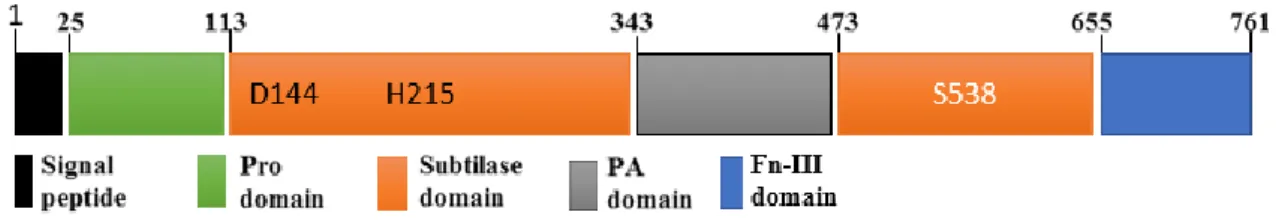
The catalytic triad first observed as Ser–His–Asp in serine proteases composed by serine, histidine and aspartate amino acids is the main characteristic of S8 clade structure because these elements are highly conserved among subtilase family. The structure of this family also usually presents a signal peptide, a pro-domain, a subtilase domain and a protease associated (PA) domain within the subtilase domain (Antão and Malcata, 2005; Dodson and Wlodawer, 1998; Siezen and Leunissen, 1997). Figure 1 represents the conserved structure of plant subtilases but some subtilases present either other domains such as the fibronectin (Fn) III-like domain (Rawling and Salvesen, 2013) or domain deletion.
PA domain in plants is within the S8 peptidase domain. The PA domain in plants is found inserted between the His and Ser active site residues within the subtilase domain (also called S8 peptidase domain), (fig.1), where it causes displacement of the reactive Ser from the catalytic triad to the C-terminal. the PA domain is found in the pyrolysin family of subtilases which includes bacterial endopeptidases, involved in immune response evasion, and plant subtilases such as cucumusin involved in plant pathogen defense and development (Siezen and Leunissen, 1997), and is also implicated in substrate determination of peptidases or form protein–protein interactions (Bruinenberg et al., 1994; Mahon and Bateman, 2000).
Figure 1: Domain architecture of plant subtilases. presenting all four domains namely, pro-domain, a
subtilase domain, a protease associated (PA) domain and the fibronectin (Fn) III-like domain and signal peptide (Adapted from Rose et al., 2010). In addition to the domain borders the figure shows three residues that constitute the active site. This figure represents the domain architecture of SlSBt3.
The additional domain, Fibronectin III (Fn-III), that is present in some subtilases (fig.1), is known to utilize short-peptide surface loops to perform its interactions with other proteins or domain in the signalling pathway cascade (Bencharit et al., 2007). In SBT3, deletion mutants lacking the entire Fn-III domain or part of it were impaired in autocatalytic processing activity and accumulated intracellularly as unprocessed zymogens and the interface of the Fn III domain and the subtilase domain is largely hydrophobic, so it helps the stabilization of the protein by shielding hydrophobic surface patches from solvent. This interaction appears to stabilize the loop system near the active site, what made the researcher consider the Fn-III domain as a required element for SBT3 activity (Cedzich et al., 2009).
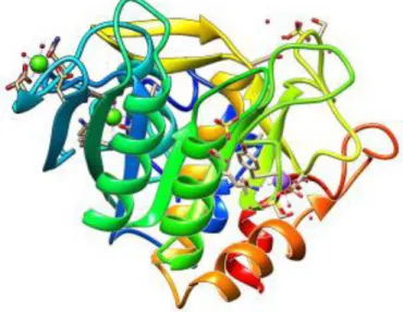
The SBT structure is generally described as a monomer (figure 2) suffering a homodimerization through PA domain mediation in order to be activated (Ottmann et
4
al., 2009; Rose et al., 2010). The PA domain interacts with the pro domain, leading to the cleavage of the N-terminal, and allowing the access of substrates to the catalytic site, for subtilase activity stimulation (Bergeron et al., 2000).
Another feature of subtilases is the apparent Ca2+ independence (Rose et al., 2010). The first subtilases were published as enzymes that theirs activity were influenced by Ca2+ presence, because in their structure there are three conserved calcium binding sites and the calcium binding is an important contribution to enzyme stability (Alexander et al., 2001). In contrast to those findings, although Ca2+-binding sites are conserved and critical for stability in other subtilases, SBT3 from tomato was found to be Ca2+-free and its thermostability is Ca2+-independent, because the activity were not influent (Ottmann et al., 2009). For further confirmation Ottmann and other co-workers performed experiment with addition of calcium ions and verified the enzyme activity, surprisingly, no calcium ions could be identified in the structure of SlSBT3 despite the fact that the general organization of the calcium binding regions is retained. In another subtilases a positively charged site chain of Lys498 mimics the calcium ion bound function (Rose et al., 2010).
Figure 2: Structure of the SlSBt3 monomer. The domains are represented in different colours. Signal peptide correspond to the red sequence, Pro domain in orange, S8 domain in yellow colour, PA domain in green and Fn-III domain in blue colour. This image obtained with the UCSF Chimera package using the following PDB code:1THM.
1.2.3. Subtilase involvement in plant-specific processes
Plants continuously face biotic and abiotic threats. Subtilases are known to participate, direct or indirectly in most cellular processes, such as, general protein turnover, the cell wall dynamic (either by cleavage of structural protein or by regulation of cell wall remodelling enzymes) (Schaller et al., 2012), specific plant development regulator (Berger and Altmann, 2000) and as a determinant host factor mediating activation of primed immune responses against threat from pathogenic microorganisms (Jones and Dangl, 2006; Ramírez et al., 2013).
Genetics approaches have identified subtilases as highly specific regulators of plant development. For example, in the Arabidopsis subtilases, experiments performed that
5
consist in mutation in SDD1 gene (stomatal density and distribution 1), confer an interruption of stomata formation pattern, resulting in clustering of guard cells and increase of stomatal density (Berger and Altmann, 2000; von Groll, 2002). Another example of gene that was found to encode a subtilase which regulate a plant development is ALE1 (abnormal leaf shape 1). This gene is involved in cuticle formation and epidermal differentiation during embryo development in Arabidopsis (Tanaka et al., 2001). How do both genes act is the question that is still without answer but it is speculated that these genes are required for the generation of peptide signals, which act non-cell autonomously to control plant development (Berger and Altmann, 2000; Tanaka et al., 2001; von Groll, 2002). The studies by Takeda et al. (2007), highlighted the participation of subtilases in symbiotic interactions. Interaction between plant roots and fungi resulting in a mycorrhiza or between plant roots nitrogen-fixing Rhizobia as nodule symbiosis.
Concerning the interaction between plants microbes, it is known that many plant-associated microbes are pathogens that impair plant growth and reproduction. Plants use two immunological mechanisms to respond to infection namely, innate and acquired immune system. The first one consists of physical and chemical barriers that are dependent on the recognition of broadly conserved molecular features, known as microbe-associated molecular patterns (MAMPs), by plasma membrane proteins known as pattern recognition receptors (PRRs). These receptors also detect a plant degradation product resulting from the action of invading pathogens, or endogenous peptides called damage-associated molecular patterns (DAMPs), and this recognition is called pattern-triggered immune response (PTI) which is characterized by production of reactive oxygen species (ROS), phosphorylation cascades, and a transcriptional reprogramming that lead to defence responses. The second line of protection, adaptive immunity, typically requires activation (Ausubel, 2005; Boller and Felix, 2009; Jones and Dangl, 2006).
In plant immunity, several studies were published in the last decade showing the involvement of subtilases in response to biotic and abiotic environment stimulus. The first evidence of subtilisin-like proteases participation in plant-pathogen interactions was shown by Granell and co-workers when studying induction of pathogenesis-related (PRs) proteins in tomato by citrus exocortis viroid, silver ion and ethephon. After inoculation of tomato with citrus exocortis viroid they identified high levels of P69 (tomato subtilase) (Granell et al., 1987), and two years later it was also implicated in tomato leaves to Phytophothora infestans (Christ and Mösinger, 1989), when the researchers were looking for the PRs involved in response P. infestans and other biotic and abiotic inducers such as salicylic acid (Jordá et al., 1999) and correlations of those response with resistance. Moreover, during their maturation, the majority of subtilases suffer glycosylation and are secreted to plant extracellular matrix (ECM) where they accumulate and presumably recognize and process substrates (Siezen and Leunissen, 1997; Taylor et al., 1997; Tornero et al., 1996, 1997; Yamagata et al., 1994), so this accumulation of subtilases in ECM of plants rise the possibility for an important subtilase involvement during pathogenesis because, ECM is the place where the first host-pathogen interaction and recognition events take place (Dixon and Lamb, 1990). Later, in Arabidopsis thaliana inoculated with oomycete H. arabidopsidis and P.
6
for immune priming in Arabidopsis. They identified an extracellular subtilase and named SBT3.3, which its loss of function result in an enhanced plant susceptibility, further substantiating its value in establishing an effective plant immune response. They also showed that the production of SBT3.3 rapidly increases during the activation of innate immunity preceding the activation of salicylic acid (SA) responsive genes, responding very rapidly to H2O2, a common ROS species generated during
PTI to activation of innate immune responses (Ramírez et al., 2013).
1.3. The link between subtilase and phytohormone signaling in grapevine response to P. viticola
Little is known about plant subtilases’ substrates. Studies in Arabidopsis and tomato have identified a prosystemin as the subtilase SBT3 substrate (Bergey et al., 1996; Ryan, 2000). Systemin was the first plant peptide hormone that was isolated from tomato (Solanum lycopersicum) by Clarence (Bud) Ryan and his group a quarter of a century ago (reviewed by Pearce et al., 1991). They found out that it was involved with
the activation of the systemic wound-induced defense response. Also, it could be several early wound responsive genes (responding within 2–4 h) involved in the octadecanoid pathway for jasmonic acid biosynthesis and of prosystemin (PS) itself or are late (8 h) responsive genes that include genes for proteinase inhibitors (I and PI-II) that interfere with digestive processes in herbivores (Bergey et al., 1996; Ryan, 2000). The prosystemin function was demonstrated with tomato plants transformed with an antisense prosystemin cDNA driven by the constitutive cauliflower mosaic virus promoter. The transformed plants were found to be severely impaired in their systemic wound response and the plants expressing the antisense gene not only accumulated low levels of proteinase inhibitors I and II in leaves of wounded plants, but lost their ability to mount inducible defences against Manduca sexta larvae (McGurl et al., 1992, 1994; Orozco-Cardenas et al., 1993).
Bergey and co-workers used the similarity of the structure of jasmonic acid and its precursor phytodienoic acid to the structures of some prostaglandins and the fact that that prostaglandins are derived from arachidonic acid released from membranes by phospholipase A2 as a base to propose a model in which wounding and systemin activated a lipase in receptor cell membranes resulting in the release of linolenic acid, the production of jasmonic acid, and the activation of proteinase inhibitor genes. In that model, oligosaccharides are localized signals, whereas systemin is the systemic signal that activates the defence signalling pathway (Figure 3) (Bergey et al., 1996; Farmer and Ryan, 1992).
7 Figure 3: Model for the activation of defensive genes in tomato plants in response to herbivore and pathogen attacks (Adapted from Farmer and Ryan, 1992).
Studies supporting the linking between salicylic acid and jasmonic acid were conducted by Doares and co-workers, where they described that the presence of salicylic acid as inhibitors of the octadecanoid pathway, culminate with deficient plant capacity to respond to a pathogen attack (Doares et al., 1995), by controlling transcriptional reprogramming of JA-induced defensive genes (Caarls et al., 2015). Those results may be related with the reason why one of the subtilase subfamily protease called phytaspase is less expressed or blocked when the octadecanoid pathway for jasmonic acid biosynthesis is inhibited (Beloshistov et al., 2017).
8
2. Objectives
In grapevine, only recently the participation of subtilases in resistance has been shown. The main goal of this work was to unravel the function of these subtilases and their involvement in grapevine resistance to P. viticola, to gain a more comprehensive knowledge on their role in plant immunity, thus contributing to the development of alternative strategies for fungal diseases control.
To achieve this goal the following tasks were performed:
• Phylogenetical analysis of the selected grapevine subtilases (based in the work of Figueiredo et al., 2016) with the subtilases previously described in other plant models as involved in plant immunity and JA signalling;
• Expression analysis of Vitis subtilases that share sequence similarity with subtilases predicted to be involved in plant defence against pathogen infection, after inoculation with P. viticola and elicitation with jasmonic acid and salicylic acid;
• Selection of resistance and JA-associated candidates;
• Gene isolation, cloning and propagation in adequate expression vectors; • Recombinant protein production and characterization.
9
3. Materials and Methods
3.1. Protein Sequence Alignment and Phylogenetic Analysis
Protein sequences from grapevine subtilases putatively involved in grapevine resistance against pathogens (selected in Figueiredo et al. 2017 and Figueiredo J., master thesis, 2016), Arabidopsis, tobacco, rice, cotton and tomato subtilases shown to participate in plant immunity were obtained from the NCBI database (February 2017) and aligned using the DNASTAR's MegAlign, version 11.1.0 (59) ( https://www.dnastar.com/t-megalign.aspx), gaps were manually checked. A maximum likelihood (ML) phylogenetic analysis was performed with MegAlign with the following parameters: protein substitution model PROTCAT; protein substitution model + BLOSUM62; bootstrap 1000 iterations with rapid bootstrap analysis. Tree was viewed and edited on MegAlign (https://www.dnastar.com/t-megalign.aspx).
3.2. Plant Material
Two Vitis vinifera genotypes, Regent and Trincadeira (tolerant and susceptible, respectively to P. viticola) were selected to assess subtilase expression after inoculation with P. viticola and elicitation with either jasmonic (JA) or salicylic acids (SA). Plant material inoculated with P. viticola and harvested at 6, 12 and 24hpi (Figure 4), as described in Figueiredo et al. (2012) was already available at the laboratory. For the elicitation experiments, wood cutting from the two genotypes were obtained at the Estação Vitivinícola Nacional, at Dois Portos, Torres Vedras, Portugal and grown in 2.5L pots in universal substrate under controlled conditions in a climate chamber (Phytoclimate 5000 EH Aralab, Lisbon) at 23 / 18 ºC (day / night), relative humidity 60% and a photosynthetic photon flux density of 300 µmol m-2 s-1. One month prior to the elicitation experiments, pots were transferred to the exterior environment at the campus of the Faculty of Science, University of Lisbon (FCUL), Portugal, and irrigated whenever necessary. Grapevine leaves were elicited with 1mM SA (0.2ml) or 1mM JA (0.2mL), (Sigma Aldrich) in 0.05% tween 20 (0.1ml) solutions. Control plants were sprayed with a 0.05% tween 20. The second and third fully expanded leaves beneath the shoot apex were harvested at 6 and 24hours post elicitation (hpe) (Figure 4), immediately frozen in liquid nitrogen and stored at −80°C. Due the damage during the storage, samples collected at 6 hpe with SA, were discarded (Figure 4). Three biological replicates were collected, being each biological replicate a pool of three leaves from three different plants.
10 Figure 4: Grapevine genotypes Inoculation with P. viticola and Elicitation with JA and SA (Jasmonic
Acid and Salicilic Acid). hpe- hours post elicitation, hpi- hour post inoculation.
3.3. RNA Extraction and cDNA Synthesis
Total RNA was isolated from frozen leaves with the Spectrum™ Plant Total RNA Kit (Sigma-Aldrich, USA), according to manufacturer's instructions. Residual genomic DNA was digested with DNase I (On-Column DNase I Digestion Set, Sigma-Aldrich, USA). RNA purity and concentration were measured at 260/280nm using a spectrophotometer (NanoDrop-1000, Thermo Scientific) while RNA integrity was verified by agarose gel electrophoresis (1.2% agarose in TBE buffer). Genomic DNA (gDNA) contamination was checked by qPCR analysis of a target on the crude RNA (Vandesompele et al. 2002). Complementary DNA (cDNA) was synthesized from 2.5µg of total RNA using RevertAid®H Minus Reverse Transcriptase (Fermentas, Ontario,
Canada) anchored with Oligo(dT)23 primer (Fermentas, Ontario, Canada), according to
manufacturer's instructions.
3.4. Quantitative Real Time PCR
Quantitative real time PCR (qPCR) experiments were carried out using Maxima™ SYBR Green qPCR Master Mix (2×) kit (Fermentas, Ontario, Canada) in a StepOne™ Real-Time PCR system (Applied Biosystems, Sourceforge, USA). A final concentration of 2.5mM MgCl2 and 0.2μM of each primer were used in 25μL volume reactions,
together with 4μL of cDNA as template. Primer sequences and reaction details are provided in Table 1. Thermal cycling for all genes started with a denaturation step at 95°C for 10minutes followed by 40 cycles of denaturation at 95°C for 15seconds and annealing at the appropriate temperature (Table 1) for 30seconds. Each set of reactions included a control without cDNA template. Dissociation curves were used to analyse non-specific PCR products (Supplementary data 1). Three biological replicates and two technical replicates were used for each sample. Gene expression (fold change) was calculated as described in Hellemans et al. (2007). The reference genes used for the normalization were the previously described in Monteiro et al. (2013). Statistical significance (p < 0.05) of gene expression was determined by the Mann–Whitney U test using IBM® SPSS® Statistics version 23.0 software (SPSS Inc., USA).
11 Table 1: Candidate reference genes and target genes primer sequences, amplicon length and qPCR analysis. ---- Discarded (low abundance transcript); *The nomenclature used in this table, is in accordance with the last update of Vitis vinifera L. subtilases (Figueiredo et al., 2017).
3.5. Cloning of the VviSBT4.19 cDNA
All the steps or procedures followed from the cloning of VviSBT4.19 cDNA to the recombinant protein production are described in the supplementary data 2.
3.5.1. Amplification of VviSBT4.19 Open Reading Frame (ORF)
VviSBT4.19 coding sequence (XM_010660203.2) was amplified with the oligonucleotides VviSBT4.19 forward (5’CCG GAA TTC ATG TGC ATA GCT TAC CTT CTA3’) and reverse (5’CAC CGC TCG AGG TGC TTG CCG CAT CAT TTA3’), containing EcoRI and XhoI restriction sites (underlined), respectively. The VviSBT4.19 coding region was amplified by PCR using Vitis vinifera cv Regent inoculated with P. viticola at 6hpi cDNA as template, with the following program: initial denaturation at 98°C for 30s, denaturation at 98°C for 10s, annealing at 56°C for 30s, extension at 72°C for 2min: 30s, 35 cycles and final extension at 72°C for 10min. The following polymerase chain reaction (PCR) mix was used: 1µl of cDNA, 0.5µl of Phusion polymerase (1.0 unit/50 µl), 2.5µl of primers (10µM), 10µl 5X Phusion HF buffer, 5µl of dNTPs ((10mM) and 28.5µl of nuclease-free water in a total of 50µl. The
12
PCR products were visualised on a 1.5% (w/v) agarose gel and the band corresponding to the VviSBT4.19 coding sequence was excised and purified using QIAquick PCR Purification Kit Protocol (QIAquick Spin Handbook 03/2008) following manufacturer’s instructions. Concentration was measured in a NanoDrop 1000 (Thermo Scientific).
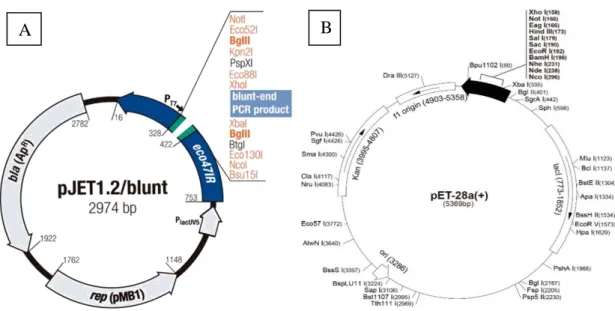
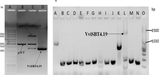
3.5.2. Bacterial strains, and vectors
E. coli TOP10 (Invitrogen, New York) and pJET1.2/blunt Cloning vector (Thermo
Scientific, Waltham, Massachusetts, USA) (Figure 4A) were used to perform DNA manipulation. E. coli BL21codon plus (Stratagene, California, USA) and pET28a(+) (Novagen, USA) (figure 4B), were used for Vitis vinifera Cucumisin (VviSBT4.19) gene expression.
Figure 5: Schematic maps of vector used for VviSBT4.19 cloning and expression. A- pJET1.2/blunt
Cloning vector contain ß-lactamase gene conferring resistance to ampicillin, Lethal gene eco47IR which enables positive selection of the recombinants, T7 RNA polymerase promoter for in vitro transcription of the cloned insert, Multiple cloning site (MCS), Insertion site Blunt DNA ends for ligation with insert. B-This vector (pET-28a(+)) contain a T7 promoter, a T7 terminator, a T7 transcription start region, His-tag coding sequences, a multiple cloning site, coding sequence of gene lacI, a pBR322 origin of replication and kanamicin resistance gene and a f1 origin of replication.
3.5.3. VviSBT4.19 cloning in pJET1.2/blunt Vector
To construct the recombinant plasmids, VviSBT4.19 gene was cloned into the pJET1.2/blunt vector using CloneJET™ PCR Cloning Kit (#K1231, #K1232), according manufacturer’s instructions, under following volumes: 6µL of nuclease-free water, 10µL of 2X reaction Buffer, 1 µL pJET1.2/blunt of 2µL of the gene and 1µL of T4 DNA Ligase. The ligation was performed at 22°C for 5min.
13
3.5.4. Preparation of chemically competent E. coli One Shot TOP10 cells
E. coli One Shot TOP10 was submitted to a protocol for competence induction and used
as host for amplification of recombinant plasmids. Cells were plated in SOB (Super Optimal Broth) medium at 37°C overnight. One colony was inoculated in 225mL of SOB medium and grown at 37°C, 170 rpm until the OD600nm reached 0.5. Cells were
kept on ice for 10 minutes and centrifuged at 1400g for 5minutes. The supernatant was discarded and the pellet resuspended in RF1 buffer (100mM RbCl2, 50mM MgCl2
(4H2O), 30mM KAC, 10mM CaCl2-2H2O, 15% (v/v) glycerol). Cells were kept on ice
for 15minutes and centrifuged at 1400g for 5minutes. The supernatant was discarded and the pellet re-suspended in RF2 buffer (100mM MOPS, 10mM RbCl2, 75mM CaCl2
-2H2O, 15% (v/v) glycerol). Cells were divided in 100µL aliquots and frozen in liquid
N2.
3.5.5. E. coli One Shot TOP10 transformation
Competent E. coli One Shot TOP10 cells were transformed with the pJET-VviSBT4.19 constructs according following procedures: 100µl of cells were thawed one ice and 10µl of ligation product was added and incubated on ice for 30 minutes, also submitted to a heat-shock for 45seconds at 42°C, without shaking, and re-incubated on ice for 2minutes. 800µL of LB culture medium was added and incubated at 37°C, 200rpm for 30minutes. Cells were centrifuged at 1200g for 2minutes at room temperature and the supernatant was discarded. The cell pellet was re-suspended in remaining medium and plated in LB agar medium supplemented with 50μg/mL of ampicillin for growth at 37°C overnight.
3.5.6. Colony PCR and pJET-VviSBT4.19 purification
The presence of the VviSBT4.19 was confirmed by colony PCR using the cell colonies grown in the agar plate as template. Colony PCR reaction conditions were: initial denaturation at 95°C for 3min, denaturation at 94°C for 30s, annealing at 60°C for 30s, extension at 72°C for 2min:30s, 35 cycles and final extension at 72°C for 10min. The volumes of PCR components of 20µl contained 0.25µl of GOTaq polymerase (5U/µl), 0.4µl of each primer (10µM), 4µl 5X Colorless GoTaq® Reaction Buffer, 2µl of dNTPs (10mM), 1.2µl of MgCl2 (1.5mM) and 11.75µl of nuclease-free water and one colony
were added in each mix. The colony PCR products were analysed in 1% agarose gel electrophoresis. Bacteria presenting the PCR band correspondent to the VviSBT4.19 insertion were inoculated in LB medium supplemented with 50μg/mL of ampicillin and incubated at 37°C, 200rpm, overnight. The constructs were extracted using NZYMiniprep Kit, according to manufacturer’s instructions. The purified products were quantified in a NanoDrop 1000 (Thermo Scientific). For further confirmation of the presence of the pET28a(+) vector and the gene, the purified plasmid were digested using EcoRI and XhoI restriction enzymes (Thermo Scientific, Waltham, Massachusetts, EUA) under following conditions: 10µL of nuclease-free water, 8µL of 10X Tango buffer, 20µL of pET28a(+) or pJET-VviSBT4.19, 1µL of each enzyme. The reaction occurred at 37°C for 2 h and the enzymes were inactivated at 80°C for 20min,
14
with subsequent analysis by 1.0% (w/v) agarose gel electrophoresis and analysed by gene sequencing using the T7 primers (STABVida Company, Caparica, Portugal)
3.5.7. Cloning in the expression vector pET28a+
The plasmid containing the VviSBT4.19 insertion was extracted from E. coli One Shot TOP10 using the NZYMiniprep Kit, per manufacturer’s instructions. Both plasmid containing the VviSBT4.19 and pET28a(+) were hydrolysed using EcoRI and XhoI restriction enzymes as previously described.
The VviSBT4.19 ORF was cloned into the expression vector (pET28a(+)) using the T4 DNA Ligase enzyme (New England Biolabs) as follows: 9µL of nuclease-free water, 2µL of 10X T4 DNA Ligase Buffer, 2µL of pET28a(+), 6µL of VviSBT4.19 and 1µL of T4 DNA Ligase. The ligation was performed at 22°C for 2hours. The plasmid and gene volume were calculated using the formula below:
𝑛𝑔 𝑖𝑛𝑠𝑒𝑟𝑡 = 𝑏𝑝 𝑖𝑛𝑠𝑒𝑟𝑡
𝑏𝑝 𝑣𝑒𝑐𝑡𝑜𝑟 𝑥 3 𝑥 𝑛𝑔 𝑣𝑒𝑐𝑡𝑜𝑟
The ligation product was used to transform E. coli BL21 and One Shot TOP10 cells using same procedures as described above.
3.5.8. Colony PCR and pET28a(+)-VviSBT4.19 purification
The transgene insertion was confirmed by colony PCR as previously described. The colony PCR products were analysed in 1% (w/v) agarose gel electrophoresis. The recombinant bacteria were inoculated in LB medium supplemented with 50μg/mL of kanamycin and incubated at 37°C, 200rpm, overnight. The constructs were extracted from bacteria using NZYMiniprep Kit, according to manufacturer’s instructions. To further confirm the presence of the pET28a(+)-VviSBT4.19, the purified DNA were digested using EcoRI and XhoI restriction enzymes, with subsequent analysis by 1.0% (w/v) agarose gel electrophoresis, as previously described. Gene sequencing using the T7 promotor and T7 terminator primers was also performed (STABVida Company, Caparica, Portugal).
3.6. Expression of VviSBT4.19 in BL21 Codon Plus and Tuner bacteria 3.6.1. Competent E. coli BL21-CodonPlus and Tuner cells (Stratagene) preparation
E. coli BL21-CodonPlus (Stratagene) are engineered to contain extra copies of genes
that encode the tRNAs that most frequently limit translation of heterologous proteins. E.
coli. Tuner (Novagen, USA) contains a mutation in the lac permease (lacZY) gene. This
enables adjustable levels of protein expression throughout all cells in a culture. The lac permease (lacY) mutation allows uniform entry of IPTG into all cells in the population, which produces a concentration-dependent, homogeneous level of induction. By
15
adjusting the concentration of IPTG, expression can be regulated from very low levels up to the robust, fully induced levels commonly associated with pET vectors.
E. coli BL21-CodonPlus and Tuner cells were submitted to protocol of Cohen et al., 1972 for competence induction and used as host for expression of recombinant plasmids. Cells were plated in solid LB (Luria-Broth) medium supplemented with 34μg/mL chloramphenicol at 37°C overnight. One colony was inoculated in 3mL of liquid LB medium supplemented with 34μg/mL chloramphenicol at 37°C overnight, with shaking (250 rpm). About 500μL of cells were inoculated in 10mL of liquid LB medium supplemented with 34μg/mL chloramphenicol and incubated at 37°C, 200rpm for 3h. Cells were centrifuged at 1500g for 7minutes. The supernatant was discarded and the pellet re-suspended in 3ml CaCl2, also incubated on ice for 20minutes and
centrifuged at 1500g for 7minutes. The supernatant was discarded, the pellet re-suspended in 1ml CaCl2 and kept on ice (1h minimum) until using for transformation.
3.6.2. Transformation of expression bacteria (BL21-CodonPlus and Tuner)
The extracted and purified positive constructs (pET28a(+)-VviSBT4.19) were used to perform the transformation of BL21-CodonPlus cells, described as a member of large group of expression bacteria.
Competent E. coli BL21-CodonPlus and Tuner cells were transformed using following procedures: 100µl of cells were thawed one ice and 100ng of pET28a(+)-VviSBT4.19 were added and incubated on ice for 30minutes, and then submitted to a heat-shock for 2 minutes at 42°C, without shaking, and re-incubated on ice for 3minutes. About 800µL of LB culture medium was added and incubated at 37 °C, 200 rpm for 1h. Cells were centrifuged at 1200g for 2minutes at room temperature and the supernatant was discarded. The cell pellet was re-suspended in remaining medium and plated in LB agar medium supplemented with 50μg/mL of kanamycin and chloramphenicol for growth at 37°C overnight. The recombinant cells presence was confirmed by colony PCR and miniprep using the cell colonies grown in the agar plate following same condition as described above.
3.6.3. Recombinant protein expression in a pET28a(+) vector in the BL21-CodonPlus cells and inTuner cells
Recombinant colonies were grown overnight in TB medium (3ml) at 37°C with shaking. The overnight culture was transferred into TB medium (10ml) containing kanamycin (50 μg/ml). The culture was incubated at 37°C until an OD600nm of 0.5-0.6
was reached. IPTG (0.1 mM) was added to induce expression and the culture was incubated for 4h at 37°C. The culture was centrifuged (2000 g, 10min, 4 °C), the pellet resuspended in 20ml of PBS-T [137 mM NaCl, 2.7 mM KCl, 10 mM Na2HPO4, 1.76
mM KH2PO4, 0.1% (v/v) Tween-20]. The suspension was incubated for 10min at RT
and stored at -20°C. After thawing, cells were disrupted by sonication on ice for 4x20s with 10s intervals and centrifuged (5000g, 10min, 4°C). The supernatant was filtered
16
through. To assess the expression and solubility of the protein, the supernatant and pellet of recombinant cells were analysed by 10% SDS-PAGE.
3.6.4. Sodium dodecyl sulfate-polyacrylamide gel electrophoresis (SDS-PAGE) and Western Blot
Protein samples were separated in a 10% reducing SDS-PAGE (using a 4% staking gel) as follows: samples (10µl) were combined with an equal volume of loading buffer [125mM Tris-HCl buffer, pH 6.8, 4% (w/v) SDS, 20% (v/v) glycerol, 10% (v/v) 2-mercaptoethanol, 0.02% (v/v) bromophenol blue], were heated for 5min and placed on ice until loaded into the gel. After sample loading, electrophoresis was conducted at 90V for 90min. The gel was stained with Coomassie blue staining solution [0.2% (w/v) Coomassie blue, 7.5% (v/v) acetic acid, 50% (v/v) ethanol] for 2h. Coomassie stained gels were destained with destaining solution [50% (v/v) methanol, 10% (v/v) acetic acid] for 2 h. The image was captured on Gel Doc™ XR+ Gel Documentation System. For western blot analysis, the Mini Trans-Blot cell (Biorad, Califórnia-EUA) was used, following the manufacturer’s instructions. To perform the blotting, aPVDF membrane previously activated in 100% methanol was used. Transfer buffer contained 3.0g/l Tris, 14.4 g/l glycine, 20% (v/v) methanol. Transfer was conducted at 90V in blotting apparatus for 70min. After protein transfer, the membrane was blocked for 1h at RT with 3% (w/v) non-fat milk in TBS-T (20mM Tris-HCl buffer, 200mM NaCl, 0.1% Tween-20, pH 7.4). The membrane was incubated overnight, at 4°C, with mouse anti-Histag primary antibody (His-probe Antibody (H-3), Santa Cruz Biotechnology, INC) diluted 1:1000 in 0.5% (w/v) TBS-T. The membrane was washed (3x10min) in TBS-T and then incubated 1h, RT, with HRP-linked secondary antibody (m-IgGκ BP-HRP) diluted 1:1000 in 3% (w/v) TBS-T, and then washed with TBS-T (3x10min). Membranes were incubated in ECL Western Blotting Detection Reagent in the dark before digital imaging which is also performed in the dark.
3.6.5. Dot Blot
ThePVDF membrane was activated in 100% methanol for 5min. The membrane and filter paper were soaked into transfer buffer (3.0g/l Tris, 14.4g/l glycine, 20% (v/v) methanol) for 5min. The filter papers were placed on a clean surface and the membrane was placed on the filter paper, to guarantee the capillarity. The boiled samples (the lysates combined with an equal volume of loading buffer [125 mM Tris-HCl buffer, pH 6.8, 4% (w/v) SDS, 20% (v/v) glycerol, 10% (v/v) 2-mercaptoethanol]) were applied on the membrane before it dries. The subsequent steps were the same as for the western blot, which include the blocking and the incubation with primary and secondary antibodies and digital imaging.
17
4. Result and discussion
4.1. Phylogenetical analysis of selected grapevine subtilases
Plants continuously face threats from quite a diversity of sources. Nowadays, there is evidence of direct or indirect participation of subtilases in the plant responses to these threats, for example as a determinant host factor mediating activation of primed immune responses against pathogenic microorganisms (Jones and Dangl, 2006; Ramírez et al., 2013). Duan and co-workers used yeast two-hybrid assay and bimolecular fluorescence complementation (BiFC) analysis to show that the cotton subtilase GbSBT1 interacts with a prohibitin (PHB)-like protein expressed in V. dahliae pathogens during infection
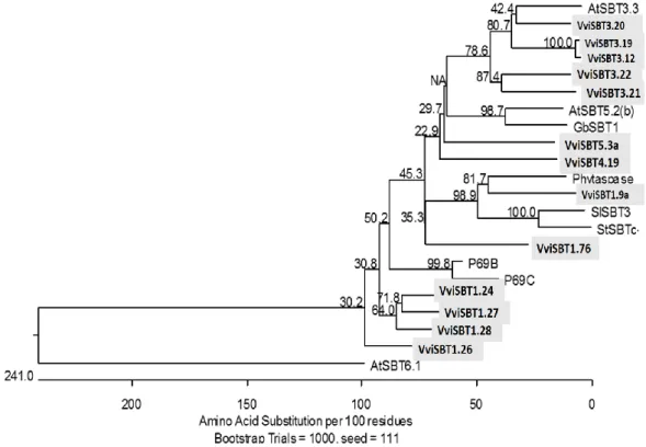
(Duan et al., 2016). Previous studies done by Ramírez and co-workers in A. thaliana also associated subtilase AtSBT3.3 with the defence response to pathogen attack (Ramírez et al., 2013) and same results were found in P69 in S. lycopersicum (Tornero et al., 1996). In grapevine, following the characterization of the grapevine subtilase gene family by our group, 13 subtilases were associated with Plasmopara viticola resistance
(Figueiredo et al., 2016). In order to understand if these 13 grapevine subtilases are related to other plant subtilases described as participating in immune priming, plant resistance and JA signalling, a phylogenetical analysis was performed. The subtilase sequences GbSBT1 from cotton (Duan et al., 2016), P69B, P69C and SISBT3 from tomato (Meichtry et al., 1999), phytaspase from tobacco (Chichkova et al., 2010), tomato (Beloshistov et al., 2017) and rice (Tripathi and Sowdhamini, 2006), StSBTc from Solanum tuberosum (Fernández et al., 2015), and AtSBT3.3, AtSBT5.2(b) and AtSBT6.1 from Arabidopsis (Rautengarten et al., 2005) were used as resistance-associated subtilases.
Figure 6: Phylogenetical analysis of 13 Vitis subtilases with other plant subtilases predicted to be involved in plant defence against pathogen infection. VviSBT3.20, VviSBT3.19 Isoform X2,
VviSBT3.12 Isoform X1, VviSBT3.22, VviSBT3.21 Isoform X1, VviSBT5.3a, VviSBT4.19 Isoform X1,
18 Vitis subtilases; SISBT3, P69B and P69C are tomato subtilases; Phytaspase is subtilase from tobacco,
StSBTc the Solanum tuberosum subtilase; AtSBT3.3, AtSBT5.2(b) and AtSBT6.1 are Arabidopsis subtilases; GbSBT1 is a cotton subtilase.
Seven grapevine SBT Protein sequences VviSBT3.19 Isoform X2, VviSBT5.3a,
VviSBT4.19 Isoform X1, VviSBT3.20, VviSBT3.21 Isoform X1,VviSBT3.12 Isoform X1, and VviSBT3.22 showed sequence similarity to the immunity related subtilases namely, cotton and Arabidopsis subtilase (Figure 5). In the phylogenetical analysis
VviSBT3.19 Isoform X2 and VviSBT3.12 Isoform X1 are closely related (fig.5), the same occurs with VviSBT3.21 Isoform X1 and VviSBT3.22, thus VviSBT3.19 Isoform X2 and VviSBT3.21 Isoform X1 were selected for further analysis.
In summary, 5 grapevine subtilases were chosen (VviSBT3.19 Isoform X2,
VviSBT5.3a, VviSBT4.19 Isoform X1, VviSBT3.20, VviSBT3.21 Isoform X1) for gene expression profiling after inoculation with P. viticola and elicitation with either JA or SA.
4.2. Expression analysis of the selected grapevine subtilases after inoculation with
P. viticola and elicitation with JA or SA
We have evaluated by qPCR the expression profiling of the selected grapevine subtilases upon both pathogen inoculation and JA or salicylic acid elicitation in two grapevine genotypes: Vitis vinifera cv Regent and cv Trincadeira, resistant and susceptible to P. viticola respectively. We have chosen these phytohormone to elicitate these cultivars because Guerreiro et al.(2016), related the activation of JA and SA pathway at early stages (6, 12, 24hpi) of inoculation with P. viticola. The employment of synergistic/antagonistic mechanisms may a boost to have depper understanding of V.
vinifera cv. Regent response to the biotrophic oomycete P. viticola.
Two (VviSBT3.19 and VviSBT3.20) of the 5 subtilases analysed did not show any results, so they were discarded duelow abundance transcript (table 1). After P. viticola inoculation, in the susceptible genotype, the expression of VviSBT5.3a decreased at 6hpi while the expression of VviSBT3.21 increased. VviSBT4.19 showed no alteration. Contrarily, the resistant genotype VviSBT5.3a, VviSBT4.19 and VviSBT3.21 were highly upregulated with the higher expression increase being of the subtilase VviSBT4.19 (figure 6A).
Cao and co-workers (2014) analysed the expression of several grapevine subtilases under different abiotic stimuli and in different tissues without stimulation. They found that the expression of the subtilase VviSBT3.21 was suppressed under abiotic stress conditions. Figueiredo and co-workers, studying the cultivar-specific kinetics of gene induction during downy mildew early infection in grapevine, showed an expression increase of VviSBT3.21 at 6hpi (Figueiredo et al., 2012). This result suggests that this subtilase may be involved in the response to biotic stimulus, but not in response against abiotic stimulus.
At 12hpi all 3 subtilases were upregulated in the susceptible genotype but the VviSBT4.19 showed high level of expression comparatively to VviSBT5.3a and
19
downregulated and in contrast VviSBT4.19 was upregulated. At 24hpi all 3 subtilases were upregulated in the susceptible genotype and resistant genotype, but with decreased fold change comparatively to 12hpi time point in the resistant genotype (figure 6A). This regulation may lead to the hypothesis that these subtilases may be participating in the early stage of pathogen infection in the resistant genotype and that a time-delay is occurring in the susceptible genotype. This delay in the response of grapevine susceptible genotypes may be associated to an attempt to establish a defense response that is not strong or fast enough to overcome the pathogen, as already described by Figueiredo and co-workers (2012). Moreover, it is speculated that these proteins may be playing an important role in the resistant cultivar once they may be participating in the regulation of biological processes such as pathogen recognition leading to further induction of defence responses (Jordá et al., 1999; Tornero et al., 1996, 1997; van der Hoorn and Jones, 2004).