BJRS
RADIATION SCIENCES
07-02A (2019) 01-11ISSN: 2319-0612
Accepted: 2018-11-02
Verification of angular dependence in MOSFET detector
C. H. Souza
a; J. M. B. Shorto
a; P. T. D. Siqueira
a; M. G. Nunes
a; I. A. Silva Jr
a; H.
Yoriyaz
aaInstituto de Pesquisas Energéticas e Nucleares (IPEN / CNEN), 05508-000, São Paulo, SP, Brazil
ABSTRACT
In vivo dosimetry is an essential tool for quality assurance programs, being a procedure commonly performed with thermoluminescent dosimeters (TLDs) or diodes. However, a type of dosimeter that has increasing popularity in recent years is the metal-oxide-semiconductor field effect transistor (MOSFET) detector. MOSFET dosimeters fulfill all the necessary characteristics to realize in vivo dosimetry since it has a small size, good precision and feasibility of meas-urement, as well as easy handling. Nevertheless, its true differential is to allow reading of dose in real time, enabling immediate intervention in the correction of physical parameters deviations and anticipation of small anatomical changes in the patient during treatment. In order for MOSFET dosimeter to be better accepted in clinical routine, information reporting performance should be available frequently. For this reason, this work proposes to verify reproducibility and angular dependence of a standard sensitivity MOSFET dosimeter (TN-502RD-H) for Cs-137 and Co-60 sources. Exper-imental data were satisfactory and MOSFET dosimeter presented a reproducibility of 3.3% and 2.7% (±1 SD) for Cs-137 and Co-60 sources, respectively. In addition, an angular dependence of up to 6.1% and 16.3% for both radioactive sources, respectively. It is conclusive that MOSFET dosimeter TN-502RD-H has satisfactory reproducibility and a considerable angular dependence, mainly for the Co-60 source. This means that although precise measurements, special attention must be taken for applications in certain anatomical regions in a patient.
1. INTRODUCTION
Cancer, malignant tumor or malignant neoplasm are names given to a same group of diseases that have as common characteristic the disordered cell growth. According to the World Health Organi-zation (WHO), more than 8.8 million people came to death due to cancer in 2015, making it the second leading cause of death worldwide [1]. National Cancer Institute (INCA), a Brazilian refer-ence in the treatment of tumors, reports that there are four main types of cancer treatment that can be used jointly or not: surgery, bone marrow transplantation, chemotherapy and radiotherapy [2]. Radiation therapy is the treatment where ionizing radiation is used to destroy tumor cells or inhibit their development [3]. An ideal radiotherapy treatment seeks to deliver the highest possible dose to tumor cells with less damage to surrounding healthy cells. Based on this principle, increasingly ad-vanced techniques and devices have been developed in the radiotherapy field, which represents an immeasurable gain for whole society. On the other hand, for increasingly elaborate procedures to be implemented, tools must be developed to ensure proper administration of dose to the patient, being one of the roles assigned to quality assurance programs in radiotherapy.
In vivo dosimetry is an indispensable tool for quality assurance programs, especially in Total Body Irradiation (TBI) treatments and dose estimation in critical structures due to uncertainties in patient positioning or in absence of accurate dose calculation systems. For this purpose, the most common-ly used dosimeters are thermoluminescent dosimeters (TLDs) and diodes [4]. However, a type of dosimeter that is increasingly being used is metal-oxide-semiconductor field effect transistor (MOSFET). MOSFET detectors meet the necessary characteristics to realize in vivo dosimetry since it has a small size, good precision, and measures feasibility, as well as easy handling. When compared to TLD, MOSFET dosimeter has advantages such as the possibility of real-time dose administration and easy data acquisition. A dosimetry by TLD tends to be a lengthy and laborious process since a post-irradiation step is required for data extraction [5]. Furthermore, when compar-ing MOSFET dosimeter to semiconductor detectors, the main advantage is its small size, becompar-ing MOSFET 100 times smaller than a diode [6]. All these features make of MOSFET dosimeter a via-ble tool for applications such as in vivo dosimetry, dosimetry of small field and brachytherapy. In addition, online dose reading allows an anticipation of small anatomical changes in the patient,
be-sides making possible a correction in the characterization of physical parameters used during irradi-ation [4] [5] [7].
MOSFET dosimeter operation is based on the variation of threshold voltage obtained when a parti-cle ionizes its sensitive volume of silicon dioxide (SiO2), producing pairs of electron-hole charge.
The sensitive volume of SiO2 for a standard sensitivity MOSFET dosimeter is 0,04 mm² area
ex-tending through a very thin layer of 0.5 µm thickness [8].
It is important to note that threshold voltage varies linearly with administered dose, and a direct conversion of voltage in dose is always possible performed when calibrated [9]. In addition, the dual MOSFET composing the integrated circuit system of the device makes its response independ-ent of room temperature [5].
Advantages presented by this radiation detector when incorporated into radiotherapy are evident. However, for MOSFET dosimeter to be better accepted in clinical routine, information related to its performance should be made available frequently. For this reason, this work aims to verify repro-ducibility and angular dependence of MOSFET dosimeter for radioactive sources of Cesium-137 and Cobalt-60.
2. MATERIALS AND METHODS
2.1. MOSFET DOSIMETRY SYSTEM
MobileMOSFET System is a dosimetry system manufactured by Best Medical Canada. It basically consists of a Reader Module TN-RD-16, a license for the mobileMOSFET Monitoring Dose Verifi-cation Software TN-RD-75, a standard sensitivity dosimeter TN-502RD and cables for connection to PC-Reader and power supply. Moreover, there is the possibility of connecting Reader-PC through a Wireless Transceiver. With exception of TN-502RD dosimeter and wireless connection, all components supplied by the manufacturer were used during irradiation. The reader was connect-ed directly to PC via 15 meters RS-232 cable that accompany dosimetry system, as can be seen in Figure 1.
Figure 1: Dosimetry System MOSFET composed of notebook (with Dose Monitoring
Soft-ware installed), reader and dosimeter MOSFET
Reading module of MOSFET dosimetry system is a device where the detector is connected for data collection. This reader is connected to a microcomputer that must have manufacturer's software installed. Reading module operates on the dual bias, which enables either a standard bias sensitivity setting or a high bias sensitivity setting. In summary, a reader configured in high bias sensitivity varies three times more its voltage than when configured in standard bias sensitivity, and is therefo-re motherefo-re ptherefo-recise [10]. Nevertheless, thetherefo-re is a "cost" for this gtherefo-reater accuracy because if voltage con-sumption of dosimeter is three times higher in high bias sensitivity, it is a consequence that useful time of detector is anticipated.
In addition to choosing a bias sensitivity of reading module, there is also another selection to be made before an irradiation is performed: the sensitivity of the detector. MOSFET dosimeter re-commended for higher dose applications, such as in radiotherapy, is TN-502RD standard sensitivity model. In addition to this model, a TN-1002RD is also available, which is a dosimeter three times more sensitive than standard sensitivity. However, its useful life is shorter and is recommended by
the manufacturer to be applied at lower doses, such as those commonly given in diagnostic radio-logy.
MOSFET dosimeter used in this work was the TN-502RD-H, a standard sensitivity model that is reinforced for greater strength and durability when compared to standard TN-502RD according to manufacturer. The reader was configured in high bias sensitivity for all experiments in order to ob-tain the highest possible precision [11].
2.2. IRRADIATION SYSTEMS: CESIUM-137 AND COBALT-60
In experiments performed with MOSFET detector, the energy deposition by source of Cs-137 is due to its emission of 0.662 MeV photons. In turn, a Co-60 source emitting 1.17 and 1.33 MeV photons. The activities of the Cs-137 and Co-60 sources were 15.16 TBq and 31.18 GBq, respectively
Both the Cs-137 and Co-60 radioactive source irradiation systems are located in the Instruments Calibration Laboratory from Nuclear and Energy Research Institute (LCI / IPEN) [12]. For experi-ments using Cs-137 source, the detector was placed on 100 cm SSD (Source-to-Surface Distance), suspended in the air for a time of 15 minutes, generating a fixed dose of 30 cGy.
Regarding the experiments with the Co-60 source, the detector was placed on a 100 cm SSD, sus-pended in the air for 16 minutes and 17 seconds, also generating a fixed dose in all irradiations.
2.3. REPRODUCIBILITY
To verify reproducibility at each detector angle at the Cs-137 source, a single standard sensitivity MOSFET dosimeter (TN-502RD-H) was irradiated three times at a constant dose for each of the five different angles (0°, 45°, 90°, 135° and 180°).
Experimental measurements on the source of Co-60 were performed after measurements with Cs-137 with the same MOSFET dosimeter. Four irradiations were done in a constant dose for each of the five different angles (0°, 45°, 90°, 135° and 180°). This procedure was repeated three times, generating a total of 12 irradiations for each angle.
2.4. ANGULAR DEPENDENCE
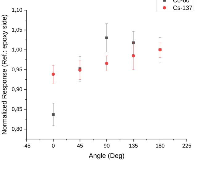
Angular dependence of the MOSFET dosimeter was evaluated by means of reproducibility measu-rements for each of the five angles in each of the radioactive sources. Average voltages were nor-malized to an angle corresponding to epoxy side (180°) as this orientation showed a greater respon-se than Kapton side (0°).
3. RESULTS AND DISCUSSIONS
Readings of TN-502RD-H MOSFET dosimeter with respect to reproducibility are shown in Tables 1 and 2 for five different angles. MOSFET dosimeter presented a reproducibility of 3.3% and 2.7% (± 1 SD) for Cs-137 and Co-60 sources, respectively, being satisfactory at all angles for both radio-active elements.
Table 1: Reproducibility of TN-502RD-H MOSFET dosimeter for Cs-137 source
Irradiation No. Angle response (mV) 0° 45° 90° 135° 180° 1 97 97 100 98 104 2 95 100 101 101 101 3 99 96 98 105 104 Mean (mV) 96.67 (± 1.9) 97.73 (± 2.0) 99.46 (± 1.4) 101.45 (± 3.3) 103.01 (± 1.4)
Table 2: Reproducibility of TN-502RD-H MOSFET dosimeter for Co-60 source Angle response (mV) Irradiation No. 0° 45° 90° 135° 180° 1 60 65 69 69 69 2 55 65 70 70 69 3 56 65 70 67 66 4 56 66 68 67 68 1 56 63 72 70 65 2 58 63 69 67 69 3 56 64 70 70 65 4 57 63 71 69 69 1 56 61 65 68 67 2 54 66 72 67 67 3 55 62 69 68 67 4 57 66 67 68 67 Mean (mV) 56.31 (± 1.5) 64.07 (± 1.6) 69.31 (± 1.9) 68.48 (± 1.3) 67.29 (± 1.5)
Figure 2 show the experimental data concerning angular dependence of TN-502RD-H MOSFET detector. Responses were normalized to 180° angle, corresponding to epoxy side with face facing the source. An angular dependence of up to 6.1% and 16.3% was found for sources of Cs-137 and Co-60, respectively.
It is also observed that sources of Cs-137 and Co-60, with mean energies of 0.66 and 1.25 MeV, respectively, presented values of angular dependence with distinct intensities, being more visible to the beam of higher Co-60 energy. This may raise the hypothesis that angular dependence of the MOSFET dosimeter also depends on the incident beam energy, results also found in literature [13]. However, it is not possible to state this categorically, since experiments were carried out on only two different radiation beam qualities to verify angular dependency so far.
Figure 2: Normalized angular dependence of TN-502RD-H MOSFET dosimeter for Cs-137 and Co-60 sources -45 0 45 90 135 180 225 0,80 0,85 0,90 0,95 1,00 1,05 1,10 Co-60 Cs-137
Norm
alized
Resp
on
se (
Ref.:
ep
oxy sid
e)
Angle (Deg)
It is also known that the manufacturer provides the sale of a build-up hood, promising to obtain electronic balance and relative anisotropy to the detector. According to the manufacturer's manual, a maximum angular dependence of up to 2% is guaranteed if this build-up layer is used [14]. Howev-er, because you did not have this accessory, it was not possible to verify that this information is cor-rect.
Scalchi et al. states that although angular dependence is not a problem for skin dosimetry in TBI treatments, an as isotropic detector as possible is essential for in vivo dose measurements in skin
since some types of radiotherapy treatments, such as for those used in breast and chest wall treat-ments, can only be performed with anisotropic detectors. This is also the case, for example, of treatments in anatomical regions with large angles involved [15].
4. CONCLUSION
It was possible to verify that the detector has a good reproducibility of measurement for both Cs-137 and Co-60 sources. Moreover, it was found to exist angular dependence of dosimeter in both energies, being much greater for source of Co-60, that emits higher energies photons, demonstrating that much attention is needed for applications in certain configurations and anatomical regions in patient in radiotherapy treatment. All this information agrees with the literature and motivates the research group to continue seeking to contribute to the understanding of this device. As a next step, new experiments should be performed for more energies in order to analyze how the response of this detector varies with energy of incident photon.
5. ACKNOWLEDGMENT
The authors would like to thank Dr. Ademir Xavier da Silva from Federal University of Rio de Janeiro (UFRJ) for the loan of the MOSFET dosimetry system, accompanying reading module, ca-bles, and all necessary set so that we could carry out experimental measurements with our MOSFET dosimeters. The authors are also grateful for the financial support of the Coordination for the Im-provement of Higher Education Personnel (CAPES) and the National Council for Scientific and Technological Development (CNPq).
REFERENCES
1. WHO – World Health Organization. World Health Organization: Who is cancer? Avai-lable at: <http://www.who.int/cancer/en/>. Last accessed: 01 Dec. 2017.
2. INCA – Instituto Nacional do Câncer. Instituto Nacional do Câncer: Tratamento do
Câncer. Available at <http://www2.inca.gov.br/wps/wcm/connect/cancer/site/tratamento> Last
accessed: 01 Dec. 2017.
3. INCA – Instituto Nacional do Câncer. Instituto Nacional do Câncer: Radioterapia. Avai-lable at <http://www.inca.gov.br/conteudo_view.asp?ID=100> Last accessed: 01 Sept. 2017.
4. SCALCHI, P., FRANCESCON, P., Calibration of a MOSFET detection system for 6-MV in-vivo dosimetry. Int. J. Radiation Oncology Biol. Phys., v. 40, p. 987-993, 1998.
5. GOPIRAJ, A.; BILLIMAGGA, R. S.; RAMASUBRAMANIAN, V. Performance characte-ristics and commissioning of MOSFET as an in-vivo dosimeter for high energy photon external beam radiation therapy. Reports of Practical Oncology & Radiotherapy, v. 13, p. 114-125, 2008. 6. BOWER, M. W. Physical anthropomorphic phantom of a one-year-old child with
real-time dosimetry, Gainesville, Florida, United States of America, 1997.
7. RAMANI, R.; RUSSEL, S.; O’BRIEN, P. Clinical dosimetry using MOSFETs, Int. J.
Ra-diation Oncology Biol. Phys., v 37, p.959-964, 1997.
8. RAMASESHAN, R.; KOHLI, K. S.; ZHANG, T. J.; LAM, T.; NORLINGER, B.; HALLIL, A.; ISLAM, M. Performance characteristics of a microMOSFET as an in vivo dosimeter in radia-tion therapy, Phys. Med. Biol., v 49, p. 4031-4048, 2004.
9. SHARP, R.E.; PATER, S.L. The use of pMOS dosimeters at megagray total doses, AEA
Technol, RADECS, 1995.
10. SOUBRA, M.; CYGLER, J.; MACKAY, G. Evaluation of dual bias dual metal oxide-silicon semiconductor field effect transistor detector as radiation dosimeter, Med. Phys., v 21, p. 567-572, 1994.
11. BEST MEDICAL CANADA. Best Medical Canada: MOSFET Dosimeters. Available at: <http://www.bestmedicalcanada.com/product_dosimeters.html> Last accessed: 01 Dec. 2017. 12. SILVA JR, I.A. Desenvolvimento e Implementação de um Sistema Automatizado para
Adequação do Processo de Calibração de Monitores de Radiação Gama, Universidade de São
Paulo, São Paulo, Brazil, 2012.
13. KUMAR, A. S. et al. Characteristics of mobile MOSFET dosimetry system for megavoltage photon beams. J. Med Phys. v. 39, p. 142-149, 2014.
14. BEST MEDICAL CANADA. Operator’s manual for Mobile MOSFET System. Ottawa, Canada, 2008.
15. SCALCHI, P. et al. Characterization of a new MOSFET detector configuration for in vivo skin dosimetry. Medical Physics, v. 32, p. 1571-1578, 2005.