UNIVERSIDADE DA BEIRA INTERIOR
Ciências
Development of intelligent vehicles for
co-delivery of anti-tumoral drugs to breast cancer
cells
João Filipe Gonçalves Marques
Dissertação para a obtenção de grau de Mestre em
Bioquímica
Orientador: Professor Doutor Ilídio Joaquim Sobreira Correia
Coorientador: Mestre Vítor Manuel Abreu Gaspar
ii
UNIVERSIDADE DA BEIRA INTERIOR
Ciências
Desenvolvimento de veículos inteligentes para
co-entrega de drogas anti-tumorais
nas células do cancro da mama
João Filipe Gonçalves Marques
Dissertação para a obtenção de grau de Mestre em
Bioquímica
Orientador: Professor Doutor Ilídio Joaquim Sobreira Correia
Coorientador: Mestre Vítor Manuel Abreu Gaspar
iv
“Cum mente et malleo”
Claude-Henri Gorceix
vi
Aknowlegments
First, I would like to thank to Professor Ilídio Correia for the possibility of developing my master thesis in his group, making every day easier because of its ability to join the group, resulting in an optimum work environment.
I thank to Master Vítor Gaspar, my co-supervisor, for his relentless support, guidance and advices, unlimited knowledge, and for being a friend. The constant discussion of ideas and issues made me grow up, and without him I would never be able to develop this work.
I would like to thank Master David Märkl and Professor Eugénia Gallardo, for providing UPLC column as well as for all the support in my work, without their help I would not have achieved my aims.
Moreover, I would like to thank to Elisabete Costa, for the endless support, help, and mainly for her friendship, being my partner every day and night, during this year.
I thank all my family, specially my father, mother, sister and brother-in-law for their unlimited support, patience, love, support, affection and most of all, for never stop believing in me.
Finally, and foremost, a very special thanks to my girlfriend, Agustina, for all her love, advices and support during my thesis development. An unending acknowledge for her patience in my busy lab days and her limitless affection and care.
viii
Abstract
Presently breast cancer arises as one of the most prevalent malignancy in women worldwide, contributing for high rates of mortality and morbidity in several million patients. Currently there is still no available cure for this disease and to further aggravate this scenario the existing treatments such as chemotherapy, are generally ineffective due to poor tumor bioavailability. Moreover, the commonly used anti-tumoral compounds also lead to the development of drug resistant malignant cells after repeated administration, a fact that originates the development of more aggressive cells that possess the capacity to metastasize and spread to healthy organs. These facts evidence the urgent need for the development of novel therapeutic approaches that improve the therapeutic outcome and patient survival. The recent developments in the field of Nanotechnology, particularly regarding the capacity to manipulate matter at the nanoscale, has brought forth the opportunity to devise novel drug delivery systems to tackle some of these issues. From this stand point, the research work presented in this thesis describes the development of a novel drug delivery system based on micellar carriers, with a core-shell structure, that are capable to simultaneously deliver multiple drugs to breast cancer cells. These nanocarriers are comprised by a hydrophilic and a hydrophobic polymer organized in a block-by-block structure that was synthesized through macromolecular chemistry. The manipulation of the various reaction conditions yielded block co-polymers with different hydrophobic chains, which influenced the available space in the nanocarrier core. The nanocarriers were formulated by co-polymer self-assembly into nanosized micelles that demonstrated the capacity to encapsulate with high efficiency, an anti-tumoral drug, Crizotinib and a potent inhibitor of the cell transporters responsible for drug resistance, Sildenafil. The drug release profile of the micellar carriers revealed a spatiotemporally controlled release that was faster for Sildenafil than Crizotinib. Moreover, the drug loaded micelles demonstrated to be highly biocompatible and accomplished uptake into adherent breast adenocarcinoma cells. This relevant finding let to the intracellular localization of both the anti-tumoral drug and the drug resistance inhibitor, and thus, improved the bioavailability of the bioactive therapeutics. Subsequently, the study of the therapeutic performance of the co-delivery systems illustrated that the simultaneous delivery of both drugs improved the anti-tumoral capacity of Crizotinib evidencing the existence of a highly synergistic effect. Strikingly, the micellar systems achieved the same anti-tumoral effect of the free drugs, using 2-fold less drug concentration. Besides indicating that the release profile maintains drug concentrations in the therapeutic window, these crucial results highlight the effect of dual drug conjugation and the use of Crizotinib as an anti-tumoral compound for breast cancer therapy and Sildenafil as a multidrug resistance inhibitor. Overall, the unique approach developed in this thesis possesses tremendous potential for a future clinical application in breast cancer patients that acquired resistance to standard therapies.
ix
Keywords:
xi
Resumo alargado
Atualmente, o cancro de mama surge como uma das neoplasias com maior prevalência no sexo feminino em todo o mundo, contribuindo para altas taxas de mortalidade e morbilidade em vários milhões de pacientes. Atualmente, ainda não se encontra disponível uma cura para esta doença e para agravar ainda mais este cenário, os tratamentos existentes geralmente baseados na quimioterapia, são extremamente ineficazes devido à baixa biodisponibilidade do fármaco no local do tumor. Além destas limitações, estes tratamentos também provocam graves efeitos secundários que afetam órgãos vitais como o fígado, coração ou rins. Este facto contribui para uma progressão desta neoplasia de forma mais acelerada e é responsável por um prognóstico bastante limitado em termos do tempo de vida das pacientes.
Não obstante, devido à necessidade de manter a concentração terapêutica dos fármacos no organismo são efetuadas múltiplas administrações em regimes que usualmente se prolongam durante meses, para promover um aumento da probabilidade de localização dos fármacos nas células alvo. Esta abordagem terapêutica é crucial para a redução da massa tumoral, no entanto, foi recentemente descoberto que este tratamento origina a formação de células tumorais mamárias com mutações que lhes permitem adquirir um fenótipo muito mais agressivo e resistente ao fármaco anti-tumoral administrado. Esta resistência é muitas vezes atribuída ao aumento da expressão de proteínas membranares que têm a capacidade de expelir os ingredientes farmacêuticos ativos para o meio extracelular, eliminando-os assim do seu local alvo. Além do desenvolvimento de resistência, é despoletada também nas células tumorais a capacidade de metastização, permitindo o alastrar da doença para os órgãos saudáveis. Estes factos evidenciam a necessidade urgente do desenvolvimento de novas abordagens terapêuticas com vista a melhorar o prognóstico clínico e a qualidade de vida do paciente.
Neste contexto, os desenvolvimentos recentes no campo da Nanotecnologia, particularmente em relação à capacidade de manipular a matéria à escala nanométrica, têm trazido a oportunidade de conceber novos sistemas de entrega de drogas para resolver alguns destes problemas. Diversos nanoveículos, com base polimérica e/ou lipídica, têm sido desenvolvidos durante as últimas décadas, tendo sido alguns deles já aprovados e usados no tratamento de doentes em unidades hospitalares. No entanto, apesar dos grandes avanços obtidos no design e produção de nano-veículos para aplicação na terapia do cancro da mama num âmbito clínico, a sua maioria apenas consegue melhorar a biodisponibilidade e biodistribuição do fármaco anti-tumoral nas células alvo. De facto, até à data, apesar da panóplia de nanotransportadores disponívels para terapia do cancro da mama muito poucos exploram a entrega de vários fármacos ou moléculas bioativas em simultâneo, como forma de combater os problemas associados à multirresistência adquirida pelas células neoplásicas.
Com o conhecimento destas limitações atuais, e tendo como objetivo principal melhorar cada vez mais a terapia contra o cancro da mama, o trabalho de investigação apresentado nesta
xii
tese descreve o desenvolvimento de um novo sistema de entrega de drogas baseado em nano-veículos micelares, com uma estrutura "núcleo-concha” que lhes permite transportar e entregar simultaneamente múltiplas drogas a células de cancro da mama. Estas micelas, são formadas por dois polímeros rearranjados numa estrutura em bloco. Cada um dos blocos é constituído por um polímero hidrofílico e outro polímero hidrofóbico, um facto que imprime características anfifilicas aos nanotransportadores produzidos com estes biomateriais. Esta estrutura única foi sintetizada por intermédio de reações químicas macromoleculares. A manipulação das diversas condições da reação originou a síntese de co-polímeros com diferentes cadeias hidrofóbicas, um factor crucial visto que este influencia o espaço disponível no núcleo da micela, assim como a sua estabilidade e capacidade de encapsulação de moléculas bioativas. Os materiais sintetizados foram caracterizados física e químicamente por intermédio de técnicas referidas nas diretivas internacionais como adequadas para caraterizar nanomateriais para aplicações biomédicas. As micelas foram posteriormente formuladas pelos co-polímeros sintetizados recorrendo a um método de formação espontânea que origina micelas nanoméricas com uma morfologia esférica, como revelado por microscopia eletrónica de varrimento.
O Crizotinib, um conhecido fármaco anti-tumoral neste momento em ensaios clínicos de fase IV e o Sildenafil, um potente inibidor dos transportadores celulares responsáveis pela resistência aos fármacos, foram escolhidos posteriormente para serem incluídos dentro das nano-micelas poliméricas. Após a otimização do processo de encapsulação foi obtida uma elevada eficácia de inclusão destes dois fármacos no interior dos transportadores micelares. O perfil de liberação destes fármacos dos transportadores micelares revelou ser controlado, sendo mais rápido para o Sildenafil do que para o Crizotinib. Além disso, as micelas produzidas demonstraram ser altamente biocompatíveis quando administradas a células humanas saudáveis. A sua internalização nas células cancerígenas da mama revelou ser muito elevada, um resultado extremamente vital pois, evidencia, a entrega intracelular de ambos os fármacos, o anti-tumoral e o inibidor da resistência celular, levando a um aumento da sua biodisponibilidade.
Consequentemente, o estudo do desempenho terapêutico destes nanotransportadores demonstrou que a co-entrega de ambos os fármacos potenciou a atividade anti-tumoral do Crizotinib, evidenciando a obtenção de um efeito sinérgico. Surpreendentemente, os sistemas micelares obtiveram um efeito anti-tumoral similar ao obtido com os fármacos na sua formulação farmacêutica livre, administrando no entanto concentrações de fármaco duas vezes menores. Estes resultados ilustram a importância do sistema desenvolvido na terapia do cancro da mama. Além deste facto, uma vez que o perfil de libertação mantém as concentrações dos fármacos dentro da janela terapêutica, estes resultados realçam o efeito da conjugação das duas drogas e a utilização do Crizotinib como um composto anti-tumoral para a terapia do cancro da mama.
xiii
Em geral, a abordagem exclusiva desenvolvida nesta tese demonstra um enorme potencial para aplicação futura em pacientes com cancro da mama, que apresentem resistência às terapias convencionais.
Palavras-chave
xiv
List of Publications
Articles in peer reviewed international journals:
Costa, C.E., Gaspar. V.M., Marques, J.G., Coutinho, P., Correia, I.J. (2013). Evaluation of Nanoparticle Uptake in Co-culture Cancer Models, PloS one. Article in press.
Gaspar V. M., Marques J. G., Sousa F., Louro R. O., Queiroz J. A. and Correia I. J. (2013). Biofunctionalized nanoparticles with pH-responsive and cell penetrating blocks for gene
Delivery, Nanotechnology (24) 275101.
Poster communications:
Gaspar, V.M., Marques, J.M., Sousa, F., Louro, R.O., Queiroz, J.A., Correia, I.J., Characterization of Chitosan Nanoparticles Uptake and Intracellular Trafficking in Cancer Cells, 11th International Conference of the European Chitin Society, 5-8 May 2013, Porto, Portugal.
Elisabete C. Costa, Vítor M. Gaspar, João F.G. Marques,Paula Coutinho, Ilídio J. Correia, Mimicking Breast Cancer Microenvironment with In Vitro Co-culture Models, Instituto Politécnico da Guarda (IPG), 3rd of May 2013, Guarda, Portugal.
Poster awards:
Costa, C. E., Gaspar. V. M., Marques, J.G., Coutinho, P., Correia, I. J., Mimicking Breast Cancer Microenvironment with In Vitro Co-culture Models, Instituto Politécnico da Guarda (IPG), 3rd of May 2013, Guarda, Portugal.
Oral Presentations:
Gaspar, V.M., Marques, J.M., Sousa, F., Louro, R.O., Queiroz, J.A., Correia, I.J., Characterization of Nanomedicines Uptake and Intracellular Trafficking in Cancer Cells, VI Symposium on Technology and Health, Instituto Politécnico da Guarda (IPG), 3rd of May 2013, Guarda, Portugal.
Index
Figure Index xix
Table Index xxi
List of abbreviations xxii
Chapter 1 25
Introduction 25
1. Cancer 26
1.1. Malignant Phenotype and Cancer Cell Hallmarks 26
1.2. Breast Cancer and its heterogeneity 28
1.3. Multidrug Resistant Cancer Cells 29
1.4. Anti-tumoral drugs used for breast cancer therapy 30
1.5. Exploring novel combinatorial drug therapies to overcome MDR and improve
anti-cancer therapy 30
2. Nanotechnology-based carriers for cancer drug delivery 33
2.1. Passive Targeting – The Enhanced permeability and Retention Effect 36 3. Organic and Inorganic biomaterials used for self-assembly drug delivery systems 37
3.1. Hydrophobic polymers used for the production of self-assembled DDS micelles
for cancer therapy 41
3.2. Hydrophilic polymers used for the production of self-assembled DDS micelles
for cancer therapy 42
Aims 44
Chapter 2 45
Materials and Methods 45
2. Materials and Methods 46
2.1. Materials 46
2.2. Methods 47
2.2.1. Synthesis of mPEG-PLA 47
2.2.2. Nuclear Magnetic Resonance 47
2.2.3. Fourier Transform Infrared Spectroscopy 48
2.2.4. Gel Permeation Chromatography 48
2.2.5. X-ray Powder Diffraction 49
2.2.6. Differential Scanning Calorimetry 49
2.2.7. Micelle self-assembly - Film Hydration Method 49
2.2.8. Determination of the Critical Micellar Concentration 50
2.2.9. Haemolysis Assay 50
2.2.10. Encapsulation Efficiency and Release Profile samples analysis. 51
2.2.11. Morphological Characterization of PEG-PLA micelles 52
2.2.12. Characterization of PEG-PLA Micelle Size and Zeta Potential 52
2.2.13. Release profile of encapsulated drugs 52
xvii
2.2.15. Flow Cytometry 53
2.2.16. Micelle uptake by Breast Cancer cells 54
2.2.17. IC50 determination and Synergic effect evaluation 54
2.2.18. Drug Loaded Nanoparticle Incubation 55
2.2.19. Apoptosis assay 55
2.2.20. Statistical Analysis 55
Chapter 3 56
Results and Discussion 56
3. Results and Discussion 57
3.1. Synthesis of PEG-PLA block co-polymers 57
3.2. NMR analysis of PEG-PLA block co-polymers 59
3.3. FTIR analysis of PEG-PLA co-polymers 62
3.4. GPC analysis of block co-polymer 63
3.5. XRD analysis of PEG-PLA co-polymers 64
3.6. DSC characterization 65
3.7. CMC Determination 67
3.8. Haemocompatibility Assay 68
3.9. Analysis of Single and Multiple Drug Loading into Micellar Carriers 70
3.10. Morphological Characterization of Nano-sized PEG-PLA micelles 73
3.11. Micelle physicochemical characterization – Size and Surface Charge 74
3.12. Evaluation of Drug Release Profile 76
3.13. PEG-PLA co-polymers biocompatibility 77
3.14. Characterization of Micelle Cellular Uptake 79
3.14.1. Flow Cytometry analysis 79
3.14.2. Determination of the Inhibitory concentration of free Crizotinib in Breast
Cancer Cells 81
3.15. Evaluation of the anti-tumoral effect of combinational drugs 82
3.15.1. Free Crizotinib and Sildenafil 82
3.15.2. Single and Multiple Drug-loaded Micelles 83
3.16. Breast cancer cell apoptosis 84
Conclusion and Future Perspectives 86
References 87
xix
Figure Index
Figure 1 – Schematic representation of the tumorigenesis process. 6
Figure 2 - Cancer hallmarks and their possible therapeutic pathways to overcome cancer. 7
Figure 3 – Molecular structure of Crizotinib (PF-2341066). 31
Figure 4 – Molecular structure of Sildenafil (PF-4540124). 32
Figure 5 – Proposed mechanism of action for Sildenafil synergic activity in conjugation with other chemotherapy drugs by blocking drug efflux from MDR transporters (ABC’s) and
increasing intracellular cyclic guanosine monophosphate (cGMP) levels. 3
Figure 6 – Evolution of drug delivery systems along time. 4
Figure 7 – Enhancement of drug concentration and bioavailability through nanoparticle
conjugation. 5
Figure 8 – Presentation of nanoparticle EPR effect and enhanced intracellular drug
concentration due to efflux pump inhibition. 36
Figure 9 – Representation of the drug-delivery systems currently available. 8 Figure 10 – Polymeric micelle structure composed with block co-polymers that self-assemble
in water solutions. 41
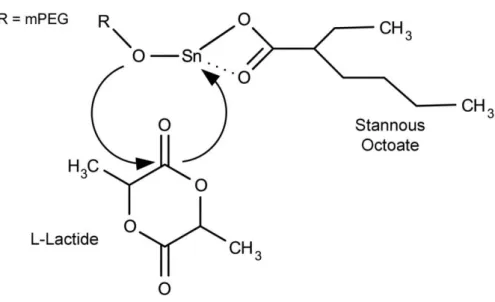
Figure 11 - Mechanism of polymerization of L-lactide. mPEG acts as macroinitiator and
Sn(Oct)2 act as a catalyst. 43
Figure 12 – COI mechanism of ROP of L-LA polymerization using mPEG as macroinitiator and
Sn(Oct)2 as catalyst. 8
Figure 13 – NMR of the synthesized mPEG and L-Lactide raw materials used in the
polymerization process. 60
Figure 14 – NMR of the synthesized PEG-PLA block co-polymers with different polymerization
times, (PL4, PL8 and PL30). 61
Figure 15 – FTIR spectra of the native materials (L-Lactide and PEG) and of the synthesized
block co-polymers (PL4, PL8 and PL30). 63
Figure 16 – Chromatograms of PEG standards (PEG 2000, PEG 4000 and PEG 8000) and of the
synthesized materials (PL4, PL8 and PL30). 63
Figure 17 – X-ray diffraction spectra of native materials (L-Lactide and mPEG) and of the
synthesized block co-polymers (PL4, PL8 and PL30). 65
Figure 18 – DSC analysis of mPEG, L-Lactide, PL4, PL8 and PL30. 66
xx
Figure 20 – Supernatants resulting from the haemolysis assay (A). On the bottom of the image two representative SEM images of RBC previously incubated with nanoparticles: (B) PL8 and
(C) PL30. 69
Figure 21 – Quantification of free heme groups (indicator of haemolysis) after incubation with
synthesized micelles (PL4, PL8 and PL30). 69
Figure 22 – Representative chromatograms of the separation of Crizotinib and Sildenafil analytes and the Internal Standard (Protriptiline) with two different mobile phases:
CH3CO2NH4 (A) and Na2HPO4 (B). 71
Figure 23 – Encapsulation efficiency of Crizotinib and Crizotinib/Sildenafil combination in PL4,
PL8 and PL30 nanoparticles. 72
Figure 24 – SEM micrographs of: PL8 micelles with ecapsulated Crizotinib (A) and Crizotinib+Sildenafil (B) respectively; and PL30 micelles with loaded Crizotinib (C) and
Crizotinib+Sildenafil (D) respectively. 73
Figure 25 – Size, PDI and zeta potential of PL micelles loaded with drugs. (A and B) PL8 loaded with Crizotinib and loaded with Crizotinib+Sildenafil, respectively; (C and D) PL30 nanoparticles loaded with Crizotinib and with Crizotinib+Sildenafil, respectively. 75 Figure 26 - Release profile of PL8 formulations at physiologic pH (pH = 7.4) (A) and at the characteristic acidic tumor pH (pH = 6.5) (B). PL8C/CS represents Crizotinib and Sildenafil
loaded PL8 micelles. 77
Figure 27 – Characterization of PL8 micelles cytotoxicity using: MCF-7 cells (A) and Fib-H (B). 78 Figure 28 – Overlaid cytometry histograms of MCF-7 cells incubated during 2 and 4 h with
RITC-micelles. 79
Figure 29 – CLSM images of MCF-7 cell with internalized PEG-PLA micelles encapsulating RITC. 80 Figure 30 – IC 50 determination of Crizotinib anti-tumoral activity in MCF-7 breast cancer cells. Blue curve represents the mathematical fitting performed for IC50 calculation. 81 Figure 31 – Evaluation of anti-tumoral activity of Crizotinib (alone) and Sildenafil (alone) and
when Sildenafil was compbined with Crizotinib after 48 h incubation. 82
Figure 32 – Evaluation of the anti-tumoral activity of Crizotinib, Sildenafil and synergic effect
between both when delivery through PEG-PLA micelles. 83
Figure 33 - CLSM images of apoptotic MCF-7 cells that were incubated 24 h with PL8CS
xxi
Table Index
Table 1 – Comparison of micelles approved or enrolled in clinical trials by FDA. 40 Table 2 - Degree of polymerization, Mn of PLA and mPEG-PLA calculated by proton peak
integration. 62
Table 3 – Calculated percentage of crystallinity of PL4, PL8 and PL30. 65
Table 4 - DSC data analysis of L-LA, mPEG, PL4, PL8 and PL30. 67
Table 5 – Calibration standards with injection on mobile phase, coefficient of variation (CV)
and bias. 97
Table 6 – Calibration standards with injection on PBS, coefficient of variation (CV) and bias. 97 Table 7 – Calibration standards with injection on water, coefficient of variation (CV)
and bias. 98
xxii
List of abbreviations
ΔH Heat Enthalpy
ABC ATP-Binding Cassettes
AKT Protein Kinase B
ANOVA One-way Aanalysis of Variance
BCRP Breast Cancer Resisting Protein
bFGF Basic Fibroblast Growth Factor
CDCl3 Deuterated Chloroform
cGMP Cyclic Guanosine Monophosphate
CLSM Confocal Laser Scanning Microscopy
CMC Critical Micellar Concentration
DDS Drug Delivery Systems
DIC Differential Interference Contrast
DLS Dynamic Light Scattering
DMEM Dulbecco’s Modified Eagle’s Medium
DNA Deoxyribonucleic Acid
DSC Differential Scanning Calorimetry
EDTA Ethylenediamine Tetraacetic Acid
EMA European Medicines Agency
EO Poly(ethylene glycol) Monomer
EPR Enhanced Permeability and Retention
ER Oestrogen Receptor
FBS Fetal Bovine Serum
FDA Food and Drug Administration
FPLC Fast Protein Liquid Chromatography
FTIR Fourier Transform Infrared Spectroscopy
GPC Gel Permeation Chromatography
HER2 Human Epidermal Growth Factor Receptor 2
hFIB Human Fibroblasts
HGF Hepatocyte Growth Factor
HPLC High Performance Liquid Chromatography
IC50 Half Maximum Inhibitory Concentration
LA Lactic Acid
MCF-7 Michigan Cancer Foundation-7
MDR Multi-Drug Resistance
MetOH Methanol
Mn Number Average Molecular Weight
MPS Mononuclear Phagocytic System
MRT Mean Residence Time
MTD Maximum Tolerated Dose
MTS 3-(4,5-dimethylthiazol-2-yl)-5-(3-carboxymethoxyphenyl)-2-(4-sulfophenyl)-2H-tetrazolium)
Mw Molecular Weight
NA Not Applicable
NMR Nuclear Magnetic Resonance
PBS Phosphate Buffer Saline
xxiii
PDE5 Phosphodiesterase 5
PDI Polydispersity Index
PEG Poly(ethylene glycol)
PEG-PCL Poly(ethylene glycol)-block-Poly(ε−caprolactone) PEG-PLA Poly(ethylene glycol)-Poly(lactic acid)
PEOz Poly(oxazoline)
PFS Patient Free Survival
PHEMA Poly (2-hydroxyethyl methacrylate)
PL Synthesized Poly(ethylene glycol)-Poly(lactic acid)
PLA Poly (lactic acid)
PTA Phosphotungstic Acid
PLGA Poly (D,L-lactide-co-glycolide)
P-gp P-glycoprotein
RBC Red Blood Cell
RITC Rhodamine B Isothiocyanate
ROI Region of Interest
ROP Ring Opening Polymerization
RT Room Temperature
SEM Scanning Electron Microscopy
siRNA Small Interfering RNA
Sn(Oct)2 Stannous Octoate
TEA Triethylamine
Tm Melting Temperature
UPLC Ultra Performance Liquid Chromatography
USA United Stated of America
VEGF Vascular Endothelial Growth Factor
25
Chapter 1
Introduction
26
1. Cancer
1.1. Malignant Phenotype and Cancer Cell Hallmarks
Cancer is a disease that is mainly characterized by an uncontrolled cell proliferation that ultimately leads to the formation of a tumor mass. Generally, healthy cells accurately control their growth by carefully delivering signalling agents to nearby cells and guaranteeing tissue homeostasis [1]. When homeostasis is affected, for example, by the accumulation of genetic mutations, cells begin to acquire a malignant phenotype (Figure 1) [2]. Age, growth signal molecules, hormones, genetic background, ionizing radiation, pollution and unhealthy working environments are the major risk factors for the development of cancer [3, 4]. These factors normally induce genetic mutations in normal cells, that often result in metabolically and physicochemical dysfunctional cells [5]. These genetic abnormalities present in tumor cells are generally correlated with the activation oncogenes (c-MET) and silencing of tumor suppressor genes (p53) [2]. This fact results in the deregulation of major signalling pathways that promote cell survival and proliferation [1]. Usually, cancer cells overexpress proteins responsible for cell cycle progression and underexpress proteins involved in growth suppression [1].
27
After acquiring the malignant phenotype, cancer cells commonly exhibit major characteristics, the so termed “Hallmarks of cancer” (Figure 2). This set of unique features ultimately differentiates malignant cells from their healthy counterparts [1].
Figure 2 - Cancer hallmarks and their possible therapeutic pathways to overcome cancer. A multi-combinational drug therapy could be an interesting approach to fight some of hallmarks described. (Adapted from [1]).
One of the most relevant hallmarks that tumor cells possess is their capacity to sustain proliferative signalling. To successfully promote sustained proliferation, cancer cells need to bypass growth suppressor supervision mechanisms, that are commonly mediated by p53 and retinoblastoma-associated proteins (Rb) [1]. Remarkably, it is currently known that approximately 50% of all cancer cells have a mutation in the p53 gene, a fact that completely eliminates its pro-apoptotic activity [1, 7]. Yet another strategy that cancer cells acquire in order to resist to cell death is the increase of expression of anti-apoptotic proteins from the Bcl family, such as Bcl-2 and Bcl-xL [1]. Moreover, the immortalized (i.e., limitless replicative potential of cancer cells), is also a hallmark that greatly impairs patient survival rates [1]. To achieve this unique characteristic cancer cells recruit telomere stabilizing proteins, such as reverse transcriptase telomerase, that maintain telomere size across all the cell division cycles [8]. Telomeres are the main actors in the unlimited proliferation and cell immortality process. They regulate the cell replication capacity by shortening at each division till restrict completely cell division, a fact that hence does not occur in most cancer cells [9]. Another important hallmark of cancer cells is their highly activated metabolic rates that constantly
28
need an uptake of nutrients in order to remain metabolically overactive [1]. To sustain the need for large amounts of nutrients, tumor cells trigger the development of new blood vessels from nearby vasculature by constantly expressing angiogenesis inducers, like vascular endothelial growth factor (VEGF) and basic fibroblast growth factor (bFGF) [10-13]. This relentless expression of VEGF induces the formation of aberrant vasculature characterized by a leaky profile due to the existence of large fenestrations (600-800nm) in the endothelial walls [14, 15]. The high abundance of rich vascular networks together with the lack of expression of cell adhesion molecules such as E-cadherin, leads to cell extravasation. These cells consequently invade and colonize other organs and tissues throughout the body, a process known as metastization [16]. These unique characteristics are common to all types of cancer and contribute for a poor patient’s clinical prognostic and disease progression.
1.2. Breast Cancer and its heterogeneity
Focusing particularly on breast cancer, this is actually the most diagnosed malignancy in women, in what concerns to solid tumors [17, 18]. This disease affected about 1.5 million of women worldwide in 2010 [17, 18] and 464.000 new cases have arisen in 2012, in the European Union, representing the third most deadliest cause among women [19]. When this disease is diagnosed at a very early stage of development, it has an excellent long-term prognosis in terms of patient free survival (PFS) rate [20]. However, in late stages of disease (stage III and IV) that often involve metastization, especially to adjacent lymph nodes, the survival rate decreases dramatically [20]. In this scenario even after mastectomy, 40% of women still develop metastasis in auxiliary lymph nodes [20]. Surprisingly, this type of tumor represents a particularly heterogeneous group with defined biological features and responses to therapy [21]. Actually, nowadays breast cancers are divided accordingly to their gene-expression subtypes into: i.) luminal (related with estrogen and progesterone receptors) ii.) human epidermal growth factor receptor 2 (HER2) and iii.) basal-like, which typically do not express HER2hormone receptors, having generally a triple negative phenotype [21-23]. HER2 are characteristic of different breast cancer subtypes. Cancers that do not have ERBB2, progesterone (PR) or estrogen (ER) receptors, are called triple negative breast cancer cells [24]. These type of cells present a constitutively enhanced Protein Kinase B (AKT) activity due to a mutation in MAGI3–AKT3 gene [21]. Currently available chemotherapies for cancer treatment are still largely limited by deleterious side effects that contribute for a limited therapeutic outcome. These side-effects are often associated with the lack of tumor cell specificity, with the consequent drug partitioning in other tissues and organs, such as the liver and lungs [20]. Such bottlenecks significantly reduce the bioavailability of chemotherapeutics at the target tumor site and are responsible for the administration of anti-tumoral drugs to patients for an extended period of time, at lower dosages (commonly between each 21 days across 6 to 8 sessions of chemotherapy [25]. Moreover, the rapid
29
systemic clearance of drugs and the difficult vascular access to lymph nodes of patients with breast cancer, contribute for the ineffectiveness of the treatments [26]. This therapeutic regime is also commonly associated with the development of drug resistant breast cancer cells.
1.3. Multidrug Resistant Cancer Cells
One of the main causes of chemotherapy failure in the improvement of PFS is the acquired drug resistance of cancer cells, after a few rounds of chemotherapy, a phenomenon that is termed multi-drug resistance (MDR) [27, 28]. MDR can result from non-cellular and cellular based mechanisms [28-30]. Furthermore, barrier mechanisms, that are not directly related to cells, such as poorly vascularized tumor regions that are protected against chemotherapy drugs also contribute to the MDR phenotype [29-31]. High interstitial pressure and the lack of microvasculature are additional barriers that affect drug extravasation into tumor tissues [32]. To overcome these issues the administration multiple chemotherapy drugs, can arise as a very promising approach [32]. In fact, since the acquired MDR by cancer cells is related to an over-expression of enzymes with specific drug metabolization or efflux capacity, the additional administration of chemical compounds that can shut-down their activity can contribute to MDR reversal [28-30]. Among the different resistance mechanisms, drug efflux pumps are currently the most extensively studied [30]. The resistance mediated by these efflux pumps can be attributed to an overwhelming decrease in drug uptake, since drug efflux to the extracellular medium is constantly promoted by P-glycoprotein (P-gp) and other ATP-binding cassettes (ABC) proteins that are over-expressed in these type of cells [30]. ABC’s are a particularly relevant superfamily of transmenbrane proteins that have the capacity to transport various kinds of substrates, such as hydrophobic drugs (Doxorubicin, Cisplatin, Paclitaxel) [30, 33]. Some types of cancer cells over-express particular types of ABC’s, such as ABCB1 (P-gp), ABCC1, and ABCG2 (also termed Breast Cancer Resisting Protein, BCRP), that are involved in the regulation of the intra cellular concentration of chemotherapy agents [27, 34]. It is described that these ABC transporters are highly expressed in inner regions of the tumors, due to the existence of hypoxic microenvironments [35]. In addition, MDR can also result from coordinated detoxification processes mediated by cytochrome P450 and deoxyribonucleic acid (DNA) repair mechanisms [36] reducing the effect of some drugs that have DNA damaging as a target (Cisplatin, Doxorubicin), however to a less extent than that dependent on ABC transporters [28]. Since the majority of efflux activity is ATP dependent, researchers have recently begun to explore more powerful P-gp inhibitors to overcome this critical hurdle [30].
30
1.4. Anti-tumoral drugs used for breast cancer therapy
The vast majority of drugs currently used for cancer therapy are severely limited by their low specificity to target tissues and cells, a fact that is responsible for deleterious side-effects [37]. A part from this, also drug physicochemical characteristics contribute for a rather ineffective biological activity. The latter is a consequence of the poor water solubility of anti-tumoral pharmaceuticals [38]. Due to this characteristic, chemotherapeutics have a narrow mean residence time (MRT) in blood circulation and a higher administered dosage is required to produce a therapeutic effect, dangerously increasing the associated toxic side-effects [38]. A particularly potent drug used for the treatment of breast cancer is Tamoxifen [39]. This drug is an antagonist of breast cancer cells oestrogen receptor (ER) and is currently used also against several other types of cancer [40]. Despite this, the effective doses that reach the tumor site limits its effectiveness [40, 41]. Other anthracycline-based therapeutics, such as Doxorubicin, Trastuzumab, Daunorubicin and Mitoxantrone are effectively used as chemotherapy agents for breast cancer [42]. However, their high cardiotoxic effects restrains their widespread use in a clinical context [40]. In addition, it has been extensively reported in the literature that after the administration of these drugs, breast cancer cells acquire an aggressive MDR phenotype, making different therapeutics ineffective [39].
Recently an interesting approach based on the use of multiple drugs has been proposed to overcome these issues [43, 44]. This unique strategy relies on the establishment of a synergistic effect by attacking various intracellular targets, with diverse drugs during the various stages of treatment, thereby improving treatment effectiveness [43]. Unique conjugation of compounds (Doxorubicin and Sildenafil [44]) has shown to reverse drug resistance to a certain extent, increasing the refractory period of the development of novel cancers [45, 46]. Nevertheless, regardless of this potential, the differences in the pharmacokinetic/pharmacodynamic profiles of each administered drug, the low stability and permeability still limits their in vivo application [45].
1.5. Exploring novel combinatorial drug therapies to
overcome MDR and improve anti-cancer therapy
Currently, the investigation of novel drug combinations that target key hallmarks in cancer cells may unlock the possibility to discover extremely valuable synergies. Such investigation of new combinations using recently approved drugs is particularly valuable since, in the last decade, several improvements have been made in the drug development pipeline, especially in drugs aimed to be used in cancer therapy.
31
Taking this into account, Crizotinib (PF-2341066), an anti-tumoral drug that has been recently approved by the United States Food and Drug Administration (FDA) for non-small lung cancer arises as an interesting bioactive molecule to be used also in breast cancer therapy [47].
Figure 3 – Molecular structure of Crizotinib (PF-2341066).
In fact, regarding the specific antitumoral activity of Crizotinib in breast cancer cells, it has been reported that this particular drug has a half maximum inhibitory concentration (IC50) of 3.34 ± 0.52 µM in Michigan Cancer Foundation-7 (MCF-7) cell line, illustrating its possible use in the treatment of this malignancy [48]. Crizotinib mechanism of action is based on its capacity to be an ATP-competitive molecule (inhibiting ATP-dependent efflux pumps), and a potent inhibitor of c-MET phosphorylation [49, 50]. c-MET is a receptor that is overexpressed in most cancers, including breast cancer cells [51, 52]. When over-expressed c-MET receptors are activated by hepatocyte growth factor (HGF) binding, a massive cellular proliferation and invasion are stimulated and these are deeply related with tumor progression and growth [53]. Complementary studies have shown that Crizotinib has the ability to induce apoptosis via Caspase-3, as well as reduce micro vessel density [49]. Furthermore, it is described that this drug also specifically inhibits the ABCB1 efflux transporter, both in vitro and in vivo [33]. The anti-angiogenic effects, some MDR reversal capacity and the direct inhibition of tumor growth render it as a potent drug with very suitable characteristics for cancer therapy if it achieves high levels of bioavailability at target cells [33, 49]. However, since Crizotinib, as other chemotherapy drugs, is rather limited in the inhibition of several other ABC transporters, it is valuable to explore its conjugation with other drugs that have a broad spectrum of ABC inhibition [54].
32
Figure 4 –Molecular structure of Sildenafil (PF-4540124).
Sildenafil, also known commercially as Viagra®, is a potent antagonist of ABC efflux pumps,
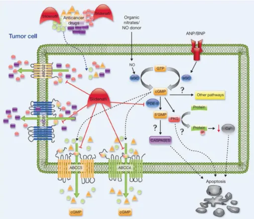
since it inhibits the action of ABCG1, ABCC4, ABCC5 and ABCG2 that are present in breast cancer cells (Figure 5) [27]. This drug is also a phosphodiesterase 5 (PDE5) inhibitor that has been widely used to treat male erectile dysfunction and pulmonary hypertension [27, 55]. Recently, its application for anti-cancer therapy has been evaluated, and the obtained results [27, 55-57], suggested that this drug has the capacity to potentiate the effect of standard chemotherapeutics, by blocking the above mentioned ABC proteins [34, 55, 56, 58, 59]. The inhibition of these ABC transporters, that are involved in the efflux of chemotherapy drugs to the extracellular medium, results in drug accumulation inside the cell cytoplasm, thus potentiating the therapeutic effects. Besides, it is described that PDE 5 expression is enhanced in diverse human carcinomas such as metastatic breast cancer, giving Sildenafil an excellent potential to be used synergistically with other potent chemotherapy drugs [60]. Interestingly, Sildenafil has also shown to protect the heart from toxic effects of chemotherapeutic drugs, such as Doxorubicin [34, 56]. The cardio protective effect is assigned to the enhanced expression of nitric oxide synthase, a enzyme that is involved in the activation of protein kinase C and G, and up-regulation of Bcl-2/Bax [56, 58, 61, 62].
33
Figure 5 – Proposed mechanism of action for Sildenafil synergic activity in conjugation with other chemotherapy drugs by blocking drug efflux from MDR transporters (ABC’s) and increasing intracellular cyclic guanosine monophosphate (cGMP) levels (Adapted from [27]).
2. Nanotechnology-based carriers for cancer
drug delivery
In the last couple of decades the technological breakthrough of manipulating matter at the nanoscale, has encouraged an emerging and compelling interconnection between Nanotechnology, Pharmacy and Medicine, that will surely remain for many years to come [63]. This close affiliation has opened up the opportunity to devise novel and effective therapeutic approaches based on miniaturized nanoscale delivery systems to treat a multitude of impairing diseases, such as cancer [64]. Nanomedicine focused on cancer therapeutics is a particularly interdisciplinary field, that gathers knowledge from Biology, Chemistry, Engineering, Physics and Medicine in order to tackle the complexity associated with cancer cells through the development of drug delivery systems (DDS), that can increase drug bioavailability in diseased tissues and also minimize deleterious side effects in healthy cells [65]. Nano-sized devices, commonly with size ranges within 1 to 100 nm [66], or 1 to 1000 nm [67], provide a unique molecular interaction with biological systems and particularly with single cells [15, 40, 68, 69]. In fact, the sub-cellular size of nanoparticles is a unique characteristic that renders them as an ideal platform to concentrate highly potent
anti-34
tumoral drugs inside malignant cells, since they are readily internalized by various cell uptake pathways [70]. This concept was initially described in the late 60’s, when liposomes where proposed as carriers for proteins and drugs, in order to enhance their delivery to a broad spectrum of pathologies (Figure 6) [71]. Since then, a wide variety of nanomaterials has been used for the manufacture of ever more evolved and efficient DDS’s [15, 45].
Figure 6 –Evolution of drug delivery systems along time (Adapted from [45]).
The use of nanoparticulated carriers changes the pharmacological properties of anti-tumoral compounds, to an extent that dramatically increases their overall therapeutic effect and prolongs PFS rates [72]. In fact, the inclusion of chemotherapeutics in DDS, utterly modulates their pharmacokinetic/pharmacodynamic profiles, especially their serum half-life and bioavailability at the target site (Figure 7) [72]. More importantly, their loading in DDS significantly reduces their toxicity, unlocking the potential to administrate pharmaceutic compounds with remarkable anti-tumoral activity [15, 45], using these nanotransporters. Furthermore, they can also contribute to reduce the frequency of administration to achieve an increased therapeutic effect, in comparison to systemic free-drug administration [15, 45].
35
Figure 7 – Enhancement of drug concentration and bioavailability through nanoparticle conjugation. (Adapted from [73]).
Nanocarriers designed for cancer therapy may be administered through various routes such as: i.) nasal airways, ii.) oral-bucal, iii.) intra-dermic and intramuscular, iv.) intra-ocular and v.) systemic injection through the blood stream [74]. The administration modality affects the overall biodistribution of the nanocarrier-drug conjugate and should be carefully chosen according to each cancer type and location [72]. Regardless of the administration route, once the nanocarriers interact with biological fluids their physicochemical properties change to such an extent that influences nanocarrier-cell interactions, and consequently internalization in target cells [74]. Opsonization process is one of the major barriers that DDS need to overcome [75]. Opsonization and consequent phagocytosis is mediated by the Mononuclear Phagocytic System (MPS). This system is comprised by immune system cells such as phagocytic cells, that are especially present in the spleen and liver. Depending on nanoparticle surface and size, opsonins, proteins present in serum, rapidly bind to nanoparticles enabling macrophages to remove these nanovehicles from blood circulation, affecting their function [75, 76]. It has been previously reported by Letchford and co-workers that nanoparticles with less than 200 nm present a decreased elimination by MPS, and accordingly longer blood circulation times [67]. This size-dependent interaction with the immune system determines the extent of nanoparticle susceptibility to phagocytosis [77]. Once nanocarriers successfully
36
evade MPS, they can fulfil their accumulation in tumor tissues. The accumulation of DDS in diseased tissues after intravenous administration is a critical parameter, since it dictates drug delivery efficiency into target cells. Presently, two major strategies can be employed to promote this accumulation in the tumor microenvironment, the so-termed active and passive targeting [78]. The latter will be particularly focused in this work.
2.1. Passive Targeting – The Enhanced permeability and
Retention Effect
Passive targeting takes advantage of the biological characteristics of the tumor microenvironment itself, namely by exploiting the existence of a highly vascularized network of leaky blood vessels that surround tumor tissues [79, 80]. The lack of lymphatic drainage also contributes for molecule retention in tumors [81]. This phenomenon is generally termed as the enhanced permeability and retention (EPR) effect and is characteristic of solid tumors (Figure 8) [82]. EPR is a probabilistic effect that depends on an extended serum half-life of molecules, such as nanoparticulated carriers, and on their capacity to extravasate through the fenestrations on the endothelial wall of blood vessels to the tumor periphery [83].
Figure 8 –Presentation of nanoparticle EPR effect and enhanced intracellular drug concentration due to efflux pump inhibition. Nanoparticles extravasate through the leaky vasculature due to the fenestrations in the endothelial wall surrounding the tumor microenvironment (Adapted from [68]).
These fenestrations are formed due to the uncontrolled angiogenesis. Such process causes high vascular density originating large gaps [68]. It is clear that free chemotherapy drugs distribute throughout the entire body will be partitioned through the various organs (e.g. the liver, kidney or lungs), but due to the EPR effect they can also be accumulated in diseased tissues, although to a far less extent than that of their initial concentration. This EPR effect
37
has provided the basis for the formulation of nanodevices suitable for anti-cancer therapy [81, 82]. Studies with animal models conclude that neutral to slightly negatively charged drug-loaded nanoparticles, with sizes below 150 nm, have the ability to accumulate into tumor tissues by the EPR effect [84]. This tumor accumulation potentiates the anti-tumoral effect, since it increases the bioavailability of chemotherapeutics at the tumor site [15]. However, despite this potential, the EPR effect still presents some limitations, especially due to the pathophysiological heterogeneity of each tumor and the high, interstitial pressure [85]. However, the increased blood flow and reduced blood pressure caused by the tumor vasculature, enhances nanoparticle extravasation into tumors [86, 87].
3. Organic and Inorganic biomaterials used for
self-assembly drug delivery systems
Among the different types of nanoparticles produced, they can be classified according to their composition into two major groups: inorganic and organic [40]. Inorganic DDS are characterized by their stability, good loading capacity and controlled release of drugs. However, some inorganic nanoparticles can have cytotoxic effects such as those associated with the accumulation of iron, silver and gold in the human body. Nevertheless, these are versatile systems that can also be employed in theranostic applications, due to their bioimaging capacity [88, 89].
Organic DDS are particularly advantageous for the formulation of anti-cancer therapeutics for breast cancer therapy, due to their high biocompatibility, biodegradability, high loading capacity and versatile chemical composition that allows their modification with bioactive macromolecules [90, 91]. Among organic DDS, amphiphilic nanoparticles present advantages over the other organic nanoparticles due to their self-assembly behaviour [92]. This particular class comprises: i.) liposomes; ii.) dendrimers; iii.) protein nanoparticles and iii.) polymeric DDS, namely micelles, polymersomes and nanocapsules among others, some of which are present in Figure 9.
38
Figure 9 –Representation of the drug-delivery systems currently available. (Adapted from [93]).
Lipid-based nanoparticles, also termed liposomes, where one of the first delivery vehicles used in medicine [15]. Liposomes are generally comprised of natural or synthetic lipids, having diverse architectures (multilamelar, core shell, rod like or star shaped), which can accommodate both hydrophilic and hydrophobic molecules [15, 94, 95] like liposome based nanoparticle designed for delivery Tamoxifen to breast cancer cells [96]. However, the use of liposomes as DDS for cancer therapy has several issues that hinder their widespread application in the clinic. Liposomes present a very rapid blood clearance, which is further accelerated after multiple administrations of liposomal formulations coated with hydrophilic polymers that are aimed to increase their blood circulation time. This phenomenon has been only recently described [97]. Opsonisation of liposomes and consequent capture by MPS [68, 94] and their cytotoxicity to healthy cells are the main disadvantages of this class of DDS. Nevertheless, some polymer-modified liposomes that take advantage of the EPR effect and prove to be non-toxic, have been developed for cancer therapy [38, 98]. Doxil®, is a FDA
approved and commercially available pegylated-liposomal formulation, that carries Doxorubicin for cancer therapy [99, 100]. Polymeric nanocarriers provide several advantages over liposomes such as better overall drug/carrier stability [101] and sustained drug controlled release [102]. Polymers used in drug delivery can be synthetic or natural [103]. The synthetic polymers have some advantages over the natural. They can provide a
39
spatiotemporally controlled drug delivery of bioactive pharmaceutics for longer periods than some natural polymers, such as chitosan or alginate that suffer from extensive swelling [103]. Among the different DDS that can be formulated with polymeric materials, micelle carriers are one of the most versatile vehicles for being applied in cancer therapy. They can be used for increasing the solubility of poor water soluble chemotherapeutic drugs, by approximately one thousand fold, a fact that renders them ideal candidates for cancer treatment [104]. Micelles are commonly formulated by two blocks of polymers, hydrophobic and hydrophilic, that are generally grafted together by chemical linkages [67]. The so termed block co-polymers are generally formed by a hydrophilic shell and a hydrophobic core, thus having an amphiphilic character (Figure 10). The hydrophobic core is used to encapsulate hydrophobic molecules, such as chemotherapy drugs. Micelle formation occurs spontaneously in water solutions, if the concentration of the amphiphilic polymer increases to a point in which the hydrophobic chains establish favourable hydrophobic interactions, among each other. This is an interesting phenomena that ultimately results in the formation of the so termed core-shell structure [67]. The minimum concentration required for a micelle self-assembly is called Critical Micellar Concentration (CMC) [67]. Micelles formed with amphiphilic co-polymers are advantageous due to the lower CMC, than those formed with the use of surfactants [67]. In this unique core-shell architecture the drug is protected from biological degradation and its deleterious side effects are significantly reduced [38]. Polymeric micelles can also be easily modified by imprinting targeting moieties on their surface, in order to increase their tissue specificity [105, 106]. Furthermore, the drug loading efficiency and the release profile can be largely improved by simply manipulating the size of hydrophobic or hydrophilic polymer backbones [38, 107]. Poly (ethylene glycol) (PEG) is one of the most used hydrophilic polymers (generally between 1 and 15 kDa) for the formation of the outer shell [38]. However, in the last decade other hydrophilic polymers have also been used to assemble polymeric micelles such as poly (2-hydroxyethyl methacrylate) (PHEMA) and poly (oxazoline) (PEOz) [108, 109]. Nowadays, several formulations of polymeric micelles are currently available for cancer therapy. These include SP1049C® a Pluronic-based micelle for
Doxorubicin delivery [101], Genexol-PM® a Poly (ethylene glycol)-Poly (lactic acid) (PEG-PLA)
40
Table 1 –Comparison of micelles approved or enrolled in clinical trials by FDA. NA (not applicable) (Adapted from [68]).
Name Formulation Diameter
(nm) t½ (h)
Clearance
(mL/min/kg) Comments Doxorubicin 0.9% NaCl NA 0.8 14.4 Small-molecule drug
SP1049C Pluronic micelle +
Doxorubicin 22–27 2.4 12.6 Micelle nanoparticle
NK911 PEG–Asp micelle +
Doxorubicin 40 2.8 6.7 Micelle nanoparticle
Doxil PEG–liposome + Doxorubicin 80–90 84.0 0.02 PEGylated liposome nanoparticle with long circulation Cremophor EL Polyethoxylated Castor Oil Taxol (Paclitaxel) NA 21.8 (20.5) 3.9 (9.2) Small-molecule drug
Genexol-PM PEG–PLA micelle +
Paclitaxel 20–50 11.0 4.8 Micelle nanoparticle
Abraxane Albumin + Paclitaxel 120 21.6 6.5
Albumin nanoparticle before injection;
status in
vivo unknown
XYOTAX PG + Paclitaxel Unknown 70–120 0.07–0.12 Polymer nanoparticle
Camptosar (prodrug of
SN-38)
0.9% NaCl NA 11.7 5.8 Small-molecule
prodrug
LE-SN-38 Liposome + SN-38 Unknown 7–58 3.5–13.6 Liposome nanoparticle
Topotecan (camptothecin
analogue)
0.9% NaCl NA 3.0 13.5 Small-molecule drug
CT-2106 PG + Camptothecin Unknown 65–99 0.44 Polymer nanoparticle
IT-101 Cyclodextrin-containing polymer + Camptothecin 30–40 38 0.03 Polymer nanoparticle with extended circulation times
41
In a recent study, a combination of two block co-polymers (poly(HEMA-co-histidine)-g-PLA and PEG-PLA) was used to specifically target HeLa cells and deliver Doxorubicin [110]. A modified poly (ethylene glycol)-block-poly (ε−caprolactone) (PEG-PCL) micelle, carrying paclitaxel, showed to be effective against MCF-7 cells and breast cancer stem cells [111].
Figure 10 – Polymeric micelle structure composed with block co-polymers that self-assemble in water solutions (Adapted from [68]).
3.1. Hydrophobic polymers used for the production of
self-assembled DDS micelles for cancer therapy
Poly (lactic acid) (PLA) and poly (D,L-lactide-co-glycolide) (PLGA) have been extensively used for the manufacture of DDS [112]. These synthetic polymers are highly biocompatible and biodegradable, since their hydrolysis yields degradation products (lactic acid or glycolic acid) that are inserted in natural metabolic pathways. Particularly, L-Lactic acid integrates the citric acid cycle [103], or is converted to glucose, in the Cori cycle [113, 114]. Moreover, since these degradation products are formed at a slow rate, they do not affect normal cell metabolism [103, 114]. These polymers have been largely tested in animals and are currently used for bone implants, sutures and other applications in humans, such as cancer therapy, as previously mentioned (Genexol-PM®) [101, 113, 115].
Lactic acid (LA) is the monomeric unit of the PLA polymer backbone [114]. This monomer can be formed by bacterial sugar conversion, which makes the LA an inexpensive raw material to obtain [114]. PLA can be produced by direct condensation or ring opening polymerization (ROP) of L-lactide, under specific conditions, yielding very homogeneous polymeric chains with low size and low polydispersity index (PDI) [116, 117]. As a hydrophobic polymer, PLA presents good hydrophobic drug encapsulation capacity, up to 10% of its weight. PCL is more
42
hydrophobic than PLA and promotes a slightly better encapsulation of hydrophobic drugs [118], but its rather slow biodegradation is a factor that can impair its biological applicability [119].
3.2. Hydrophilic polymers used for the production of
self-assembled DDS micelles for cancer therapy
PEG is a widely investigated synthetic polymer approved by the FDA and by the European Medicines Agency (EMA) for nanocarriers production to be used for drug delivery applications due to its unique and versatile physicochemical characteristics [93, 120]. In fact, PEG is so versatile that it can be chemically synthesized with a plethora of pendant groups (e.g. OH, -NH2, -COOH, -SH, -Maleimide, -NHS esters, among others) that unlock the possibility to graft PEG chains to virtually any other polymers including PLA and PLGA [121]. Moreover, the relative low cost of PEG polymers and their capacity to endow DDS with stealth ability in serum, as discussed henceforth, account for its ever growing use [93, 122]. PEG is also biocompatible, being generally considered to have low toxicity in comparison with cationic Liposomal formulations. Presently, PEG is used as an excipient in several intravenous medicines such as Ativan®, or in daily products such as toothpaste [123]. In fact, the World
Health Organization as set as limit a daily dose of 10 mg/Kg for administration via oral route, although only for PEGs with a molecular weight (Mw) up to 10 kDa [123].
The latter parameter is crucial since PEG Mw and PDI plays an important role in envisioned biomedical applications [93]. The molar mass of PEG used in pharmaceutical and biomedical applications commonly ranges from 400Da to 50kDa [93]. Longer PEG chains have been conjugated to low-Mw drugs and other small molecules, like small interfering RNA (siRNA) and oligonucleotides, slowing down their clearance [93]. PEGs with 1 kDa to 5 kDa are more suitable for DDS production [93, 101]. PEG chains with this length range are very flexible, and the polymer can acquire various conformations which is very important for evading interaction with blood components such as opsonins [93]. PEG chains create a water barrier on nanoparticle surface that prevents opsonins adhesion [67]. Less opsonin coating results in lower immunological response and elimination by MPS is therefore reduced [93]. This protective character is only possible due to the steric hindrance effects that PEG promotes, functioning as an actual shield for protein or cell adsorption to DDS [124]. PEG also reduces enzymatic degradation and has the ability to shield the polymer from cationic charges that could contribute for erythrocyte lysis [93]. Regarding the PDI parameter, the polymer should have a PDI lower than 1.1 to ensure homogeneity in biological responses [125]. Due to these valuable characteristics PEG-based micelles have been widely used in self-assembled DDS formulations with core-shell structures, since the PEG moiety is highly hydrophilic [126].
43
PEG-PLA nanoparticles are widely used to deliver hydrophobic drugs due to their self-assembly capacity, high stability, relatively small size (20 to 200 nm), safe administration in humans and high drug loading capacity [93, 127]. The hydrophobic interaction between drugs and the hydrophobic moiety of the amphiphilic co-polymer governs the encapsulation stage [128]. A PEG-PLA micelle loaded with Paclitaxel (Genexol-PM) is currently approved in South Korea, for cancer therapy [129]. Moreover, PEG-PLA nanocarriers are also under stage II clinical trial, in United Stated of America (USA) for Doxorubicin delivery [15, 68]. Since this drug is considered to be more suitable for breast cancer therapy than standard Cremophor EL, a castor oil pegylated nanoparticle with encapsulate paclitaxel [130]. The high dose required for intravenous injection of Cremophor EL (26 mL per injection for treatment of an average weight patient) elicits side effects [104]. Besides, Genexol-PM proved in clinical trials with breast cancer patients, to have better therapeutic efficiency, since it presented a higher maximum tolerated dose (MTD), and improved pharmacokinetic profile, delaying the time of disease progression in 9 months [130].
Synthesis of block co-polymer PEG-PLA can be performed by ROP between PEG, which acts as macro initiator of the reaction and L-lactide [131]. Commonly tin(II), zinc and aluminium salts, such as stannous octoate (Sn(Oct)2), are used to catalyze the ROP reaction, as shown in
Figure 11 [132, 133]. The length of polymer can then be controlled by changing the weight ratio between the initiator and L-lactide monomer [126].
Figure 11 - Mechanism of polymerization of L-lactide. mPEG acts as macroinitiator and Sn(Oct)2 act as a
catalyst (Adapted from [131]).
The amphiphilic character of PEG-PLA allows its self-assembly in water [134]. Various methods can be used to assemble PEG-PLA micelles, such as: i.) direct dissolution in water, ii.) film rehydration, that consists in using an organic volatile solvent followed by evaporation-solubilisation cycles [135] and iii.) sonication [118], with this method yielding the highest encapsulation efficiency of hydrophobic drugs [118].
44
Aims
The global aim of this thesis was to develop a new approach for breast cancer therapy through the synthesis of micellar nanovehicles capable of co-delivery of two drugs to breast cancer cells. The specific aims of this research include the:
Synthesis and characterization of amphiphilic block co-polymers capable of self-assembling into nanosized micelles;
Characterization of the physicochemical properties of the self-assembled micelles;
Evaluation of multi-drug loading efficiency by produced micelles and investigation of their release profile;
Assessment of micelle uptake by breast cancer cells;
Study of the anti-tumoral potential of free drug pharmaceutical formulations;
Evaluation of the anti-tumoral and synergic effect through micellar vehicle delivery.
45
Chapter 2
46
2. Materials and Methods
All methods performed in this thesis are according to the directives set forth by the International Standards Organization (ISO) in the following standard guidelines: i.) ISO/Technical Report (TR) 13014-2013 - Nanotechnologies guidance on physicochemical characterization of engineered nanoscale materials for toxicological assessment; and ii.) ISO 10993-5:2009 - Biological evaluation of medical devices - Part 5: Tests for in vitro cytotoxicity.
2.1. Materials
Metoxy Poly(ethylene glycol) (mPEG) 2000 was obtained from Nanocs (New York, USA). Acetone, Acetonitrile (HPLC-grade), Dichloromethane, Dulbecco’s Modified Eagle’s Medium (DMEM), Methanol (MetOH) (High Performance Liquid Chromatography (HPLC)-grade), Resazurin, Rhodamine B Isothiocyanate (RITC), Toluene, were acquired from VWR Internacional (Carnaxide, Portugal). Antibiotic–Antimycotic (penicillin and streptomycin), Cacodylate, Cellulose dialysis membrane, Collagen type I, Fetal bovine serum (FBS), Glutaraldehyde, Paraformaldehyde, Phosphoric acid, and Triton X-100 were obtained from Sigma–Aldrich (Sintra, Portugal). MCF-7 (ATCC® HTB-22) mammary gland adenocarcinoma cell
line was obtained from ATCC (Middlesex, United Kingdom) and primary normal human dermal fibroblasts (hFIB) from Promocell (Heidelberg, Germany). Rompun (Xylazine) was purchased from Bayer Health Care (Carnaxide, Portugal) and Imalgene (Ketamine) was obtained from Merial Laboratories, (Lyon, France). Stannous Octoate was purchased from (Cymit Quimica, Barcelona, Spain). All the glassware used in polymer synthesis was borosilicate 3.3 supplied by Afora SA (Spain). PF-02341066 (Crizotinib) and PF-4540124 (Sildenafil) were purchased from Tocris Bioscience (Nortpoint, United Kingdom). CellEventTM Caspase-3/7® and Hoechst
33342® where obtained from Invitrogen (Carlsbad, USA). L-Lactide monomer and the Pyrene
fluorescent probe were acquired from TCI (Tokyo Chemical Industry, Co., LTD., Japan). 3-(4,5-dimethylthiazol-2-yl)-5-(3-carboxymethoxyphenyl)-2-(4-sulfophenyl)-2H-tetrazolium) (MTS) was obtained from Promega (Madison, WI, USA). Phalloidin CruzFluor® 647 was obtained
from Santa Cruz Biotechnology (Santa Cruz Biotechnology, Santa Cruz, Canada). All the used salts were of analytical grade and used without further purification.

![Figure 6 – Evolution of drug delivery systems along time (Adapted from [45]).](https://thumb-eu.123doks.com/thumbv2/123dok_br/18179864.874399/34.892.146.750.274.597/figure-evolution-drug-delivery-systems-time-adapted.webp)


![Table 1 – Comparison of micelles approved or enrolled in clinical trials by FDA. NA (not applicable) (Adapted from [68])](https://thumb-eu.123doks.com/thumbv2/123dok_br/18179864.874399/40.892.79.815.136.1131/table-comparison-micelles-approved-enrolled-clinical-applicable-adapted.webp)
![Figure 10 – Polymeric micelle structure composed with block co-polymers that self-assemble in water solutions (Adapted from [68])](https://thumb-eu.123doks.com/thumbv2/123dok_br/18179864.874399/41.892.191.790.226.561/figure-polymeric-structure-composed-polymers-assemble-solutions-adapted.webp)