ABSTRACT
Objective: To evaluate the impact of thoracic radiotherapy on respiratory function and exercise capacity in patients with breast cancer. Methods: Breast cancer patients in whom thoracic radiotherapy was indicated after surgical treatment and chemotherapy were submitted to HRCT, respiratory evaluation, and exercise capacity evaluation before radiotherapy and at three months after treatment completion. Respiratory muscle strength testing, measurement of chest wall mobility, and complete pulmonary function testing were performed for respiratory evaluation; cardiopulmonary exercise testing was performed to evaluate exercise capacity. The total radiotherapy dose was 50.4 Gy (1.8 Gy/fraction) to the breast or chest wall, including supraclavicular lymph nodes (SCLN) or not. Dose-volume histograms were calculated for each patient with special attention to the ipsilateral lung volume receiving 25 Gy (V25), in absolute and relative values, and mean lung dose. Results: The study comprised 37 patients. After radiotherapy, significant decreases were observed in respiratory muscle strength, chest wall mobility, exercise capacity, and pulmonary function test results (p < 0.05). DLCO was unchanged. HRCT showed changes related to radiotherapy in 87% of the patients, which was more evident in the patients submitted to SCLN irradiation. V25% significantly correlated with radiation pneumonitis. Conclusions: In our sample of patients with breast cancer, thoracic radiotherapy seemed to have caused significant losses in respiratory and exercise capacity, probably due to chest wall restriction; SCLN irradiation represented an additional risk factor for the development of radiation pneumonitis.
Keywords: Breast neoplasms; Radiotherapy; Radiation pneumonitis; Respiratory function tests; Exercise test.
Impact of thoracic radiotherapy on
respiratory function and exercise capacity
in patients with breast cancer
Milena Mako Suesada1,a, Heloisa de Andrade Carvalho2,b,
André Luis Pereira de Albuquerque1,c, João Marcos Salge1,d,
Silvia Radwanski Stuart2,e, Teresa Yae Takagaki1,f
Correspondence to:
Heloisa de Andrade Carvalho. InRad, Travessa da Rua Dr. Ovídio Pires de Campos, 75, Portaria 3, CEP 05403-010, São Paulo, SP, Brasil. Tel.: 55 11 2661-6722. Fax: 55 11 3885-7036. E-mail: [email protected]
Financial support: None.
INTRODUCTION
In breast cancer, postoperative thoracic radiotherapy is widely used in order to reduce the risks of loco-regional recurrence and to improve overall survival.(1,2) However,
irradiation of thoracic structures involves risks, primarily to the lungs.
Radiation pneumonitis (RP) is the most significant adverse effect and generally appears between one
and four months after radiotherapy completion.(3,4)
The etiology and physiopathology of RP are related to a cytokine-mediated signal cascade that causes early damage of the cells in the alveolar space, progressing
to an acute exudative inflammatory process. Clinical
symptoms include cough, low-grade fever, dyspnea, fatigue, and pleuritic chest pain.(3-5)These symptoms
can be reflected by changes on pulmonary function tests (PFTs) results, with reduced FVC, FEV1, TLC, and DLCO. (3,4) Radiologic evaluation of the lung toxicity has
usually been made by chest X-rays. However, HRCT has
proven to be sensitive in detecting early changes, but it is not used as part of routine follow-up.(6,7)
Radiotherapy-induced injury may also acutely lead to systemic impairment, frequently referred to as a diminished exercise capacity and worsening of quality of life.(8-10)Those changes have been evaluated by means of
specific questionnaires, but the objective quantification of
such changes, to our knowledge, has not been previously performed. In breast cancer, this should be carefully considered due to improvement of prognosis and life expectancy in these patients. Therefore, it is essential to quantify and investigate exercise limitation after thoracic radiotherapy in order to identify the involved mechanisms.
The purpose of the present study, therefore, was to quantify the acute impact of thoracic radiotherapy on respiratory function and exercise capacity in patients with breast cancer at three months after irradiation.
METHODS
The study was approved by the institutional research ethics committee, and giving informed consent was mandatory for enrollment. Inclusion criteria were
confirmed histological diagnosis of breast cancer, indication 1. Divisão de Pneumologia, Instituto do
Coração – InCor – Hospital das Clínicas, Faculdade de Medicina, Universidade de São Paulo, São Paulo (SP) Brasil. 2. Departamento de Radiologia e
Oncologia / Radioterapia, Instituto de Radiologia – InRad –Faculdade de Medicina, Universidade de São Paulo, São Paulo (SP) Brasil.
a. http://orcid.org/0000-0002-7665-9704 b. http://orcid.org/0000-0003-0979-7768 c. http://orcid.org/0000-0003-3486-5240 d. http://orcid.org/0000-0001-5121-0129 e. http://orcid.org/0000-0002-0504-776X f. http://orcid.org/0000-0003-2277-2100
Submitted: 9 May 2017. Accepted: 13 February 2018.
for postoperative irradiation, in accordance with the institutional routine protocol, and a period of at least four weeks with no treatment before radiotherapy was required. All of the patients had to be fully treated in the institution and were consecutively selected over a one-year period. Patients were excluded if they presented with metastatic disease, concomitant respiratory disease, neuromuscular or rheumatologic disease, or cognitive disorders hindering the completion
of PFTs or exercise tests. The patients underwent HRCT
of the chest, complete PFT, cardiopulmonary exercise
testing (CPET), respiratory muscle strength testing,
and measurement of chest wall mobility. In addition,
the Medical Research Council (MRC) dyspnea scale
was also applied to evaluate respiratory symptoms during activities of daily living over the study period.(11)
All of the measurements were conducted a few days (an average of 2 to 5 days) prior to the beginning of radiotherapy and at three months after the completion of treatment.
Radiotherapy
Radiotherapy was delivered using a 3D conformal technique. The whole breast or chest wall was irradiated
with opposed tangential fields and, when indicated,
additional irradiation of supraclavicular lymph nodes
(SCLNs) was performed with a direct anterior field.
Patients were treated with 6 MV photon beams and a total dose of 50.4 Gy (28 × 1.8 Gy; 5 days/week). Dose-volume histograms for the heart, lungs, and contralateral breast were calculated with no tissue
heterogeneity correction (Eclipse planning system;
Varian®, Palo Alto, CA, USA). The ipsilateral lung volume
that received at least 50% of the dose (25 Gy = V25),
corresponding to the limits of the fields (or the actual
irradiated lung volume) and the mean lung dose were correlated with the functional test results. V25 was
calculated as absolute (cm3) and relative (%) values. Respiratory function evaluation
Complete PFTs and DLCO were performed with a body plethysmograph (Elite DX; MedGraphics Corp., Saint Paul, MN, USA). The following parameters were measured: FVC, FEV1, inspiratory capacity (IC), TLC, RV, maximal voluntary ventilation, and DLCO.
Measurements were expressed as absolute volumes and percentages of the predicted values for the Brazilian population(12-14) and performed according to
the guidelines of the American Thoracic Society (ATS)/
European Respiratory Society.(15)
Respiratory muscle strength was assessed by MIP
and MEP in accordance with the ATS guidelines.(16)
These variables were measured by using a pressure
transducer (OEM Medical, Marshalltown, IA, USA). Chest cirtometry was used in order to evaluate
chest wall mobility at the level of axillary level and at the level of the xiphoid process.(17) Patients were
asked to exhale up to RV and then inhale up to TLC, and the difference between the two measurements
was calculated.
Exercise capacity evaluation
A maximal incremental cycle ergometer protocol was conducted in accordance with the ATS recommendations(18) using a CardiO
2 System ®
(MedGraphics). The following parameters were
determined: oxygen uptake (VO2; mL·min
-1); carbon dioxide output (VCO2; mL·min
-1); respiratory exchange
ratio; minute ventilation (VE, L·min-1); tidal volume
(VT, mL); and RR (breaths/min). The mean VO2 for the last 15 seconds of the ramp was considered as
the peak oxygen uptake (VO2peak). During the test,
12-lead electrocardiography, blood pressure, and pulse oximetry values were monitored. In addition, dyspnea
and leg fatigue were assessed by the modified Borg
scale every two minutes.(19)
Dynamic relationships were determined to evaluate
metabolic (∆VO2/∆workload [W]; mL·min -1·W-1), cardiovascular (∆HR/∆VO2;beats·min
-1·L·min-1), and respiratory (∆VE/∆VCO2; L·min
-1·L·min-1) responses,
as previously described.(20) Ventilatory reserve was
calculated using the VE at exercise cessation/maximal voluntary ventilation ratio as a reference.
HRCT
HRCT was performed with 1-mm slices at increments
of 10 mm from the apex to the base of the lungs during maximal inspiration without the use of i.v. contrast. The scans were analyzed and scored in accordance with the Schratter-Sehn et al. scoring system.(6)Briefly, this
score ranges from zero (no changes) to 5 according to the severity of radiological abnormalities. A single radiologist, specializing in lung diseases, evaluated
and classified all of the scans in accordance with the
protocol.
Classification of RP and radiation dermatitis
RP was graded in accordance with the toxicity criteria
by Cox et al.,(21) and radiation dermatitis was classified
in accordance with the criteria for skin adverse events described by Freedman et al.(22) The two systems are
based on the severity of respiratory and dermatological symptoms after radiotherapy, respectively.
Statistical analysis
The analyses of variations between the respiratory and exercise test results obtained before and after radiotherapy were performed using the Student’s t-test.
ANOVA was used in order to analyze the variance in HRCT scan results, in severity of RP, and in severity
of radiation dermatitis in relation to changes in PFT
and CPET variables after radiotherapy completion. The differences in changes observed in respiratory
and exercise capacity variables between the group
submitted to SCLN irradiation and that submitted to
chest wall/breast radiotherapy only were analyzed
with the Student’s t-test. The differences in chest wall
for independent samples. The significance level was set at 5% (p ≤ 0.05). Statistical analysis of the data
was performed with the IBM SPSS Statistics software
package, version 20.0 (IBM Corporation, Armonk, NY, USA).
RESULTS
The study comprised 40 female patients; of those, 2 were excluded because they developed pleural metastasis, and 1 was excluded because she missed
the third-month evaluation. Therefore, the final sample
comprised 37 patients. The mean age of the study population was 53.5 ± 10.9 years. Seven patients (18.9%) had a smoking history, but only 1 was a
current smoker during the study period. No significant
changes in the mean body mass index were observed between the two time points studied, that is, before radiotherapy and at three months after radiotherapy (28.2 ± 4.4 kg/m2 vs. 28.2 ± 4.8 kg/m2). Table 1 presents the affected breasts and the characteristics
of treatment prior to radiotherapy. The mean time between chemotherapy and radiotherapy was 107.0
± 81.8 days. SCLNs were irradiated in 20 (54%) of
the cases. At three months after radiotherapy, 29 (78%) of the patients had developed RP and 33 (89%) presented with skin toxicity according to the adopted
scales (Table 2). Worsening of respiratory symptoms was also observed according to MRC scale. Before
radiotherapy, 30 (81.2%) of the patients reported no symptoms (zero score), and 7 (18.9%) had a score of 1. After radiotherapy, the number of patients with no symptoms decreased to 8 (21.6%), 24 (64.9%) had a score of 1, and 5 (13.5%) had a score of 2.
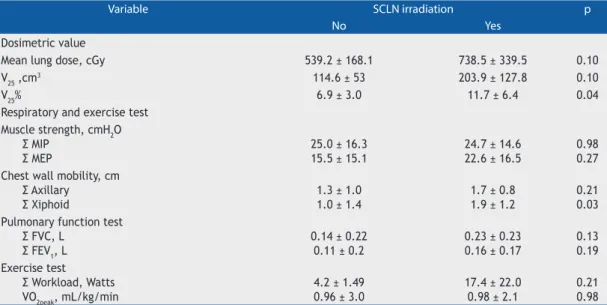
Table 3 shows a comparison of mean lung dose (MLD) and V25 (in absolute and relative values) between
patients who underwent SCLN irradiation or and those who did not. Only V25%showed a significant difference
between the two groups of patients.
At 3 months after radiotherapy, we found that there
was a significant loss in MIP and MEP (p < 0.0001 for both), as well as significant decreases in all chest
wall mobility at the axillary level and at the level of
the xiphoid process (p < 0.0001). Regarding PFTs, except for DLCO, a significant decrease in FVC, FEV1, TLC, and IC was detected after radiotherapy (Table 4).
Maximal CPET results showed that there was a significant decrease in workload and VO2peak after radiotherapy (p < 0.05). Significant reductions were
also detected in VE, VT, and respiratory exchange ratio but not in RR (Table 4). All of those changes were observed only at peak exercise.
The dynamic relationships showed significant changes
in metabolic response, and there was a tendency toward a decrease in the respiratory response when compared with the pre-radiotherapy condition (Table 4). All of the patients used an average of 40% of ventilatory reserve at peak exercise before and after radiotherapy.
All of the respiratory parameters, as well as those obtained from exercise testing, showed to be decreased
when patients submitted to SCLN irradiation were included; however, the only significant decrease was
found for chest wall mobility at the level of the xiphoid process (p = 0.03; Table 3).
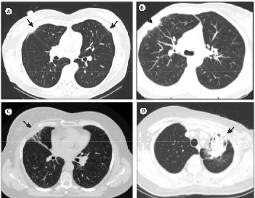
After radiotherapy, changes were identified in 87% of the HRCT scans (Figure 1), and the distribution
according to the adopted scoring system(6) is presented
in Table 5. Those changes were more prominent in the
patients submitted to SCLN irradiation.
Higher HRCT scores were significantly correlated with greater losses of FVC (p = 0.01). None of the
patients had grade 3 or higher radiation dermatitis,
and the presence of skin symptoms was significantly
correlated with reductions in chest wall mobility at the level of the xiphoid process (p = 0.05).
Patients submitted to mastectomy showed greater losses in chest wall mobility when compared with the other patients (p = 0.06). Grade 2 radiation dermatitis was more prevalent in the former group of patients (51% vs. 33% in those submitted to breast conserving surgery).
DISCUSSION
The present study reports the results of a prospective
analysis of the early effects of thoracic radiotherapy
on respiratory function at rest and during exercise
in patients treated for breast cancer. Changes in PFT results and radiological findings after irradiation in
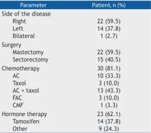
Table 1. Affected breast and delivered treatments prior
to radiotherapy in the study sample (N = 37).
Parameter Patient, n (%)
Side of the disease Right Left Bilateral
22 (59.5) 14 (37.8) 1 (2.7) Surgery
Mastectomy Sectorectomy
22 (59.5) 15 (40.5) Chemotherapy
AC Taxol AC + taxol FAC CMF
30 (81.1) 10 (33.3) 3 (10.0) 13 (43.3)
3 (10.0) 1 (3.3) Hormone therapy
Tamoxifen Other
23 (62.1) 14 (37.8) 9 (24.3)
AC: adriamycin + cyclophosphamide; FAC: fluorouracil + doxorubicin + cyclophosphamide; and CMF: cyclophosphamide + methotrexate + fluorouracil.
Table 2. Incidence of radiation pneumonitis and radiation dermatitis at three months after radiotherapy in the study
sample (N = 37).
Grade 0 1 2
Radiation pneumonitis 8
(21.6%) 19 (51.3%)
10 (27.1%)
Radiation dermatitis 4
(10.8%) 16 (43.3%)
breast cancer patients have been described in previous studies.(3,4,5,7) In our study, irradiation has led to
negative effects on exercise capacity, respiratory muscle
strength, and chest wall mobility. To our knowledge,
these findings have never been published and reveal
a physiological approach to cancer-related fatigue, present in more than 30% of breast cancer patients at the completion of therapy.(23) Those symptoms may limit
activities of daily living and enhance muscle atrophy, contributing to impairment of physical performance. The reported incidence of RP in breast cancer patients submitted to radiotherapy varies from 4.5% to 80% in prospective studies,(24-29) and these results are related
to the irradiated lung volumes,(25,26) MLD,(6,27) age,(4)
performance status,(28) use of chemotherapy,(27) and
use of tamoxifen.(29) In the present study, we observed
Table 3. Comparisons of dosimetric values, as well as of respiratory and exercise test results, between patients who
underwent supraclavicular lymph node irradiation and those who did not.
Variable SCLN irradiation p
No Yes
Dosimetric value
Mean lung dose, cGy 539.2 ± 168.1 738.5 ± 339.5 0.10
V25 ,cm3 114.6 ± 53 203.9 ± 127.8 0.10
V25% 6.9 ± 3.0 11.7 ± 6.4 0.04
Respiratory and exercise test Muscle strength, cmH2O
Σ MIP
Σ MEP 25.0 ± 16.315.5 ± 15.1
24.7 ± 14.6 22.6 ± 16.5
0.98 0.27 Chest wall mobility, cm
Σ Axillary
Σ Xiphoid 1.3 ± 1.01.0 ± 1.4
1.7 ± 0.8 1.9 ± 1.2
0.21 0.03 Pulmonary function test
Σ FVC, L Σ FEV1, L
0.14 ± 0.22 0.11 ± 0.2
0.23 ± 0.23 0.16 ± 0.17
0.13 0.19
Exercise test Σ Workload, Watts
VO2peak, mL/kg/min
4.2 ± 1.49 0.96 ± 3.0
17.4 ± 22.0 0.98 ± 2.1
0.21 0.98
SCLN: supraclavicular lymph node; V25: ipsilateral lung volume receiving 25 Gy; and VO2peak: oxygen uptake at peak exercise.
Table 4. Variation in respiratory muscle strength, chest wall mobility, pulmonary function testing, and cardiopulmonary exercise testing before and after three months of radiotherapy.
Variable Pre-RT Post-RT p
Muscle strength, cmH2O
Σ MIP
Σ MEP −95.6 ± 22.4100.0 ± 23.0
−71.8 ± 14.7
80.9 ± 16.8
0.0001 0.0001 Chest wall mobility, cm
Σ Axillary
Σ Xiphoid 4.1 ± 0.93.0 ± 1.7
2.5 ± 0.7 1.6 ± 1.4
0.0001 0.0001
MVV, L/min 124.0± 33.6 111 ± 32.6 0.0001
Pulmonary function test
Σ FVC, L Σ FEV1, L
Σ IC, L Σ TLC, L
Σ DLCO, mL/min/mmHg
3.0 ± 0.8 2.4 ± 0.6 2.3 ± 0.5 4.7 ± 1.0 21.4 ± 4.5
2.8 ± 0.7 2.2 ± 0.6 2.1 ± 0.6 4.5 ± 0.9 21.13 ± 4.5
0.0001 0.0001 0.008
0.01 0.56 Cardiopulmonary exercise test
Workload, Watts Σ VO2peak, mL/kg/min
Σ VEpeak, L/min
Σ RERpeak
Σ RRpeak, breaths/min
Σ VTpeak , L
96.5 ± 30 16.8 ± 3.0 51.4 ± 14.0
1.2 ± 0.1 35.6 ± 6.6
1.4 ± 0.3
88.0 ± 20.8 15.6 ± 3.9 45.0 ± 11.9
1.1 ± 0.1 34.7 ± 5.9
1.2 ± 0.2
0.04 0.04 0.01 0.009
0.46 0.003 Dynamic relationship
Σ Metabolic (∆VO2/∆W)
Σ Respiratory (∆VE/∆VCO2)
Σ Cardiovascular (∆HR/∆VO2)
9.2 ± 1.6 33.1 ± 5.0 73.6 ± 18.6
10.0 ± 1.5 31.4 ± 5.5 75.2 ± 18.2
0.02 0.07 0.50
RT: radiotherapy; MVV: maximal voluntary ventilation; IC: inspiratory capacity; VO2peak: oxygen uptake at peak exercise; VEpeak: minute ventilation at peak exercise; RERpeak: respiratory exchange ratio at peak exercise; and
a 78% incidence of RP, 51% and 27% being classified
as grade 1 and grade 2, respectively. The incidence of
RP in the present study correlated with the MRC scale
results, reinforcing that the severity of respiratory symptoms correlated with decreased activities of daily living and exercise tolerance.
The majority of the patients (81%) included in the present study had been previously treated with
chemotherapy, which prevents the analysis of differences
between patients treated with chemotherapy and
those who were not. Chemotherapy by itself may cause pulmonary toxicity, among other side effects.
Therefore, a higher risk of RP might be present when chemotherapy is associated with radiotherapy.(30)
We found that the patients who used tamoxifen prior
to radiotherapy (61%) showed more relevant losses in chest wall mobility than those who did not use it. However, changes in muscle strength and pulmonary function parameters were similar in both groups.
In the present study, the high incidence of radiation dermatitis (grade 1, in 43% of the patients; and grade 2, in 46%) corroborates other studies.(22,31) Both radiation
dermatitis grades and treatment with SCLN irradiation were significantly correlated with the decrease in
chest wall mobility at the level of the xiphoid process. These results strongly suggest that the loss of chest wall mobility might be associated with the severity
of skin toxicity and SCLN irradiation. However, these
Table 5. Classification of HRCT scans after radiotherapy in the overall study population (N = 37), as well as in patients
who underwent supraclavicular lymph node irradiation and those who did not, according to the scoring system by Schratter-Sehn et al.(6),a
HRCT scan score Overall SCLN irradiation
No (n = 17)
Yes (n = 20)
0 5 (13.5%) 3 (17.7%) 2 (10%)
1 17 (45.9%) 9 (52.9%) 8 (40%)
2 8 (21.6%) 3 (17.6%) 5 (25%)
3 5 (13.5%) 2 (11.8%) 3 (15%)
4 0 (0.0%) 0 (0.0%) 0 (0.0%)
5 2 (5.4%) 0 (0.0%) 2 (10%)
SCLN: supraclavicular lymph node. aScore: 0 = no changes; 1 = septal thickening, reticular subpleural opacities; 2 = subpleural thickening > 2 cm parallel to the chest wall; 3 = parenchymal bands ≥ 2.5 cm from the lung toward
the pleural surface; 4 = honeycombing aspect, cystic areas (> 1 cm diameter), thickened walls; 5 = ground glass opacities, acute radiological changes.
Figure 1. HRCT scans showing features of radiation pneumonitis. In A, HRCT scan scored as 1 in a patient submitted
findings might not only be related to SCLN irradiation
but also to the fact that patients with more advanced tumors had been submitted to mastectomy or to more
extended axillary surgery, and, consequently, SCLN
irradiation had been prescribed.
Dunlap et al.(32) suggested that chest wall toxicity
might affect muscles, connective tissue, neurovascular
bundle, and bones. In addition, Kinsella et al.(33)showed
that doses over 60 Gy correlated with symptoms of nerve dysfunction, including paresthesia, weakness, and pain. Those studies reinforce the hypothesis that radiotherapy for breast cancer treatment might ultimately engender chest wall restriction, which would diminish the ability of muscles to contract and generate
power, eventually leading to muscle weakness. Our patients had decreases in MIP and MEP, which could
be also found in pulmonary function impairment.
Inspiratory muscle weakness affects the maximal IC effort, causing reductions in TLC and IC. Conversely, expiratory muscle weakness affects the maximal expiratory effort, leading to an increase in RV.
A few studies(3,4,7) have demonstrated gas exchange,
PFT, and radiological abnormalities due to irradiation
of breast cancer. In the present study, DLCO was the only variable with no significant change, unlike in other studies in the literature. Erven et al.(34) suggested
that TLC was the most affected parameter and that
that reduction was probably related to decreased parenchymal elasticity in the irradiated portion of the
lung, which could be due to fibrosis and could explain further reductions in DLCO. Our results indicate that the reduction in TLC was probably related to changes
in respiratory muscle strength and chest wall mobility.
Furthermore, the HRCT scans showed a high incidence
of parenchymal changes after radiotherapy; however,
they were mostly confined to the pulmonary parenchyma near the pleural surface and chest wall—HRCT scan
score of 1-2 in 66% of the cases, and only 2 (5.4%) of the patients presented with a score of 5. The low incidence of severe parenchymal changes probably
preserved DLCO in our patients, but this fact also limited
the statistical power of the analysis of severe events.
As expected, the inclusion of the SCLN irradiation
in the treatment was related to greater severity of RP and radiological changes. The loss of chest wall mobility at the level of the xiphoid process was also
significantly higher in those patients. Once again, this finding might be confused with the type of surgery
performed in more advanced cases.
Likewise, V25% was significantly higher in patients
submitted to SCLN irradiation treatment. V25 has been previously described as a predictor of lung toxicity in a study of radiotherapy dose escalation for lung cancer. (35) However, V
20% values higher than 30% were also defined as causing a higher risk of RP, resulting
in radiological and functional impairment.(36) We have
chosen V25 in order to correlate our results with lung
volumes, which are included in the limits of the fields for radiotherapy in breast cancer patients. Only V25%
correlated with functional/radiological impairment, in
agreement with the already defined constraints that are
related to the proportion of the irradiated lung volume that receives a certain dose.(37) However, minimizing
V25 to 100 cm3 or less may also be a strategy to reduce
pulmonary toxicity(4) due to breast cancer irradiation.
The MLD was not related to any abnormality, probably due to the small amount of lung tissue irradiated in breast cancer patients.
Few investigations have evaluated the exercise capacity as a predictor of RP. Miller et al.(38) used the
six-minute walk test in patients with lung cancer and found that good functional capacity and good PFT results prior to radiotherapy apparently reduced the
risk of RP. In our study, CPET was chosen for this
evaluation, because it allows a global assessment of integrative exercise responses.(18) We observed a
significant decrease in workload, in VO2peak, in VE, and
in VT and respiratory exchange ratio at peak exercise.
Normally, VO2 increases almost at the same pace as does workload, and this relationship (∆VO2/∆W) reflects the efficiency of the metabolic component linked to skeletal muscles. A high ∆VO2/∆W ratio after radiotherapy might reflect a great overload related
to excessive work done by the ventilatory muscles, restricted chest wall mobility, and reduced expansion of VT during exercise. Lower ventilation and VT reinforce chest wall restriction after the intervention, which is in line with the lower cirtometry and lung volume results found in our study. Despite this possible overload of
ventilatory muscles, a definitive ventilatory limitation
was not found. Finally, we cannot rule out an additional component of peripheral muscle limitation, since
maximal VO2 was reduced and leg fatigue by the modified Borg scale was increased after radiotherapy.
Decreased exercise performance, with no ventilatory or cardiovascular limitation, indicates that our patients presented lower exercise tolerance at three months after radiotherapy. These findings reinforce our
qualitative MRC scale results and could be part of the
so-called cancer-related fatigue(39-41); however, further
studies are needed to improve the determination of the mechanisms involved.
The fact that the present study was designed to
evaluate the early effects of radiation may represent a limitation, because these effects may or may not be
reversible. Late reactions are expected to be present at least six months after the end of the treatment and
may be reflected as fibrosis, with or without symptoms
or clinical manifestations.(5,34) Therefore, a long-term
follow-up period is needed to assess the outcome of
these patients and the impact of our findings on their
quality of life.
In conclusion, thoracic radiotherapy for breast cancer
can acutely lead to significant impairment in functional
capacity and exercise performance. The negative impact on the respiratory system was characterized by muscle weakness and restriction of chest wall mobility that could cause decreases in PFT results. There were
was correlated with a higher risk of RP. Finally, the
inclusion of SCLN irradiation in the treatment fields
represents a potential risk factor for the development of RP with functional repercussions.
REFERENCES
1. Overgaard M, Hansen PS, Overgaard J, Rose C, Andersson M, Bach F, et al. Postoperative radiotherapy in high-risk premenopausal women with breast cancer who receive adjuvant chemotherapy. Danish Breast Cancer Cooperative Group 82b Trial. N Engl J Med. 1997;337(14):949-55. https://doi.org/10.1056/ NEJM199710023371401
2. Minor GI, Yashar CM, Spanos WJ Jr, Jose BO, Silverman CL, Carrascosa LA, et al. The relationship of radiation pneumonitis to treated lung volume in breast conservation therapy. Breast J. 2006;12(1):48-52. https://doi.org/10.1111/j.1075-122X.2006.00180.x
3. Jaén J, Vázquez G, Alonso E, León A, Guerrero R, Almansa JF. Changes in pulmonary function after incidental lung irradiation for breast cancer: a prospective study. Int J Radiat Oncol Biol Phys. 2006;65(5):1381-8. https://doi.org/10.1016/j.ijrobp.2006.03.008
4. Krengli M, Sacco M, Loi G, Masini L, Ferrante D, Gambaro G, et al. Pulmonary changes after radiotherapy for conservative treatment of breast cancer: a prospective study. Int J Radiat Oncol Biol Phys. 2008;70(5):1460-7. https://doi.org/10.1016/j.ijrobp.2007.08.050
5. Chopra RR, Bogart JA. Radiation therapy-related toxicity (including pneumonitis and fibrosis). Hematol Oncol Clin North Am. 2010;24(3):625-42. https://doi.org/10.1016/j.hoc.2010.03.009
6. Schratter-Sehn AU, Schurawitzki M, Zach M, Schratter M. High-resolution computed tomography of the lungs in irradiated breast cancer patients. Radiother Oncol. 1993;27(3):198-202. https://doi. org/10.1016/0167-8140(93)90074-I
7. Järvenpää R, Holli K, Pitkänen M, Hyödynmaa S, Rajala J, Lahtela SL, et al. Radiological pulmonary findings after breast cancer irradiation: a prospective study. Acta Oncol. 2006;45(1):16-22. https://doi. org/10.1080/02841860500334921
8. Whelan TJ, Levine M, Julian J, Kirkbride P, Skingley P. The effects of radiation therapy on quality of life of women with breast carcinoma: results of a randomized trial. Ontario Clinical Oncology Group. Cancer. 2000;88(10):2260-6. https://doi.org/10.1002/(SICI)1097-0142(20000515)88:10<2260::AID-CNCR9>3.0.CO;2-M
9. Markes M, Brockow T, Resch KL. Exercise for women receiving adjuvant therapy for breast cancer. Cochrane Database Syst Rev. 2006;18(4):CD005001. https://doi.org/10.1002/14651858. CD005001.pub2
10. Dimeo F, Schwartz S, Wesel N, Voigt A, Thiel E. Effects of an endurance and resistance exercise program on persistent cancer-related fatigue after treatment. Ann Oncol. 2008;19(8):1495-9. https://doi.org/10.1093/annonc/mdn068
11. Kovelis D, Segretti NO, Probst VS, Lareau SC, Brunetto AF, Pitta F. Validation of the Modified Pulmonary Functional Status and Dyspnea Questionnaire and the Medical Research Council scale for use in Brazilian patients with chronic obstructive pulmonary disease. J Bras Pneumol. 2008;34(12):1008-18. https://doi.org/10.1590/S1806-37132008001200005
12. Neder JA, Andreoni S, Castelo-Filho A, Nery LE. Reference values for lung function tests. I. Static volumes. Braz J Med Biol Res. 1999;32(6):703-17. https://doi.org/10.1590/S0100-879X1999000600006
13. Pereira CA, Sato T, Rodrigues SC. New reference values for forced spirometry in white adults in Brazil. J Bras Pneumol. 2007; 33(4):397-406. https://doi.org/10.1590/S1806-37132007000400008
14. Neder JA, Andreoni S, Peres C, Nery LE. Reference values for lung function tests. III. Carbon monoxide diffusing capacity (transfer factor). Braz J Med Biol Res. 1999;32(6):729-37. https://doi. org/10.1590/S0100-879X1999000600008
15. Miller MR, Hankinson J, Brusasco V, Burgos F, Casaburi R, Coates A et al. Standardisation of spirometry. Eur Respir J. 2005;26(2):319-38. https://doi.org/10.1183/09031936.05.00034805
16. American Thoracic Society/European Respiratory Society. ATS/ERS Statement on respiratory muscle testing. Am J Respir Crit Care Med. 2002;166(4):518-624. https://doi.org/10.1164/rccm.166.4.518
17. Kakizaki F, Shibuya M, Yamazaki T, Yamada M, Suzuki H, Homma I. Preliminary Report of the Effects of Respiratory Muscle Stretch Gymnastics on Chest Wall Mobility in Patients With Chronic Obstructive Pulmonary Disease. J Cardiopulm Rehabil. 1999;19(6):390-1. https://doi.org/10.1097/00008483-199911000-00015
18. American Thoracic Society/American College of Chest Physicians. ATS/ACCP Statement on cardiopulmonary exercise testing. Am J Respir Crit Care Med. 2003;167(2):212-77.
19. O’Donnell DE. Breathlessness in patients with chronic airflow limitation. Mechanisms and management. Chest 1994;106(3):904-12.
20. Neder JA, Nery LE, Peres C Whipp BJ. Reference values for dynamic responses to incremental cycle ergometry in males and females aged 20 to 80. Am J Resp Crit Care Med. 2001;164(8 Pt 1):1481-6. https://doi.org/10.1164/ajrccm.164.8.2103007
21. Cox JD, Stetz J, Pajak TF. Toxicity criteria of the Radiation Therapy Oncology Group (RTOG) and the European Organization for Research and Treatment of Cancer (EORTC). Int J Radiat Oncol Biol Phys. 1995;31(5):1341-6. https://doi.org/10.1016/0360-3016(95)00060-C
22. Freedman GM, Li T, Nicolaou N, Chen Y, Ma CC, Anderson PR. Breast intensity-modulated radiation therapy reduces time spent with acute dermatitis for women of all breast sizes during radiation. Int J Radiat Oncol Biol Phys. 2009;74(3):689-94. https://doi.org/10.1016/j. ijrobp.2008.08.071
23. Evans ES, Battaglini CL, Groff DG, Hackney AC. Aerobic exercise intensity in breast cancer patients: a preliminary investigation. Integr Cancer Ther. 2009;8(2):139-47. https://doi. org/10.1177/1534735409335506
24. Kubo A, Osaki K, Kawanaka T, Furutani S, Ikushima H, Nishitani H. Risk factors for radiation pneumonitis caused by whole breast irradiation following breast-conservative surgery. J Med Invest. 2009;56(3-4):99-110. https://doi.org/10.2152/jmi.56.99
25. Kahan Z, Csenki M, Varga Z, Szil E, Cserhati A, Balogh A, et al. The risk of early and late lung sequelae after conformal radiotherapy in breast cancer patients. Int J Radiat Oncol Biol Phys. 2007;68(3):673-81. https://doi.org/10.1016/j.ijrobp.2006.12.016
26. Rancati T, Wennberg B, Lind P, Svane G, Gagliardi G. Early clinical and radiological pulmonary complications following breast cancer radiation therapy: NTCP fit with four different models. Radiother Oncol. 2007;82(3):308-16. https://doi.org/10.1016/j. radonc.2006.12.001
27. Rodrigues G, Lock M, D’Souza D, Yu E, Dyk JV. Prediction of radiation pneumonitis by dose - volume histogram parameters in lung cancer--a systematic review. Radiother Oncol. 2004;71(2):127-38. https://doi.org/10.1016/j.radonc.2004.02.015
28. Hernberg M, Virkkunen P, Maasilta P, Keyriläinen J, Blomqvist C, Bergh J, et al. Pulmonary toxicity after radiotherapy in primary breast cancer patients: results from a randomized chemotherapy study. Int J Radiat Oncol Biol Phys. 2002;52(1):128-36. https://doi.org/10.1016/ S0360-3016(01)01760-6
29. Azria D, Gourgou S, Sozzi WJ, Zouhair A, Mirimanoff RO, Kramar A, et al. Concomitant use of tamoxifen with radiotherapy enhances subcutaneous breast fibrosis in hypersensitive patients. Br J Cancer. 2004;91(7):1251-60. https://doi.org/10.1038/sj.bjc.6602146
30. Segawa Y, Takigawa N, Kataoka M Takata I, Fujimoto N, Ueoka H. Risk factors for development of radiation pneumonitis following radiation therapy with or without chemotherapy for lung cancer. Int J Radiat Oncol Biol Phys. 1997;39(1): 91-8. https://doi.org/10.1016/ S0360-3016(97)00297-6
31. Osako T, Oguchi M, Kumada M, Nemoto K, Iwase T, Yamashita T. Acute radiation dermatitis and pneumonitis in Japanese breast cancer patients with whole breast hypofractionated radiotherapy compared to conventional radiotherapy. Jpn J Clin Oncol. 2008;38(5):334-8. https://doi.org/10.1093/jjco/hyn030
32. Dunlap NE, Cai J, Biedermann GB, Yang W, Benedict SH, Sheng K, et al. Chest wall volume receiving >30Gy predicts risk of severe pain and/or rib fracture after lung stereotactic body radiotherapy. Int J Radiat Oncol Biol Phys. 2010;76(3):796-801. https://doi.org/10.1016/j. ijrobp.2009.02.027
33. Kinsella TJ, Sindelar WF, Deluca AM, Pezeshkpour G, Smith R, Maher M, et al. Tolerance of peripheral nerve to intraoperative radiotherapy (IORT): clinical and experimental studies. Int J Radiat Oncol Biol Phys. 1985;11(9):1579-85. https://doi.org/10.1016/0360-3016(85)90209-3
Y. Changes in pulmonary function up to 10 years after locoregional breast irradiation. Int J Radiat Oncol Biol Phys. 2012;82(2):701-7. https://doi.org/10.1016/j.ijrobp.2010.12.058
35. Armstrong JG, Zelefsky MJ, Leibel SA, Burman C, Han C, Harrison LB, et al. Strategy for dose escalation using 3-dimensional conformal radiation therapy for lung cancer. Ann Oncol. 1995;6(7):693-7. https:// doi.org/10.1093/oxfordjournals.annonc.a059286
36. Blom Goldman U, Anderson M, Wennberg B, Lind P. Radiation pneumonitis and pulmonary function with lung dose-volume constraints in breast cancer irradiation. J Radiother Pract. 2014;13(2):211-217. https://doi.org/10.1017/S1460396913000228
37. Marks LB, Bentzen SM, Deasy JO, Kong FM, Bradley JD, Vogelius IS, et al. Radiation dose-volume effects in the lung. Int J Radiat Oncol Biol Phys. 2010;76(3 Suppl):S70-6. https://doi.org/10.1016/j. ijrobp.2009.06.091
38. Miller KL, Kocak Z, Kahn D, Zhou SM, Baydush A, Hollis D, et al. Preliminary report of the 6-minute walk test as a predictor of radiation-induced pulmonary toxicity. Int J Radiat Oncol Biol Phys. 2005;62(4):1009-13. https://doi.org/10.1016/j.ijrobp.2004.12.054
39. Mock V, Pickett M, Ropka ME, Muscari Lin E, Stewart KJ, Rhodes VA, et al. Fatigue and quality of life outcomes of exercise during cancer treatment. Cancer Pract. 2001;9(3):119-27. https://doi. org/10.1046/j.1523-5394.2001.009003119.x
40. Griffith K, Wenzel J, Shang J, Thompson C, Stewart K, Mock V. Impact of a walking intervention on cardiopulmonary fitness, self-reported physical function, and pain in patients undergoing treatment for solid tumors. Cancer 2009;115(20):4874-84. https:// doi.org/10.1002/cncr.24551