UNIVERSIDADE DE LISBOA
Faculdade de Medicina Veterinária
IN VITRO COMPARATIVE STUDIES ON NEW TARGETS FOR CANCER THERAPY, AND CHARACTERIZATION OF THEIR BIOMOLECULAR EFFECTS
ANA MAFALDA FONSECA RASTEIRO
CONSTITUIÇÃO DO JÚRI
Doutor António José de Freitas Duarte Doutor Mário António Pereira da Silva Soares de Pinho
Doutora Berta Maria Fernandes Ferreira São Braz
ORIENTADOR Doutor Mauro Dacasto
COORIENTADORA Doutora Berta Maria Fernandes Ferreira São Braz
2015
UNIVERSIDADE DE LISBOA
Faculdade de Medicina Veterinária
IN VITRO COMPARATIVE STUDIES ON NEW TARGETS FOR CANCER THERAPY, AND CHARACTERIZATION OF THEIR BIOMOLECULAR EFFECTS
ANA MAFALDA FONSECA RASTEIRO
DISSERTAÇÃO DE MESTRADO INTEGRADO EM MEDICINA VETERINÁRIA
CONSTITUIÇÃO DO JÚRI
Doutor António José de Freitas Duarte Doutor Mário António Pereira da Silva Soares de Pinho
Doutora Berta Maria Fernandes Ferreira São Braz
ORIENTADOR Doutor Mauro Dacasto
COORIENTADORA Doutora Berta Maria Fernandes Ferreira São Braz
2015 LISBOA
i
ACKNOWLEDGEMENTS
To Prof. Dr. Mauro Dacasto, my supervisor, for enthusiastically receiving me in his research team for this training period and for providing me the essential tools, experience, insight and support for the development of this dissertation.
To Prof. Dr. Berta São Braz, for kindly accepting to be my co-supervisor. For all the support, dedication and guidance over the past year and in the elaboration of this work.
To Dr. Lara Zorro Shahidian, for her kindness and availability, and for the previously developed work which was essential for the elaboration of this dissertation.
To Dr. Eleonora Zorzan, for the crucial knowledge, materials, and experience provided. For all the help, patience, availability, and encouraging words during this training period and the writing of this dissertation.
To Dr. Nuno Coelho, for helping me to adjust to a new environment, for the insight and materials provided, and for his companionship.
To Dr. Federico Bonsembiante and Dr. Maria Elena Gelain for kindly assisting me in the elaboration of this dissertation, and for all the enjoyable moments spent together.
To Prof. Dr. Mery Giantin, for the encouragement and valuable insight. To the fellow colleagues of the research team, Dr. Rosa Lopparelli, Dr. Vanessa Zancanella, Dr. Ramy Elgendy, and to Dr. Fabiana Sansonetti and Dr. Roberta Tolosi for welcoming me so well (and sweetly!), and helping me growing as a person and as a student. Grazie!
To the remaining staff of the Department of Comparative Biomedicine and Food Science of the University of Padua, for their support.
To Aislinn Cullen for the last minute help!
To Ana Carolina, Beatriz, Carina, Júlia, Melody, Vanessa, and all my remaining friends who encouraged me and pushed me to go forward throughout this challenging journey.
To my family, for having always been there with me, and to whom I cannot be grateful enough.
iii
ABSTRACT
In vitro comparative studies on new targets for cancer therapy, and characterization of
their biomolecular effects
KIT is a type III receptor tyrosine kinase encoded by the KIT proto-oncogene. Mutations in KIT and dysregulation of KIT receptor have been implicated in the pathogenesis of several diseases, including human gastrointestinal stromal tumours and canine mast cell tumours, being this latter one of the most frequent cutaneous neoplasms in the dog. Despite the relatively successful use of tyrosine kinase inhibitors in the treatment of this disease, drug resistance frequently develops over time.
G-quadruplexes are DNA secondary structures; potential G-quadruplex-forming sequences have been identified in the promoter region of several human oncogenes that are associated with cell growth and proliferation, including human KIT and, recently, its canine counterpart. Increasing evidence points to the potential role of G-quadruplexes in regulation of gene expression. Accordingly, the selective targeting and stabilization of G-quadruplexes by small molecule inhibitors has been studied as a novel anticancer approach.
In the present dissertation, the time and dose-dependent effects of three candidate small molecule inhibitors on the expression of KIT, as well as of other proto-oncogenes comprising G-quadruplexes within their promoter regions were assessed. Two disubstituted amido-anthraquinones (compounds 1 and 2), and one bisantrene-analogue (compound 3) were studied, in two human cell lines (MCF7 and HGC27).
The obtained results suggest that compound 1 was the most effective in stabilizing KIT G-quaduplex structures and inhibiting mRNA expression; furthermore, the transcriptional effect was confirmed at the protein level. The most pronounced modulation at the protein level was achieved by compound 3. Compound 2 did not cause significant changes in the expression of any of the investigated proto-oncogenes.
All in all, the direct targeting of KIT proto-oncogene G-quadruplexes with compounds 1 and 3 might be a promising treatment alternative to the tyrosine kinase inhibitors, in both human and canine KIT-related tumours and, particularly, canine mast cell tumours.
Key words: canine mast cell tumour, G-quadruplex, HGC27, KIT, MCF7, small molecule
v
RESUMO
Estudos comparativos in vitro de novos alvos para terapia do cancro, e caracterização dos seus efeitos biomoleculares
O KIT é um recetor de tirosina quinase tipo III codificado pelo proto-oncogene KIT. A ocorrência de mutações no KIT e a desregulação do recetor KIT têm sido implicadas na patogénese de diversas doenças, incluindo os tumores do estroma gastrointestinal no homem e os mastocitomas caninos, sendo estes últimos uma das neoplasias cutâneas mais frequentes no cão. Os inibidores da tirosina quinase têm sido usados com relativo sucesso no tratamento desta doença, contudo com o tempo desenvolve-se, frequentemente, resistência a estes fármacos.
Os G-quadruplexes são estruturas secundárias de ADN. Na região promotora de vários oncogenes humanos, que estão associados a crescimento e proliferação celular e que incluem o KIT humano e, recentemente, o seu congénere canino, têm sido identificadas potenciais sequências formadoras de G-quadruplex. Evidências crescentes apontam para o papel potencial dos G-quadruplexes na regulação da expressão génica. Nesse sentido, a atuação seletiva e estabilização de G-quadruplexes por meio de pequenas moléculas inibidoras tem sido estudada como uma nova abordagem anti tumoral.
Na presente dissertação foram avaliados os efeitos, dependentes do tempo e da concentração, de três moléculas possivelmente inibidoras na expressão do KIT, bem como de outros proto-oncogenes que contêm G-quadruplexes nas suas regiões promotoras. Assim, foram estudadas duas amido-antraquinonas dissubstituídas (compostos 1 e 2), e um análogo do bisantreno (composto 3), em duas linhas celulares humanas (MCF7 e HGC27). Os resultados obtidos sugerem que o composto 1 foi o mais eficaz na estabilização de estruturas G-quadruplex do KIT e na inibição da expressão de ARNm, tendo o efeito transcricional sido confirmado a nível proteico. A modulação mais acentuada a nível proteico foi alcançada pelo composto 3. O composto 2 não causou alterações significativas na expressão de qualquer um dos proto-oncogenes avaliados.
Em conclusão, a atuação direta dos compostos 1 e 3 sobre os G-quadruplexes no proto-oncogene KIT poderá ser uma promissora alternativa de tratamento aos inibidores da tirosina quinase em tumores humanos e caninos associados ao KIT, e em mastocitomas em particular.
Palavras-chave: G-quadruplex, HGC27, mastocitoma, MCF7, pequenas moléculas
vii
TABLE OF CONTENTS
ACKNOWLEDGEMENTS ... i
ABSTRACT ... iii
RESUMO ... v
TABLE OF CONTENTS ... vii
LIST OF FIGURES ... viii
TABLE OF ABBREVIATIONS, ACRONYMS, AND SYMBOLS ... ix
1. TRAINING PERIOD ACTIVITIES... 1
2. INTRODUCTION ... 3
2.1. G-QUADRUPLEX ... 3
2.1.1. G-quadruplex structure ... 3
2.1.2. G-quadruplex presence and role in biological systems ... 4
2.1.2.1. G-quadruplex in telomeres ... 5
2.1.2.2. G-quadruplex in gene promoter regions ... 5
2.1.2.2.1. G-quadruplex in the promoter region of human KIT ... 7
2.1.2.2.2. G-quadruplex in the promoter region of canine KIT ... 8
2.2. RECEPTOR TYROSINE KINASES ... 9
2.3. KIT PROTO-ONCOGENE AND THE KIT RECEPTOR ... 9
2.3.1. KIT activation mechanism ... 10
2.3.2. KIT distribution and functions ... 11
2.3.3. Human KIT mutations and related diseases ... 11
2.3.4. Canine KIT mutations and related diseases... 12
2.4. CANINE MAST CELL TUMOURS ... 13
2.4.1. Mast cell biology and function... 13
2.4.2. CMCTs and KIT ... 13
2.4.3. CMCTs epidemiology... 14
2.4.4. CMCTs diagnosis and clinical presentation ... 14
2.4.5. CMCTs therapies ... 16
2.5. TYROSINE KINASE INHIBITORS ... 17
2.5.1. TKIs in human medicine ... 17
2.5.1.1. Imatinib ... 17
2.5.1.2. Sunitinib ... 18
2.5.2. TKIs in veterinary medicine ... 19
2.5.2.1. Toceranib ... 19
2.5.2.2. Masitinib... 20
2.5.3. TKI resistance ... 21
2.6. G-QUADRUPLEXES AS POTENTIAL ANTICANCER DRUG TARGETS ... 21
2.6.1. Small molecule inhibitors ... 22
2.7. HGC27 AND MCF7 CELL-LINES ... 24
2.8. QUANTITATIVE REAL-TIME POLYMERASE CHAIN REACTION ... 24
2.9. FLOW CYTOMETRY ... 25
3. AIMS OF THE PRESENT STUDY ... 27
viii
LIST OF FIGURES
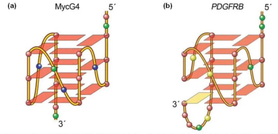
Figure 1 – Schematic structure of a G-tetrad involving four coplanar guanines, with a monovalent cation (M+) located at the centre (a) (taken from Zhang et al., 2014); schematic representation of a G4 motif formed by the stacking of three G-tetrads and stabilised by monovalent cations (b) (modified from Murat & Balasubramanian, 2014)...4 Figure 2 – Sequence and folding topologies of the major G4s formed in MYC gene promoter, MycG4 (a), and in PDGFRB gene promoter (b) (modified from Chen & Yang, 2012 and Onel et al., 2014)...7 Figure 3 – Folding topology (a), and schematic representation (b) of human KIT 1 G4
(modified from Qin & Hurley, 2008, and Amato et al., 2011)...8 Figure 4 – Sequences involved in the formation of the two G4s present in the promoter regions of human and canine KIT (Rankin et al., 2005, and Da Ros et al., 2014)...8 Figure 5 – Schematic representation of the structure of KIT receptor. D1-D5 corresponds to the five Ig-like domains (modified from Liang et al., 2013)...10 Figure 6 – Schematic representation of the activation of the KIT receptor by the SCF: on the
left, a model of KIT dimer with two free molecules; on the right, the D4-D4 and D5-D5 domains become reoriented, in close proximity in actual SCF-induced KIT dimer
(modified from Liang et al.,
2013)...11 Figure 7 – Representation of the basic structure of a RTK. TKI binding results in blocking of ATP binding, autophosphorilation and activation of downstream signalling (modified
from Bavcar & Argyle,
ix
TABLE OF ABBREVIATIONS, ACRONYMS, AND SYMBOLS
A Adenine
a.u. Arbitrary unit
A260/230, A260/280 Absorbance ratios
AgNOR Argyrophilic nucleolar organizing region ALL Acute lymphoblastic leukaemia
AML Acute myeloid leukaemia
ANOVA Analysis of variance
AQ Anthraquinone
ATP Adenosine 5’-triphosphate
B2M Beta-2-microglobulin
BCL2 B-cell CLL/lymphoma 2
BCR–ABL Breakpoint cluster region-abelson
bp Base pair(s)
C Citosine
C1, C2, C3 Compound 1, compound 2, compound 3 CD Cluster of differentiation
CD117 Cluster of differentiation 117
cDNA Complementary DNA
cm Centimetre
cm2 Square centimetre
CMCT Canine mast cell tumour
CML Chronic myeloid leukaemia
Co. Company
CO2 Carbon dioxide
CP, Ct Crossing point, cycle threshold
D Domain
DFSP Dermatofibrosarcoma protuberans
DMSO Dimethyl sulfoxide
DNA Deoxyribonucleic acid
dNTP Deoxynucleoside triphosphate DVM Doctor of Veterinary Medicine e.g. Exempli gratia/for example
ECVCP European College of Veterinary Clinical Pathology
ECVPT European College of Veterinary Pharmacology and Toxicology EDTA Ethylenediaminetetraacetic acid
x
EMEA European Agency for the Evaluation of Medicinal Products EMEM Eagle’s Essential Medium
FACS Fluorescent-activated cell sorters
FAK Focal adhesion kinase
FAM Fluorescein amidite
FBS Foetal bovine serum
FC Flow cytometry
FGF Fibroblast growth factor
FGFR Fibroblast growth factor receptor FGFR3 Fibroblast growth factor receptor 3
FL Fluorescence
FLT3 Fms-related tyrosine kinase 3
FNA Fine needle aspirate
FSC Forward scatter channel
G Guanine
g Gram
g Relative centrifugal force
G4 G-quadruplex
GAPDH Glyceraldehyde-3-phosphate dehydrogenase GIST Gastrointestinal-stromal tumour
GUSB Glucuronidase, beta
Gy Gray
h Hours
H2O Water
HES Advanced hypereosinophilic syndrome
HKG Housekeeping gene
HPRT1 Hypoxanthine phosphoribosyltransferase 1
HUGO Human Genome Organisation
IC50 Inhibitory concentration 50 ICC Interstitial cells of Cajal
Ig Immunoglobulin
Inc. Incorporated
ITD Internal tandem duplication
K+ Potassium ion
kg Kilogram
KIT V-kit Hardy-Zuckerman 4 feline sarcoma viral oncogene homolog KRAS Kirsten rat sarcoma viral oncogene homolog
xi LCK Lymphocyte-specific kinase
Ltd. Limited company
LYN Lck/Yes-related protein
m Metre
M Molar
M+ Monovalent cation
m2 Square metre
MCT Mast cell tumour
MFI Mean fluorescent intensity
mg Milligram
min Minute
mL Millilitre
mM Millimolar
mRNA Messenger RNA
MSc Master of Science
MYC V-myc avian myelocytomatosis viral oncogene homolog
n, N Number
Na+ Sodium ion
NCBI National Center for Biotechnology Information
ND Naphthalene diimide
NEAA Non-essential amino acids
ng Nanogram
NHE Nuclease Hypersensitive Element
nm Nanometre
NMR Nuclear magnetic resonance
nt Nucleotide
P/S Penicillin/streptomycin PBS Phosphate-buffered saline
PCR Polymerase chain reaction
PDGF Platelet-derived growth factor
PDGFA Platelet-derived growth factor alpha polypeptide PDGFRA Platelet-derived growth factor receptor alpha
PDGFRB Platelet-derived growth factor receptor, beta polypeptide
PE Phycoerythrin
PhD Doctor of Philosophy
PQS Putative G4-forming sequence Primer F Primer forward
xii
Prof. Professor
qPCR Quantitative real-time polymerase chain reaction
RNA Ribonucleic acid
RPMI Roswell Park Memorial Institute medium
Rq Relative quantification
RT Reverse transcription
RTK Receptor tyrosine kinase
RT-PCR Real-time reverse transcription PCR
S.A. Anonymous society
SCF Stem cell factor
SD Standard deviation
sec Second
SMI Small molecule inhibitor
ss Single-stranded
SSC Side scatter channel
T Timine
T/E Trypsin-EDTA
T0, T6, T12, T24 Time points (zero, six, twelve and twenty four hours post-treatment, respectively)
TERT Telomerase reverse transcriptase TKI Small molecule tyrosine kinase inhibitor TSS Transcription start site
U Unit
UK United Kingdom
UPL Universal ProbeLibrary
USA United States of America
UV Ultraviolet
VEGF Vascular endothelial growth factor
VEGFR2 Vascular endothelial growth factor receptor 2
WT Wild-type % Percent < Lower than > Higher than ∆ Delta, change ® Registered trademark ™ Trademark °C Celsius µg Microgram
xiii
µL Microlitre
µM Micromolar
1
1. TRAINING PERIOD ACTIVITIES
As part of the final year of my Integrated Master’s Degree in Veterinary Medicine, I was enrolled in a training period of, approximately, seven months, at the Department of Comparative Biomedicine and Food Science, University of Padua, Italy. The training period took place under the Erasmus Plus Program, and was supervised by Prof. Mauro Dacasto and co-supervised by Prof. Berta São Braz.
The aforementioned Department is involved in the teaching and research on fields related to veterinary medicine, animal science and food safety. The main topics of the research activities within this department include basic science applied to veterinary medicine, pathology and comparative medicine, as well as genetic variation in natural and cultivated populations, food safety and quality and consumer health, and bioethics. Specifically, I joined the activities of the research group on Veterinary Pharmacogenetics and Toxicogenomics, which was composed by:
- Mauro Dacasto (Full Prof., DVM, PhD, Dipl. ECVPT); - Mery Giantin (Associate Prof., DVM, PhD);
- Rosa Lopparelli (Technician, MSc in Agricultural Sciences, PhD);
- Vanessa Zancanella (Post-doctoral researcher, MSc in Biotechnology for Food Science, PhD);
- Eleonora Zorzan (PhD student, MSc in Biotechnology for Food Science); - Ramy Elgendy (PhD student, MSc in Veterinary Pharmacology, DVM).
I worked mostly with Eleonora Zorzan and Nuno Coelho (a post-graduate fellow researcher, DVM). Furthermore, I was involved in collaborative activities with two members of the veterinary pathology group, namely Maria Elena Gelain (DVM, PhD, Dipl. ECVCP) and Federico Bonsembiante (PhD student, DVM).
Overall, I was involved in the activities related to the work developed for this dissertation, which are described over the following chapters, and also in concurrent research projects, mostly related to veterinary oncology and anti-cancer chemotherapy. In particular, I was given the possibility to acquire both theoretical and practical skills on general laboratory maintenance and basic biomolecular techniques. With regard to these latter ones, techniques of cell culture handling and maintenance, total DNA (deoxyribonucleic acid) and RNA (ribonucleic acid) extraction, quantitative and qualitative assessment of the extracted nucleic acids, reverse transcription (RT), quantitative real-time polymerase chain reaction (qPCR), gel agarose electrophoresis, and flow cytometry (FC). Furthermore, I developed skills on data handling and basic statistical tools.
I also took an active part in the research group’s Journal Club meetings, which consisted in weekly scientific discussions coordinated by a member of the group who presented an article related to the ongoing research projects, and conducted a subsequent critical discussion.
3
2. INTRODUCTION
2.1. G-QUADRUPLEX
2.1.1. G-quadruplex structure
The well known double-helix structure of DNA, in which two complementary strands are held together in base pairs (bp), was first described by Watson and Crick more than six decades ago. It was later found, however, that DNA sequences can adopt alternative secondary structures such as the so-called G-quadruplexes (or “G4”), discovered by Gellert and his co-workers in 1962. Since then, they have gained increased attention for their emerging role in biological systems, and have been subject to numerous in vitro and in vivo studies. More recently, G4s have also been found to form in certain RNA sequences (Sundquist & Heaphy, 1993; Christiansen, Kofod & Nielsen, 1994; Biffi, Di Antonio, Tannahil & Balasubramanian, 2014).
G4s are formed in specific guanine-rich (“rich”) sequences, and consist of stacked G-tetrads (or G-quartets). G-G-tetrads are square-planar platforms, each one being composed by four guanines (G) connected by cyclic Hoogsteen hydrogen bonding (figure 1), which is different from the Watson-Crick hydrogen bonding of the double-helix model (Yang & Okamoto, 2010). These structures are further stabilized by monovalent cations (e.g. Na+, K+) that occupy the central cavities of the stacks and are coordinated by the strong negative electrostatic potential of eight guanine’s carbonyl oxygen atoms, neutralizing this repulsion (Düchler, 2012; Bidzinska, Cimino-Reale, Zaffaroni & Folini, 2013). The exact location of the cations depends on their nature and, consequently, different structures may be adopted. Given that K+ and Na+ are the main cations occurring in vivo, G4 formation is favoured under physiological conditions (Yang & Okamoto, 2010).
In each G-tetrad, the guanine residues can assume either a syn or anti glycosidic conformation, according to the glycosidic torsion angles, which determines the orientation of the four stranded G-tracts or legs within a G4. The legs may be parallel or anti-parallel to each other, and be arranged in all possible combinations. They are connected by loops of residual sequences that can be displayed externally, laterally or diagonally (Zhang, Wu & Zhang, 2014).
Depending on the number of separate strands that compose them, G4s can be classified as one (unimolecular or intramolecular), two (bimolecular), three (trimolecular), or four (tetramolecular) stranded (Zhang et al., 2014).
G4 have been found to be present in a wide variety of structural conformations, more or less complex. Their diversity is essentially determined by parameters such as the oligonucleotide sequence, the guanine glycosidic torsion angles, the number and orientation of the strands,
4
the number of stacked G-tetrads, the type of loop, groove and cation and the presence of binding ligands (Bidzinska et al., 2013; Zhang et al., 2014).
The formation of G4s and their structures can be investigated by means of several techniques, namely nuclear magnetic resonance (NMR) spectroscopy and X-ray crystallography. Biophysical techniques such as circular dichroism, fluorescence, and ultraviolet (UV) spectroscopies can also provide valuable insights into G4 conformation (Fernando et al., 2006; Onel, Lin & Yang, 2014). In vivo techniques have included the use of specific antibodies against G4s, and radiolabeled or fluorescent G4 ligands (Yang & Okamoto, 2010).
Figure 1 – Schematic structure of a G-tetrad involving four coplanar guanines, with a monovalent cation (M+) located at the centre (a) (taken from Zhang et al., 2014); schematic representation of a G4 motif formed by the stacking of three G-tetrads and stabilised by monovalent cations (b) (modified from Murat & Balasubramanian, 2014).
2.1.2. G-quadruplex presence and role in biological systems
DNA G4 structures with biological relevance were first found to form in eukaryotic chromosomal telomeric DNA (Henderson et al., 1987; Sundquist & Klug, 1989). Since then, numerous putative G4-forming sequences (PQSs) have been identified in eukaryotic telomeres and other DNA tandem repeats, in other regions including human gene promoters, immunoglobulin switch regions (S regions) as well as in RNA 5’ untranslated regions (5’UTRs) (Bagaut & Balasubramanian, 2012; Düchler, 2012; Maizels & Gray, 2013; Zhang et al., 2014). Bioinformatic analyses have, indeed, revealed about 400,000 PQSs in the human genome (Bidzinska et al., 2013).
The production of antibodies directed against telomeric G-quadruplex DNA in ciliates provided evidence for the in vivo presence of G-quadruplexes (Schaffitzel et al., 2001). Other than telomeres, these structures have been visualized in human cells at different sites on chromosomes, also through G4-specific antibodies, and their frequency was shown to increase in the presence of a G4-interactive compound (Siddiqui-Jain & Hurler, 2013; Onel et
5
al., 2014). Furthermore, many proteins can interact with G4s, which may involve binding and stabilization of these structures or, conversely, their unwinding and destabilization (Onel et al., 2014).
The distribution and concentration of G4s in biologically significant regions such as the telomeres and oncogene promoter regions has suggested an influence of these structures on gene metabolism and other important biological processes such as DNA replication, transcriptional regulations and genome stability (Zhang et al., 2014).
2.1.2.1. G-quadruplex in telomeres
Telomeres are DNA-protein complexes which protect the chromosome ends from degradation, and are known to play an important role in cancer, ageing and overall genetic stability. Human telomeric DNA consists of thousands of tandem repeats of the G-rich d(TTAGGG) sequence that end in a single-stranded G-rich 3´ overhang up to about 600 bases long (Maizels & Gray, 2013; Onel et al., 2014).
Under physiological ionic conditions, telomeric DNA G-rich sequences are capable of forming G-quadruplexes, which have been found to be highly polymorphic in their structures (Düchler, 2012). As an example, a 22-mer telomeric DNA was found to form an antiparallel structure in Na+ solution (Wang & Patel, 1993), and a parallel structure in the crystalline form in the presence of K+ (Parkinson, Lee, & Neidle, 2002). Furthermore, Xu and co-authors (2006) suggested that, in K+ solution, the same d[AG3(T2AG3)3] sequence could form a (3+1) or hybrid G -quadruplex. On the basis of the three different results with the same sequence, it was assumed that the formation of several polymorphic G-quadruplex structures was mainly attributable to the different conditions used for their formation, including cation type and molecular crowding environment (Zhang et al., 2014).
The presence of G4 structures within telomeres may influence the control of telomere maintenance, and, consequently play an important role in cancer and ageing (Bidzinska et al., 2013; Maizels & Gray, 2013).
2.1.2.2. G-quadruplex in gene promoter regions
A number of PQSs has been identified upstream of gene transcription start sites (TSSs), in the promoter regions of human oncogenes associated with cell growth and proliferation, including: the v-myc avian myelocytomatosis viral oncogene homolog (MYC), the Kirsten rat sarcoma viral oncogene homolog (KRAS), B-cell CLL/lymphoma 2 (BCL2), vascular endothelial growth factor (VEGF), platelet-derived growth factor receptor, beta polypeptide (PDGFRB), platelet-derived growth factor alpha polypeptide (PDGFA), telomerase reverse transcriptase (TERT) and the v-kit Hardy-Zuckerman 4 feline sarcoma viral oncogene
6
homolog (KIT) (Düchler, 2012; Onel et al., 2014). Nearly half of all known genes in the human genome harbour PQSs sequences within 1,000 nucleotides upstream of the TSS (Bryan & Baumann, 2011). Furthermore, these structures seem to be underrepresented at tumour suppressor genes level (Bochman, Paeschke, & Zakian, 2012). This evidence suggests that G4s may play a functional role in gene regulation (Bryan & Baumann, 2011). A requirement for promoter sequences is that the G-quadruplex must be generated in a duplex DNA region, rather than single-stranded DNA as in the case of telomeres (Chen & Yang, 2012).
The promoter G4-forming sequences are considerably more diverse than the telomeric repeating tandems, and often contain more than four G-tracts. As a consequence, a certain sequence can give rise to multiple G4s from the combinations of several G-tracts, or different loop isomers by utilizing different guanines on one G-tract. Three-tetrad G4s are the most widespread (Onel et al, 2014). Parallel-stranded G4 structures have been found to be common in the human promoter sequences. In fact, a particular G3NG3 motif (N being a loop residue), which forms a robust parallel-stranded structure motif with a single-nucleotide (nt) strand-reversal loop, has been found to be widely prevalent in promoter sequences. Therefore, this motif may have been naturally selected as a basis for promoter G4 formation (Chen & Yang, 2012; Onel et al., 2014).
MYC is the most extensively studied system of promoter G4 formation (Chen & Yang, 2012). It encodes for a transcription factor that regulates the expression of several genes, and is one of the most common oncogenes found to be altered in cancer (Bidzinska et al., 2013). The G4-forming region of the MYC promoter is a 27-nucleotide Nuclease Hypersensitive Element (NHE) III1 sequence which regulates most of the gene’s transcriptional activity (Chen & Yang, 2012; Onel et al., 2014). This sequence contains five consecutive runs of guanine (“G-runs”), and may fold into several G4s and loop isomers with different stabilities. The major G4 formed in K+ solution is a three tetrads parallel-stranded structure involving four consecutive three-G-runs with two G3NG3 single-nt strand-reversal loops (MycG4, figure 2). The G3NG3 parallel-stranded structural motif was found in all of the MYC G4s and loop isomers (Chen & Yang, 2012; Onel et al., 2014).
In one study, a single point mutation which destabilizes a G4 in the NHE III1 region resulted in a significant increase in basal transcriptional activity of this gene (Siddiqui-Jain, Grand, Bearss & Hurley, 2002); conversely, the induction of G4 formation by a bile acid-amino acid conjugate resulted in down-regulation of transcription (Tian et al., 2010).
These and several other evidence point to a potential regulatory role of this G4 as a MYC repressor element to transcriptional activation of the gene (Siddiqui-Jain et al., 2002).
The major G4 formed in the PDGFRB promoter sequence is another example of a three-G-tetrad parallel-stranded motif. However, instead of the four runs of consecutive guanines, a characteristic “broken” G-strand structure made up by guanines from two different G-runs is
7
adopted, with three single-nt loops (figure 2) (Chen & Yang, 2012). This represents only an example of the possible variations in parallel G4s structures.
Figure 2 – Sequence and folding topologies of the major G4s formed in MYC gene promoter, MycG4 (a), and in PDGFRB gene promoter (b) (modified from Chen & Yang, 2012 and Onel et al., 2014).
2.1.2.2.1. G-quadruplex in the promoter region of human KIT
In the promotorial region of human proto-oncogene KIT, two G-rich sequences capable of forming G4s have been identified: KIT 1 (Rankin et al., 2005) and KIT 2 (Fernando et al., 2006) (figure 4). They are located within a nuclease hypersensitive region, and are separated by 31 bp. KIT 1 is positioned between -87 and -109 bp, and KIT 2 between -140 and -160, both upstream of the TSS (Rankin et al., 2005; Fernando et al., 2006). Both sequences have been shown to form G4s under physiological conditions, which have been characterized by means of NMR and X-ray crystallography (Bryan & Baumann, 2011).
The quadruplex formed in KIT 1, in K+ solution, has a parallel fold with four loops: two single-nt double-chain reversal loops that bridge three G-tetrad layers, a two-single-nt loop which connects two adjacent corners, and a long five-nt lateral stem loop that allows the two terminal Gs to be inserted back to the G-tetrad core; unusually, despite the presence of four three-G-runs, one isolated non-G-tract guanine (G10) participates in the core of stacked G-tetrads (Phan, Kuryavyi, Burge, Neidle & Patel, 2007; Wei, Husby & Neidle, 2014) (figure 3). This peculiar “snapback” parallel-stranded G-quadruplex also contains a G3NG3 motif which can serve as a core structure (Yang & Okamoto, 2010).
Regarding KIT 2, it appears to form an intramolecular parallel-stranded G4 in K+ solution, in which the four runs of three Gs are connected by three double-chain reversal loops: two single-nt loops and a five bp intermediate loop sequence. Moreover, other structures may arise from KIT 2 such as a parallel dimeric form G4 structure, illustrating its highly polymorphic folding behaviour (Fernando et al., 2006; Hsu et al., 2009; Kuryavyi, Phan, & Patel, 2010).
8
Figure 3 – Folding topology (a), and schematic representation (b) of human KIT 1 G4 (modified from Qin & Hurley, 2008, and Amato et al., 2011).
2.1.2.2.2. G-quadruplex in the promoter region of canine KIT
Very recently, Da Ros and co-authors (2014), verified that, similar to what has been described for the human KIT proto-oncogene, two PQSs occur in the canine KIT promotorial region (figure 4).
Canine KIT 1 (“d_kit1”) is positioned from 117 to 138 bp, and KIT 2 (“d_kit2”) from 154 to -174 bp, both upstream the ATG (the positions were not referred to the TSS, because the 5’UTR of the canine KIT gene has not yet been fully characterized) (Da Ros et al., 2014). Canine KIT 1 was found to share a common overall parallel G4 folding with its human counterpart, in a K+ containing solution, evidence that is supported by the high degree of sequence homology between the two sequences (Da Ros et al., 2014).
On the other hand, human and canine KIT 2 sequences differ from each other to a greater extent. Two isoforms of canine KIT 2 were identified and, although a defined structure was not yet attributed, the multiplicity of folded structures that human KIT 2 can form in solution may also occur in the other species counterpart (Da Ros et al., 2014).
The potential functional role of the aforementioned G4 motifs may be of important relevance, not only for the canine, but also as a comparative model for human disease (Da Ros et al., 2014).
Figure 4 – Sequences involved in the formation of the two G4s present in the promoter regions of human and canine KIT (Rankin et al., 2005; Da Ros et al., 2014).
9
2.2. RECEPTOR TYROSINE KINASES
Receptor tyrosine kinases (RTKs) are a family of cell transmembrane receptors for extracellular signalling molecules, including growth factors and hormones (Sun & Bernards, 2014). They are activated through binding of aforementioned molecules and are essential regulators of critical cellular processes, such as proliferation and differentiation, cell survival and metabolism, cell migration and cell cycle control (Lemmon & Schlessinger, 2010).
A typical RTK consists of an extracellular ligand-binding domain, a single transmembrane domain, and an intracellular tyrosine kinase domain (Sun & Bernards, 2014). The RTK family can be further divided into 20 subfamilies, according to their structural characteristics, with each family sharing a homologous domain that specifies its catalytic tyrosine kinase function (Zwick, Bange & Ullrich, 2001). Members of the type III RTK are characterized by having five extracellular immunoglobulin (Ig)-like domains and a kinase insert sequence of 70-100 amino acids located in the middle of the intracellular kinase domain. This family includes receptors KIT, the platelet-derived growth factor receptor alpha (PDGFRA) and PDGFRB (Lennartsson & Rönnstrand, 2012).
2.3. KIT PROTO-ONCOGENE AND THE KIT RECEPTOR
The KIT receptor – also referred to as stem cell factor receptor, KIT or cluster of differentiation 117 (CD117) –, is a type III RTK otherwise known as receptor for stem cell factor (SCF) (Letard et al., 2008). KIT is encoded by the KIT proto-oncogene, which was first identified and sequenced in humans by Yarden and co-authors in 1987, shortly after the discovery of the feline retrovirus Hardy-Zuckerman 4 feline virus.
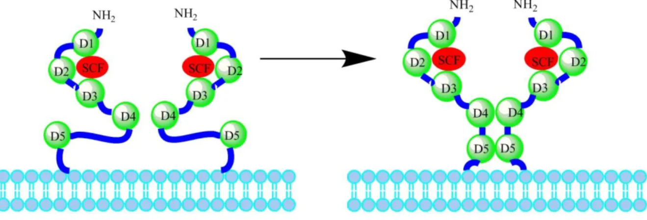
The KIT proto-oncogene is located in the chromosome 4 of the human genome (Yarden et al., 1987), and in the chromosome 13 of the dog’s genome (Reimann-Berg, Escobar & Nolte, 2012). In both species, it consists of 21 exons which encode the protein-tyrosine kinase receptor. This transmembrane receptor comprises an extracellular domain composed of five Ig-like domains (D1-D5) encoded by exons 1-9, a transmembrane domain (exon 10), and an intracellular domain (exons 11-21). The intracellular domain is further divided into a negative regulatory juxtamembrane domain and a cytoplasmatic tyrosine kinase domain that is split by an 80 amino acids kinase insert sequence into the adenosine 5’-triphosphate (ATP)-binding and phosphotransferase lobes, ending with a COOH-terminal tail (figure 5) (Letard et al., 2008; Lennartsson & Rönnstrand, 2012).
10
Figure 5 – Schematic representation of the structure of KIT receptor. D1-D5 corresponds to the five Ig-like domains (modified from Liang et al., 2013).
2.3.1. KIT activation mechanism
The ligand for the KIT receptor is SCF, also known as mast cell growth factor, KIT ligand or steel factor (Liang et al., 2013). It is a hematopoietic growth factor expressed by fibroblasts and endothelial cells, playing an important role in promoting proliferation, migration, survival, and differentiation of hematopoietic progenitors, melanocytes, and germ cells (Lennartsson & Rönnstrand, 2012). The SCF is expressed in a wide range of tissues such as brain, endothelium, gametes, heart, kidney, lung, melanocytes, skin, liver, thymus and bone marrow stromal cells (Takeuchi et al., 2010).
SCF is a homodimer and exists as membrane-bound and soluble forms, both capable of binding and activating KIT’s intrinsic tyrosine kinase activity (Masson & Rönnstrand, 2009). Each SCF molecule can bind to one molecule of KIT through contacts with the first three Ig-like domains (D1-D2-D3) of the receptor’s extracellular region, which have a complementary shape and charge to allow tight binding of SCF (Lemmon & Schlessinger, 2010; Lennartsson & Rönnstrand, 2012).
When association with the SCF takes place in two KIT monomers, their D1-D2-D3 regions are kept structurally unaltered but the two receptors are drawn closer to each other to form a dimer, leading to a reorientation on D4 and D5 (figure 6) (Lemmon & Schlessinger, 2010). Through homotypic interactions between D4 and D5 across the dimer interface, the two KIT molecules become properly oriented for activation, with the transmembrane regions getting close(r) to each other, as well as the intracellular tyrosine kinase domains, enabling their activation and following transphosphorylation along the receptor, which leads to initiation of downstream signal transduction (Lemmon & Schlessinger, 2010; Lennartsson & Rönnstrand, 2012).
11
Figure 6 – Schematic representation of the activation of the KIT receptor by the SCF: on the left, a model of KIT dimer with two free molecules; on the right, the D4-D4 and D5-D5 domains become reoriented, in close proximity in actual SCF-induced KIT dimer (modified from Liang et al., 2013).
2.3.2. KIT distribution and functions
The KIT receptor plays an important role in several cellular processes including cell survival, and normal growth, development, maintenance and proliferation. KIT-dependent cell types include hematopoietic stem and progenitor cells, but this receptor is also normally expressed in high numbers in multiple types of differentiated cells such as mast cells, interstitial cells of Cajal (ICC), melanocytes and germ cells (Amagai et al, 2015; Fernando et al., 2006). In addition to these functions, in mast cells, KIT is also involved in fibronectin adhesion, chemotaxis, and degranulation (Webster, Kiupel & Yuzbasiyan-Gurkan, 2006).
In adult dogs, KIT is expressed in the Purkinje cells of the cerebellum, in ICC, in the luminal epithelial cells of the mammary gland, endometrial epithelial cells and in mast cells (Costa, 2015).
2.3.3. Human KIT mutations and related diseases
Dysregulation of KIT receptor has been identified in several human and canine diseases, mostly associated with mutations in its codifying gene.
Loss-of-function mutations can cause piebaldism, an autosomal dominant condition that affects mammals and is characterised by the presence of white spots of amelanotic regions on the skin (Roberts & Govender, 2015).
A number of different mutations have been implicated in the pathogenesis of a variety of cancers and are known to promote tumorigenesis, which can induce the activation of the receptor, even in the absence of SCF stimulation (Bavcar & Argyle, 2012).
In humans, gain-of-function mutations in KIT can be found in gastrointestinal-stromal tumour (GIST), systemic mastocytosis, mast cell leukaemia, acute myeloid leukaemia (AML), nasal
12
T-cell lymphomas, seminoma, dysgerminoma, and melanoma (Lemmon & Schlessinger, 2010; Bavcar & Argyle, 2012; Roberts & Govender, 2015).
These mutations include missing mutation, point mutation, duplication and insertion and are often located in key regions within the KIT protein: in the extracellular domain, specifically the membrane proximal Ig-like domain (D5, exon 8 and exon 9), and especially in the juxtamembrane and the tyrosine kinase domains (exons 11 and 17, respectively) (Lemmon & Schlessinger, 2010; Liang et al., 2013; Roberts & Govender, 2015).
As mentioned before, KIT signalling is required for the differentiation, survival and function of mast cells. A critical role for mutations in the juxtamembrane and tyrosine kinase domains in mast cells tumorigenesis was revealed, both in vitro and in vivo (Kitayama et al., 1995; Hashimoto et al., 1996). Mutations in the extracelular domain have also been found in mast cell malignances, as well as AML and GIST. They are mostly found in the D5 Ig-like domain (exons 8 and 9) and are thought to stabilize intermolecular interactions between two neighbouring KIT D5 domains, promoting constitutive receptor-mediated dimerization by enhancing the affinity of adjacent domains, causing KIT autophosphorylation. Whether these mutations directly promote tumorigenesis in vivo and their exact mechanism has remained unclear (Lemmon & Schlessinger, 2010; Liang et al., 2013; Amagai et al., 2015). However, Amagai and his co-workers (2015) have very recently demonstrated for the first time that a point mutation in the extracellular domain of KIT leads to mast cells proliferation via ligand independent auto-dimerization, both in vitro and in vivo, by using a murine progenitor mast cell line, in which a KIT mutant derived from a canine mast cell tumour was introduced. Furthermore, the paracrine or autocrine activation of KIT have been implicated in many other human malignancies, such as ovarian cancer and small cell lung cancer (Liang et al., 2013).
2.3.4. Canine KIT mutations and related diseases
In dogs, several KIT mutations have been identified in mast cell tumours (MCTs) which, alike humans, lead to uncontrolled signalling (Bavcar & Argyle, 2012). These mutations are similar to those found in human GISTs (Marech et al., 2014).
Approximately 30% of canine mast cell tumours (CMCTs) present KIT mutations, which are more frequently localized in the juxtamembrane domain (exon 11), and mainly consist of internal tandem duplications (ITDs) (Marech et al., 2014; Bonkobara, 2015). These mutations result in constitutive activation of the receptor in the absence of ligand binding, playing a key role in CMCTs pathogenesis (Bavcar & Argyle, 2012; Marech et al., 2014). Mutations have also been found in exons 2, 6, 7, 8, 9, 15 and 17. Mutations in the extracellular domain, namely in D5 (exons 8 and 9) are less frequent, compared with those in exon 11, even though a significant number of mutations have been identified (Letard et al., 2008; Takeuchi et al., 2013). Overall, mutations in other exons are infrequent (<3%) (Bonkobara, 2015).
13
2.4. CANINE MAST CELL TUMOURS
2.4.1. Mast cell biology and function
Mast cells are a heterogeneous cell population which arises as precursors in the bone marrow and then migrates to the peripheral tissues, where it undergoes final differentiation and maturation, under the influence of SCF and other local cytokines (Blackwood et al., 2012; Urb & Sheppard, 2012). Although they are present in most organs and tissues of the body, it is in areas of environment interface that mast cells are normally more abundant, such as the skin, and mucosal surfaces including the lungs and the gastrointestinal tract. (Blackwood et al., 2012).
Mast cells play an important role in the mediation of hypersensivity, allergy and inflammatory responses (Rogers, 2010; Blackwood et al., 2012). They contain intracytoplasmatic granules that harbour a variety of bioactive constituents and mediators which are released to the extracellular compartment upon mast cell activation by physical or chemical trauma or when stimulated by immune mechanisms (Welle, Bley, Howard & Rüfenacht, 2008). These substances include the vasoactive amines histamine and serotonin, enzymes such as acid hydrolases and phospholipase A, proteoglycanes such as heparin, cytokines and growth factors (Rogers, 2010). The granules can be metachromatically stained with cationic dyes, which allow mast cell identification (Blackwood et al., 2012).
2.4.2. CMCTs and KIT
Mast cells can undergo neoplastic transformation in solitary and multiple cutaneous MCTs, as well as in visceral or systemic mastocytosis (Welle et al., 2008).
Several factors may be involved in the development of CMCTs (Welle et al., 2008). As mentioned before, although not entirely elucidated, mutations in the KIT proto-oncogene leading to altered expression of the receptor and loss of normal regulation have been implicated in the pathogenesis of the disease. The constitutive activation of KIT caused by those mutations may lead to degranulation and secretion of aforementioned substances including numerous pro-angiogenic factors such as VEGF, PDGF, and fibroblast growth factor (FGF), which may favour tumour development (Marech et al., 2014). In addition, these activating mutations have been associated with a higher histological grade of CMCTs and poor prognosis (Bavcar & Argyle, 2012; Marech et al., 2014). Aberrant KIT localization and KIT mutations have been associated with an increased expression of both argyrophilic nucleolar organizing regions (AgNORs) and Ki-67, which are markers of increased cellular proliferation (Webster, Yuzbasiyan-Gurkan, Miller, Kaneene & Kiupel, 2007).
14
2.4.3. CMCTs epidemiology
MCTs are one of the most frequent cutaneous neoplasms in the dog, representing up to 21% of all canine skin tumours, being diagnosed more frequently than human MCTs (Marech et al., 2014; Amagai et al., 2015).
The average age for CMCTs onset is 8.5 to 9.5 years, even if it can be developed at any age. No gender predilection has been reported. Even though any dog can be affected, certain breeds show higher predisposition, namely most of the brachycephalic breeds and Golden Retrievers. While Boxers are at increased risk for developing the disease, a well-differentiated form with a more favourable prognosis is often reported. Multiple cutaneous tumours are common, occurring in up to 15% of CMCTs. In Pugs, they frequently arise, although typically benign (Rogers, 2010).
2.4.4. CMCTs diagnosis and clinical presentation
CMCTs clinical appearance is highly variable and can mimic other cutaneous tumours and non-neoplastic conditions. For that reason, they must be considered in the differential diagnosis of any skin mass (Welle et al., 2008). CMCTs may develop anywhere in the body and are usually cutaneous (dermal) or subcutaneous (Blackwood et al., 2012). A solitary, non-painful mass is the most commonly found form of CMCTs (Rogers, 2010). These tumours can also be highly variable in their biological behaviour and progression. Although not frequently, MCTs can metastasise to draining lymph node(s), liver, spleen and bone marrow and can also cause local cutaneous “satellite lesions”. Well-differentiated cutaneous MCT are characterized as slow growing, hairless and solitary lesions. Poorly differentiated lesions, on the other hand, exhibit rapid growth, are ulcerated and pruritic, with occasional small satellite lesions in their proximities (Blackwood et al., 2012). Both disseminated mastocytosis without an associated cutaneous MCT and tumours of gastrointestinal origin are quite uncommon (Rogers, 2010).
Systemic signs including anorexia, erythema, abdominal discomfort, edema, gastrointestinal ulceration, vomiting, and melaena are less common and are most frequently associated with visceral forms of MCT and with a poorer prognosis, and/or with paraneoplastic disease due to the release of bioactive constituents from mast cell granules (Welle et al., 2008; Rogers, 2010; Blackwood et al., 2012).
Definitive diagnosis of CMCTs can be achieved through cytology from fine needle aspirate (FNA) and/or histopathology. Immunohistochemical techniques may help in differentiating certain susbsets of MCTs (Rogers, 2010; Blackwood et al., 2012).
Histological examination is also an important tool for CMCTs grading and prognosis (Sabattini, Scarpa, Berlato & Bettini, 2015). Various histological grading systems have been
15
proposed. Among them, the classification by Patnaik et al. (1984) is the most widely used, and divides CMCTs into three grades: grade I (well-differentiated) are mostly benign and slowly developed; grade III (poorly differentiated) exhibit aggressive growth, are locally invasive, more likely to metastasise, with a high recurrence potential, and frequently result in death; grade II (intermediately differentiated) tumours’ behaviour is, however, more difficult to predict, accounting for more subjective and inconsistent classifications (Blackwood et al., 2012; Sabattini et al., 2015). A new grading system was proposed by Kiupel and co-authors (2011), consisting in a two-tier classification into high or low grade MCTs. It has improved prediction of metastasis and/or new tumour development and tumour mortality and survival (Takeuchi et al., 2013; Sabattini et al., 2015).
Histological grading alone hasn’t been suitable for accurately predicting the biological behaviour and treatment response of each tumour, and supplementary prognostic markers have been investigated. KIT mutation status has been identified as a useful prognostic and therapeutic marker. KIT protein may present different expression patterns in CMCTs that can be immunohistochemically characterized and have been correlated with histopathological grading and prognosis in some studies, but not consistently (Morini, Bettini, Preziosi & Mandrioli, 2004; Costa-Casagrande et al., 2013; Takeuchi et al., 2013; Sailasuta et al., 2014; Patruno et al., 2014). Other prognostic markers have been investigated, such as: cellular proliferation markers (e.g. mitotic index, AgNOR, immunohistochemical markers Ki-67 and proliferating cell nuclear antigen), angiogenesis markers, and DNA mutational and ploidy analysis (Blackwood et al., 2012; Costa, 2015; Fonseca-Alves, 2015; Sabattini et al., 2015). Complementary to histological grading, clinical staging is recommended to define the nature and extent of the disease, especially after diagnosis of a poorly differentiated tumour or if an extensive or expensive treatment is planned, and should include, at the very minimum, FNAs of draining lymph nodes and abdominal ultrasound. In cases where nodal metastasis is present, full staging is required with abdominal ultrasound (along with spleen and liver aspiration) and, eventually, bone marrow aspiration and lung radiographs. FNAs and buffy coat smears examination must be carefully interpreted, as small numbers of normal mast cells can be seen in these preparations, particularly with concurrent inflammation. The value of buffy coat smears has been questioned in cases of CMCTs, and has, progressively, being less adopted (Blackwood et al., 2012).
A clinical staging system was proposed by the World Health Organization in which: Stage 0 is usually assigned to single tumours that are incompletely excised from the dermis without regional lymph node involvement; Stages I and II comprise single tumours confined to the dermis without or with regional lymph node involvement, respectively; in Stage III are included multiple dermal tumours, or large infiltrating tumours with or without regional lymph node involvement; and stage IV includes any tumour with distant metastasis or recurrence
16
with metastasis (Rogers, 2010). However, stage and prognosis do not directly correlate in all clinical situations (Blackwood et al., 2012).
2.4.5. CMCTs therapies
The conventional choices for CMCTs therapy are, as in most types of neoplasia, surgery, radiotherapy, chemotherapy, or a combination of these (Rogers, 2010; Couto, 2014). When making therapeutic decisions, important factors such as mass number and location, tumour grade, and presence or absence of metastasis, but also these tumours’ unpredictable biological behaviour should be considered (Rogers, 2010).
Complete surgical excision is the treatment of choice for localized, nonmetastatic CMCTs. (Rogers, 2010). The use of radiotherapy should be restricted to cases where surgery cannot achieve local control. About two thirds of dogs with a grade I or II localized MCT treated with radiotherapy alone are cured (Couto, 2014). It is more frequently performed after incompletely resected tumours, as a postoperative adjuvant therapy. It can also be used to treat local/regional nodal metastases (Blackwood et al., 2012).
The use of chemotherapy treatment is, usually, only palliative (Couto, 2014). It can be considered in the management of CMCTs for dogs with grade III tumours, tumours where grade II/III features are suggested by the pathologist, and grade II tumours with risk factors for reduced survival such as high proliferation marker values; and/or poor tumour location and metastasis. It can be performed after excision of the MCT, as well as in dogs with non excisable tumours, in attempt to treat, delay or prevent disseminated metastases (Blackwood et al., 2012; Garrett, 2014). Likewise, it can be used prior to surgery or radiotherapy, in order to reduce tumour burden and improve chances of complete excision, or to facilitate mass irradiation (Blackwood et al., 2012). Moreover, it may be considered for residual microscopic disease, in the impossibility of further surgery and if radiation therapy is not available (Blackwood et al., 2012).
Various chemotherapeutic drugs and protocols can be used, as single agents, in alternation or in combination with others. The use of prednisone and vinblastine together is the most popular used combination; lomustine is often added as well, or used as a single agent; combination of cyclophosphamide, prednisone and vinblastine has also been widely used. Other drugs include hydroxyurea (Welle et al., 2008; Blackwood et al., 2012; Couto, 2014). In dogs with nonresectable or metastatic MCTs, the response rate is, overall, of 30 to 35%, independently of the drug(s) used (Couto, 2014).
Attention should be paid to the potential toxic effects of these agents when choosing a chemotherapeutic intervention. Both lomustine and vinblastine can cause myelosuppression, thus complete blood counts must be performed prior to each dose administration (Blackwood
17
et al., 2012). Vinblastine is also a perivascular irritant, while lomustine can be hepatotoxic, requiring routine monitoring of liver enzymes (Blackwood et al., 2012; Garrett, 2014).
Supportive therapy aimed at decreasing the effects of histamine release and/or gastrointestinal signs includes H1 and H2 receptor antagonists, proton-pump inhibitors and gastro-protectants (Rogers, 2010; Blackwood et al., 2012).
Several additional treatment modalities for CMCTs have been proposed over the past few years. These include, for example, electrochemotherapy, which combines the administration of chemotherapeutic drugs (cisplatin or bleomycin) with application of electric pulses to the tumour to increase the cellular uptake of the drug, and immunotherapy based on interleukin-2 administration, which are currently under development and testing (Blackwood et al., interleukin-201interleukin-2; Costa, 2015). Since the discovery of mutations involvement in KIT receptor dysregulation in CMCTs, veterinary oncology research focused on targeted therapy based on inhibiting KIT and other related RTKs has intensified. Indeed, this new approach has been increasingly adopted in clinical practice (Bonkobara, 2015).
2.5. TYROSINE KINASE INHIBITORS
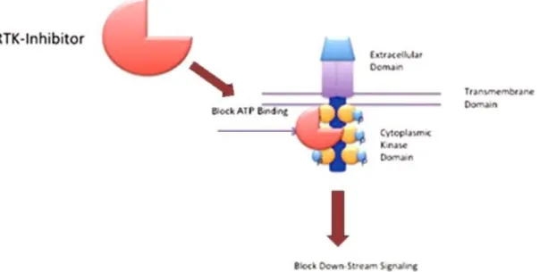
Despite the existence of several strategies for targeting protein kinases, the most successful approach has been the use of a class of drugs termed “small molecule tyrosine kinase inhibitors” (TKIs) (London, 2009). They most often act as ATP competitive inhibitors by blocking the ATP-binding sites of tyrosine kinases, which prevents receptor auto-phosphorilation and activation of downstream signal transducers, resulting in interruption of a survival/growth signal that is essential for survival of the tumour cell (figure 7) (London, 2009; Bavcar & Argyle, 2012). Their specificity is variable, with some TKIs able to target many different RTKs (Costa, 2015).
2.5.1. TKIs in human medicine
2.5.1.1. Imatinib
Imatinib (imatinib mesilate - Glivec, Novartis Europharm Limited, Camberley, United Kingdom) was the first TKI approved for use in human patients (Costa, 2015). Although originally designed to target the breakpoint cluster region-abelson (BCR–ABL) fusion protein in chronic myeloid leukaemia (CML), it was later shown to also inhibit the receptor tyrosine kinases KIT and platelet-derived growth factor (PDGFR) through binding to the receptors’ ATP-binding pockets (London, 2009; Bonkobara, 2015; Webster, 2015).
Imatinib considerably improved treatment for CML in humans (Bonkobara, 2015). Then, imatinib was gradually and successfully used to treat GIST (a tumour frequently with KIT
18
mutations). Its anti-tumour activity has been further demonstrated against other tumours with kinase mutations (Joensuu et al., 2001; Bonkobara, 2015). With regard to GIST, a control rate of 80 to 90% has been reported in patients treated with imatinib, with most of them showing partial remission and 12% complete remission (Liang et al., 2013).
At present, imatinib is considered the first-line anticancer drug for patients with unresectable or metastatic GIST (Roberts & Govender, 2015). Other therapeutic indications include, aside from CML, Ph+ acute lymphoblastic leukaemia (ALL), advanced hypereosinophilic syndrome (HES) and dermatofibrosarcoma protuberans (DFSP). The most common side effects are weight increase, neutropenia, thrombocytopenia, headache, nausea, vomiting, diarrhoea, (European Medicines Agency [EMA], 2013).
Figure 7 – Representation of the basic structure of a RTK. TKI binding results in blocking of ATP binding, autophosphorilation and activation of downstream signalling (modified from Bavcar & Argyle, 2012).
2.5.1.2. Sunitinib
Sunitinib (sunitinib malate - Sutent, Pfizer Limited, Kent, United Kingdom) is a second generation of multi-target TKIs (Liang et al., 2013). Like Imatinib, it works by sitting in the ATP-binding pocket of several RTKs, including KIT, VEGFR (vascular epithelium growth factor receptor), PDGFR, FLT3 (fms-related tyrosine kinase 3), and FGFR (fibroblast growth factor receptor); owing to this, sunitinib is active against several cancers, mostly preventing angiogenic and proliferative effects (London, 2009; Lemmon & Schlessinger, 2010; Roberts & Govender, 2015).
It has been successfully used in the treatment of renal cell carcinoma and imatinib-resistant GIST (Lemmon & Schlessinger, 2010). When treated with sunitinib, 61% of imatinib-resistant patients showed disease regression or stable disease lasting longer than four months;
19
moreover, 40% of renal cell carcinoma patients where interleukin-2 and/or interferon therapy had failed achieved a partial response, whereas an additional 25% experienced stable disease (London, 2009).
Currently sunitinib is indicated for the treatment of unresectable or metastatic GIST and as a second-line drug for imatinib-resistant GISTs. It has also been availed of in the treatment for metastatic renal cell carcinoma, and pancreatic neuroendocrine tumours. The most common side effects include fatigue, gastrointestinal disorders such as diarrhoea, indigestion and vomiting, respiratory signs (such as shortness of breath and cough), and skin disorders (discoloration, rash), among others (EMA, 2014).
2.5.2. TKIs in veterinary medicine
Tow orally administrated TKIs have been approved for use in veterinary medicine: toceranib (active substance toceranib phosphate – Palladia, Zoetis Belgium S.A., Louvain-la-Neuve, Belgium) and masitinib (in the form of masitinib mesylate – Masivet, AB Science S.A., Paris, France).
2.5.2.1. Toceranib
Toceranib is active against KIT, vascular endothelial growth factor receptor 2 (VEGFR2) and PDGFR, both of which are involved in tumour angiogenesis and metastasis (Costa, 2015). It has been approved for the use in recurrent non-resectable grade II and III cutaneous MCTs in dogs (EMA, 2013). In addition, it appears to have biological activity against mammary carcinoma, soft tissue sarcoma and anal gland adenocarcinoma, among others (Bavcar & Argyle, 2012).
Toceranib’s safety and efficacy were first studied in dogs against several malignancies, including sarcomas and carcinomas, MCTs, melanomas and myeloma (London et al., 2003; Costa, 2015). Dogs with KIT-mutated MCTs showed the highest response rate and the adverse reactions were, mostly, gastrointestinal. The maximum tolerated dose was found to be 3.25 mg/kg, every other day. Toceranib was, subsequently, tested in dogs with grade II or III MCTs with or without lymph node metastases, in a placebo-controlled double-blinded study (London et al., 2009). Toceranib-treated animals showed a 37.2% response rate versus 7.9% in those treated with a placebo. Yet, when considering all dogs that received toceranib following placebo, an overall response rate of 42.8% was recorded (n = 145). MCTs with KIT mutations, interestingly, showed a better response rate than those carrying the wild-type (WT) gene (69.0% against 36.8%) (London et al., 2009).
The most frequently reported clinical side effects for toceranib are diarrhoea, anorexia, lethargy, vomiting, neutropenia, lameness and weight loss (London et al., 2009; EMA, 2013).
20
Recent studies have investigated the combination of toceranib with conventional chemotherapy, corticosteroids and radiotherapy, to obtain better treatment outcomes. The combination of toceranib with vinblastine was examined by Robat and co-authors (2012). The maximum tolerated doses were 1.6 mg/m2 of vinblastine every other week, which was concurrent with 3.25 mg/kg of toceranib every other day, with an overall response rate of 71.0% (superior to the one showed by drugs given as single agents). Moreover, toceranib, together with prednisone and radiotherapy (eight gray, once per week, for three weeks), was used for treatment of non-resectable MCTs, resulting in a 76.4% overall response rate with no added toxicity compared with radiotherapy alone (Carlsten et al., 2012).
2.5.2.2. Masitinib
Masitinib is an oral potent and selective TKI that mostly targets KIT, either its WT or the constitutively activated mutated forms (in the juxtamembrane region); it exerts some actions on PDGFRA/PDGFRB as well as, though to lower extent, the fibroblast growth factor receptor 3 (FGFR3), the lymphocyte-specific kinase (LCK), the Lck/Yes-related protein (LYN), and, finally, with the weakest effect on the focal adhesion kinase (FAK) (Marech et al., 2014).
Masitinib is approved for the use in canine recurrent nonresectable grade II and III MCTs, particularly those harbouring KIT mutations (Costa, 2015).
The safety and efficacy of masitinib in 200 dogs with recurrent or non-resectable grade II or III nonmetastatic MCTs was assessed in a double-blinded, placebo controlled study (Hahn et al., 2008). At an oral daily dose of 12.5 mg/kg, this TKI prolonged overall time to tumour progression from 75 days (placebo) to 118 days, with a more evident effect when used as first-line treatment. Masitinib did not improve overall survival time, compared with placebo, except in dogs with KIT mutations. Subsequently, in a long-term follow-up study with 132 dogs, treatment with masitinib resulted in higher survival rates, compared to placebo, at 12 and 24 months (39.8% versus 15.0%, at 24 months) (Hahn et al., 2010). More recently, in a study comprising 12 dogs (five of which with metastatic disease) that failed to respond to previous chemotherapy, the use of masitinib as a rescue drug showed an overall response rate of 25%. However, when used as first-line treatment in other 14 dogs (six of which with metastatic disease), masitinib resulted in a 57% overall response rate (Smrkovski, Essick, Rohrbach, & Legendre, 2013).
Overall, masitinib is well tolerated, although some side effects have been reported, the most common of which were diarrhoea, vomiting, hair loss, edema, and neutropenia. Renal disorders with protein losing nephropathy and haemolytic anaemia have been described also (Blackwood et al., 2012; European Agency for the Evaluation of Medicinal Products [EMEA], 2009).
21
2.5.3. TKI resistance
In spite of the benefits of TKIs in the treatment of several cancers, often providing excellent initial results, key difficulties frequently develop with time, including the acquisition of drug resistance whose mechanisms remain only partly understood (London, 2013).
The mechanism of resistance to imatinib in GISTs may be attributed to: (a) primary resistance, for the most part due to the absence of WT allele; (b) the occurrence of KIT gene secondary mutations, e.g. in the primitive mutated exon, or secondary loop activation mutations; (c) KIT gene amplification in a large number; (d) lack of blood concentration of imatinib (Liang et al., 2013; Roberts & Govender, 2015).
Regarding masitinib, transcriptomic and proteomic studies of a KIT-mutant canine MCT-derived cell line treated with this compound, resulted in profound transcriptional changes in treated cells. About 16% of the canine genes were transcritptionally regulated with a large decrease of cell metabolism and proliferation. On the other hand, numerous genes involved in pro-proliferative pathways were up-regulated; as a whole, these could have allowed treated cells to keep proliferating and, in turn, help explaining masitinib resistance mechanisms (Klopfleisch et al., 2012).
As mentioned above, the mechanisms responsible for KIT and other RTKs resistance to certain inhibitors while remaining sensitive to others are currently under investigation. This approach might result in the development of more effective agents, such as new TKIs, as well as alternative approaches for defeating this problem (Costa, 2015). One of the approaches being investigated is based on the potential selective stabilization of the previously described G4 structures in the KIT promoter region, thus directly acting at the gene level (Bidzinska et al., 2013).
2.6. G-QUADRUPLEXES AS POTENTIAL ANTICANCER DRUG TARGETS
As the physiological role of G4s is being increasingly investigated, and particularly those formed in telomeres and oncogenic promoter regions, a growing body of evidence points towards the potential of such structures as attractive targets for anticancer therapies (Bidzinska et al., 2013). In particular, G4 targeting in promoter regions represents a potential means for modulating oncogene transcription (Onel et al., 2014). Accordingly, research on the identification, design and development of small molecular inducers and stabilizers of G4s has gained a growing interest as a novel promising anticancer approach (Zhang et al., 2014).