Like Promoter Independent of Bile Acids and Farnesoid X
Receptor
Dita Cebecauerova´1,2*, Sandra S. Strautnieks1, Jane A. Byrne1, Milan Jirsa2, Richard J. Thompson1*
1Institute of Liver Studies, King’s College London School of Medicine, at King’s College Hospital, London, United Kingdom,2Laboratory of Experimental Hepatology, Institute for Clinical and Experimental Medicine, Prague, Czech Republic
Abstract
Background:Mutations inATP8B1gene were identified as a cause of lowc-glutamyltranspeptidase cholestasis with variable phenotype, ranging from Progressive Familial Intrahepatic Cholestasis to Benign Recurrent Intrahepatic Cholestasis. However, only the coding region ofATP8B1 has been described. The aim of this research was to explore the regulatory regions, promoter and 59untranslated region, of theATP8B1gene.
Methodology/Principal Findings: 59Rapid Amplification of cDNA Ends using human liver and intestinal tissue was performed to identify the presence of 59untranslated exons. Expression levels ofATP8B1transcripts were determined by quantitative reverse-transcription PCR and compared with the non-variable part ofATP8B1. Three putative promoters were examinedin vitrousing a reporter gene assay and the main promoter was stimulated with chenodeoxycholic acid. Four novel untranslated exons located up to 71 kb upstream of the previously published exon 1 and twelve different splicing variants were found both in the liver and the intestine. Multiple transcription start sites were identified within exon23 and the proximal promoter upstream of this transcription start site cluster was proven to be an essential regulatory element responsible for 70% of totalATP8B1transcriptional activity.In vitroanalysis demonstrated that the main promoter drives constitutiveATP8B1gene expression independent of bile acids.
Conclusions/Significance:The structure of theATP8B1gene is complex and the previously published transcription start site is not significant. The basal expression ofATP8B1is driven by a housekeeping-like promoter located 71 kb upstream of the first protein coding exon.
Citation:Cebecauerova´ D, Strautnieks SS, Byrne JA, Jirsa M, Thompson RJ (2012)ATP8B1 Gene Expression Is Driven by a Housekeeping-Like Promoter Independent of Bile Acids and Farnesoid X Receptor. PLoS ONE 7(12): e51650. doi:10.1371/journal.pone.0051650
Editor:Andrea Dardis, University Hospital S. Maria della Misericordia, Italy
ReceivedSeptember 22, 2012;AcceptedNovember 2, 2012;PublishedDecember 10, 2012
Copyright:ß2012 Cebecauerova´ et al. This is an open-access article distributed under the terms of the Creative Commons Attribution License, which permits unrestricted use, distribution, and reproduction in any medium, provided the original author and source are credited.
Funding:Supported by EASL Sheila Sherlock Post-doc Fellowship (DC) and Institutional grant IKEM MZO 00023001 (MJ). The funders had no role in study design, data collection and analysis, decision to publish, or preparation of the manuscript.
Competing Interests:The authors have declared that no competing interests exist.
* E-mail: [email protected] (DC); [email protected] (RJT)
Introduction
Mutations in ATP8B1 (18q21-q22) cause variable cholestatic phenotypes ranging from progressive to benign recurrent forms (Progressive Familial Intrahepatic Cholestasis type 1, PFIC1, formerly Byler disease, and Benign Recurrent Intrahepatic Cholestasis type 1, BRIC1; OMIM 211600, 243300) [1,2]. ATP8B1 deficient patients suffer from intrahepatic cholestasis, often accompanied with extrahepatic symptoms including diar-rhoea, pancreatitis and hearing problems. Milder phenotype presents with recurrent attacks of cholestasis typically without permanent liver damage [3]. Serumc-glutamyltranspeptidase (c
-GT) activity and cholesterol concentrations are normal.
The ATP8B1/FIC1 (Familial Intrahepatic Cholestasis 1) protein, a member of the P4-type ATPases subfamily, is widely expressed in epithelial tissues [4,5,6] and is considered a phosphatidylserine flippase, translocating phosphatidylserine from the outer to the inner leaflet of the plasma membrane [6,7]. The ATP8B1 disease mechanism is, however, poorly understood. In vivo experiments using ‘‘Byler‘‘ Atp8b1G308V/G308V mice or
ATP8B1 deficient hepatocytes demonstrated defective membrane order due to the impaired flippase activity of ATP8B1 [8,9]. A more recent study [10] challenged the proposed mechanism and on ATP8B1-depleted Caco-2 cells demonstrated an unimpaired flippase activity, with profound disorganization of apical actin cytoskeleton and loss in microvilli. Since ATP8B1 deficiency is primarily characterised by cholestasis, some studies attempted to attribute the phenotype to a defective farnesoid X receptor (FXR) signalling pathway [11,12,13]. Others [14] suggested that impaired FXR activity is secondary to cholestasis and, as such, is not responsible for the PFIC1/BRIC1 phenotype. Unperturbed activity of FXR and its target genes was observed in ATP8B1-depleted Caco-2 cells using small hairpin RNA and small interfering RNA respectively [9,10], which suggests an unimpaired FXR signalling pathway in PFIC1/BRIC1 patients.
important biologically and clinically, our knowledge of its regulatory regions remains limited. Our aim was to characterise the transcriptional control of theATP8B1gene by identifying its promoter and 59untranslated (59UTR) regions, and to search for putative regulatory sites in any newly discovered parts of the gene.
Results
The 59UTR of ATP8B1 Comprises Four Novel Exons and Spans a 71 kb Genomic Region
59RACE using RNA from a number of different human tissues including liver, small intestine, large intestine and pancreas, revealed four novel untranslated exons located 30, 70, 71 and 72 kb upstream of the known exon+1 (Ex+1). These new exons have been designated exons21 to24 (Ex21 to Ex24) and their lengths and positions are summarized in Fig. 1A. Six different splicing variants comprising different combinations of the novel untranslated exons were detected (Fig. 1B). Due to the existence of two donor splice sites (tandem acceptors) at the 59end of Ex+1, two different ways of splicing the untranslated exons to Ex+1 are possible (Fig. 1C). Indeed, two different variants for each splicing event including Ex+1, differing from each other by only 3 bases (CAG), were observed. This subtle change represents a further source of 59UTR variability which generates, in total, twelve
ATP8B1mRNA isoforms (Fig. 1D).
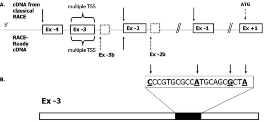
Using commercially available First ChoiceH RACE-Ready cDNA from human liver and intestinal tissue, several putative transcription start sites were identified: One each at the beginning of the novel UTR exons depicted in Fig. 2A, except Ex21, and several alternative transcription start sites within Ex 23. A transcription start site cluster was located in the region between nucleotides 135 and 115 upstream from the 39end of Ex 23 (Fig. 2B). This region contains a putative initiator sequence (Inr) [16,17]. RACE-Ready experiments also identified two other novel exons in the vicinity of Ex 23 and Ex 22, designated Ex-3b (Chr18: 55470138-55470074; GRCh37/hg19) and Ex22b (Chr 18: 55468948-55468914; GRCh37/hg19) (Fig. 2A). Ex24, which was identified by classical RACE above, was not detected using RACE-Ready cDNA. The transcription start site (TSS) at the beginning of Ex21 was only found by RACE, while using RACE-Ready cDNA Ex 21 was a part of transcripts initiating further upstream of Ex21.
Consistent Expression Pattern of the Novel ATP8B1 mRNA Isoforms in the Liver and Intestine
To define the biological relevance of the different ATP8B1
transcripts we used 7 normal human livers and 3 normal human intestinal samples in qRT-PCR experiments with individually-designed probe/primer sets covering all identified alternative exon-exon boundaries found by 59RACE (Tab. S1). The results are presented in Fig. 3, where the diagrams represent probe/ primer sets and Latin numbers relevant splicing variant(s) detected. The results show the abundance of each 59UTR splicing variant relative to the protein coding region, represented by the Ex +1/+2 boundary. qRT-PCR demonstrated the prevalent expres-sion of transcripts containing Ex 23 directly spliced to Ex +1; these splicing variants, Ex23/Ex+1 and Ex 23/CAG/Ex +1 (Fig. 3) comprise almost 2/3 of totalATP8B1expression, whilst the alternative splicing event, Ex23/Ex21 that in fact comprises two mRNA isoforms: Ex23/Ex21/Ex+1 and Ex23/Ex21/CAG/ Ex+1, comprises less than 10% of the total transcripts. Splicing variants Ex 21/Ex +1 and Ex 21/CAG/Ex +1 account for almost 26% but splicing events including Ex22 account for less than 4% of the total transcripts. The expression level of Ex24/Ex
+1 (Fig. 3, variants I and II), found by classical 59RACE but not RACE-Ready cDNA, varied significantly amongst the samples, ranging from complete absence to 3.4% of total gene expression. The expression levels of the two small rarely detected exons, located 91 bp and 215 bp downstream from Ex23 and Ex22 respectively (Ex23b and Ex 22b, Fig. 2A), and identified only using RACE-ready cDNA, were below 1% in all experiments (data not shown). Additional qRT-PCR experiments on a limited (n = 3) number of human intestinal samples did not exhibit any significant difference compared to the expression pattern detected in liver, with Ex 23 proving to be the most prevalent 59UTR exon expressed in both, liver and intestinal tissues (data not shown).
Searching of the NCBI EST database (National Centre for Biotechnology and Information, http://www.ncbi.nlm.nih.gov/) revealed one ATP8B1 transcript with Ex 23 spliced to Ex +1 without the CAG triplet (GenBank accession: DR005588.1, GRCh37/hg19). This transcript does not include the protein coding Ex+2, and thus Ex+1 must be spliced directly to Ex+3. The resultant predicted protein sequence would have a premature stop codon (TAA) at position 62, which would not give a viable ATP8B1 protein. We searched for the existence of Ex+1/Ex+3 splicing event by PCR using cDNA templates prepared from liver and intestinal RNA. qRT-PCR using the specifically-designed probe for the Ex+1/Ex+3 boundary demonstrated 20-fold lower expression of this transcript in normal human liver and intestinal tissue compared to the Ex+1/Ex+2 splicing variant (data not shown). The biological significance of the low abundance transcript therefore remains unclear.
Also, our results did not confirm the existence of previously identified 909 bp-longATP8B159exon [15].
In Silico Identification of the Putative Promoters in ATP8B1 Gene
On the basis of the 59RACE and qRT-PCR results, we predicted the major promoter region of ATP8B1 to be located upstream of the cluster of TSSs within Ex23 (P3, Fig. 4) and two weaker promoters to be upstream of Ex22 and21 (P2 and P1, Fig. 4). In agreement with our hypothesis, in silico analysis employing three independent promoter prediction programs located putative promoter regions within a CpG island 70–72 kb upstream of the protein coding Ex +1 corresponding to the chromosomal location of the novel Ex 23 (Fig. S1). Computer analysis did not predict the presence of promoter upstream of Ex 21.
Experimental Validation of the Major Promoter of the ATP8B1 Gene
In order to identify whether the major promoter of ATP8B1
resides upstream of Ex23, twelve promoter constructs (Prom 1– 12, Fig. 4) utilising the luciferase reporter gene system were prepared: Six comprised the sequences upstream of the TSS cluster in Ex23, whilst three focused on the region upstream of Ex24 and three represented the regions upstream of Ex22 and Ex21. The promoter sequences cloned ranged from 434 bp to 3379 bp in length.
The luciferase assay results, summarised in graph on Fig. 4, demonstrate the highest relative promoter activity for the short 434 bp construct (Prom 1) which is situated immediately upstream of the major TSS cluster in Ex 23. Assessment of the longer promoter constructs ranging from 747 bp up to 3379 bp (Proms 2–6) exhibited approximately 65% reduced promoter activity compared to the Prom 1 construct. Only minimal differences in the reporter assay were observed among Proms 2–6 constructs
relative to each other. Removal of the 434 bp region upstream of Ex23 resulted in a complete absence of luciferase activity in all constructs tested (Proms 7–9; Fig. 4), thus emphasizing the importance of the proximal P3 promoter in driving luciferase activity.
A putative TSS in Ex-1, not predictedin silico, was detected only by classical 59RACE. Nevertheless, the 40 kb distance between the main promoter and Ex21 in combination with the qRT-PCR results suggested the presence of an alternative regulatory region in the vicinity of Ex 21. Constructs, which included the putative
Figure 1. The heterogeneity of 59UTR ofATP8B1gene.(A) Length and position of novel untranslated exons ofATP8B1gene. (B) Six identified
alternatively spliced variants are indicated by diagonal lines. (C) The existence of two acceptor splice sites CAGCAG (tandem acceptors) at the 59 boundary of the first translated exon (Ex+1) ofATP8B1allows the generation of two different splice forms for each combination of upstream exons
promoter upstream of Ex21 and also upstream of Ex22 (Proms 10–12), showed only basal transcription activity which was less than 10% that of the Prom 1 construct upstream of Ex23 (Fig. 4).
The 59UTR of ATP8B1 is Highly Conserved between Human, Mouse and Rat
The sequences of mouse and ratAtp8b159UTR were obtained from Ensembl database (http://www.ensembl.org/index.html) and aligned with nucleotide sequences of newly identified human UTR exons using the ClustalW (http://www.ebi.ac.uk/clustalw)
program. A high degree of conservation was found in the region corresponding to Ex23 and Ex24: 83% and 82%, respectively, for a human-mouse alignment and 66% and 69%, respectively, for a human-rat alignment (Fig. 5).
Transcription Factor Binding Sites Present in the ATP8B1 Promoter Region
No consensus TATA or CAAT boxes were found in the proximal P3 promoter of theATP8B1gene. On the other hand, several putative binding sites for non-specific, general transcription
Figure 2. Transcription start sites (TSSs) identified within 59UTR ofATP8B1gene.(A) Transcription ofATP8B1gene originates from multiple TSSs (indicated by arrows); two additional TSS were identified at the beginning of rarely used exons, adjacent to Ex23 and22 respectively (dashed boxes). Sixteen different TSSs were found within the 509 bp-long Ex23 with the main cluster located between nucleotides 135 and 115 upstream from the 39end of Ex23 (B).
doi:10.1371/journal.pone.0051650.g002
Figure 3. Relative expression levels of different splicing forms assessed by qRT-PCR.Diagram of individually designed probes (for probe and primer sequences see Table S1) used to evaluate the expression levels of twelve identified splicing variants ofATP8B159UTR. Tested splicing variants are indicated by Latin numbers on the left, average expression levels for each transcript from normal liver tissues (n = 7) are shown on the right. The expression levels are presented as a relative value normalised to the expression of the protein coding region represented by Ex+1/Ex+2
boundary.
doi:10.1371/journal.pone.0051650.g003
factors (Sp1, AP-2, NFkB) were identified in that DNA sequence.
No liver- or intestine-specific transcription factor binding sites were found in the vicinity of the TSS cluster of Ex23 (Fig. 5). A putative FXR binding site was identified using only the MatInspector computer analysis software, and this was 807 bp upstream of Ex 24. However, this site, GAGTGAcTGACCA, does not correspond to any known consensus FXR binding sequence and the sequence is not conserved between human, mouse and rat.
Influence of Bile Acids on Promoter P3 Activity
To investigate the effect of bile acids on P3 promoter activity, HepG2 cells were transfected with the Prom 3, Prom 4, and Prom 6 ATP8B1 luciferase constructs, which comprise the main promoter P3, and then incubated in the presence or absence of CDCA and TCA for 24 hours. Prior to the commencement of the luciferase experiments, we evaluated the expression levels of endogenous ATP8B1, ABCB11, SHPand CYP7A1 by qRT-PCR before and after CDCA treatment to assess the normal response of HepG2 cells to bile acid stimulation. While ATP8B1 levels remained constant,ABCB11andSHPmRNAs were up-regulated and CYP7A1 mRNA down-regulated in the presence of 50 and 100mM CDCA respectively (data not shown), proving HepG2 as
a suitable model to assess the response of theATP8B1promoter constructs to bile acids.
In agreement with the unchanged mRNA expression of
ATP8B1 under CDCA stimulation, none of the luciferase constructs tested demonstrated a significant change in promoter activity in HepG2 cells after CDCA (Fig. 6A) or TCA (not shown) treatment. Since HepG2 cells do not express the NTCP, all experiments were repeated in a HepG2 cell line stably expressing rat Ntcp (rNtcp-HepG2 cells), that were in addition co-transfected with vectors expressing human FXR and RXRa. To minimise the
effect of bile acids present in fetal calf serum, the cells were cultured in medium containing charcoal-stripped fetal bovine serum with minimal content of bile acids [13]. In agreement with the previous experiments, no significant change in ATP8B1
promoter activity was observed (Fig. 6B). These results indicate no direct link between FXR, bile acids and theATP8B1 major promoter.
Discussion
In this study we demonstrate that ATP8B1 expression is regulated by a highly structured 59UTR which spans 71,964 kb and comprises four untranslated exons located a considerable distance upstream of the first protein coding exon. These exons are
Figure 4. Functional analysis ofATP8B1promoter regions.HepG2 cells were transiently transfected with luciferase reporter gene constructs containing 12 different fragments of putativeATP8B1promoters. Nine luciferase constructs (Prom 1 to Prom 9) were designed to comprise the
putative dominant promoter P3, two constructs covered promoter P1 (Proms 11 and 12) and one covered promoter P2 (Prom 10). The position of the tested fragments are indicated by horizontal double arrow lines. The number in brackets next to the construct name represents its size (bp). Prom 3 and Prom 4 were designed to include/exclude a putative FXR/RXR binding site indicated by black oval. Antisense construct encodes the same region as Prom 5, but in antisense orientation. Putative promoters (P1–P3) are depicted as horizontal thick arrows. Transcriptional activity for each construct was measured in relative light units per second (RLU/s) and corrected for the transfection efficiency using the internal controlRenillapRL-TK expression plasmid. The data shown are calculated from 3–5 independent experiments and related to the pGL3 Basic activity.
alternatively spliced. The main TSS cluster was located within nucleotides 135 to 115 upstream from the 39end of Ex23. This TSS cluster contains a putative initiator element (Inr) [17,18] and is encompassed by Sp1 binding sites (Fig. 5). The previously published TSS [1,15] is not significant. Transcription ofATP8B1
is driven by three newly identified promoters (P1, P2 and P3; Fig. 4) In liver, the promoters P1 and P2 play only minor role under physiological conditions. Promoter P3 was identified as the essential regulatory element responsible for 70% of totalATP8B1
gene expression. The 434 bp part of P3 (Prom 1 construct in Fig. 4) upstream of the main TSS cluster promiscuously serves both as exonic and promoter region and represents the main driving force of ATP8B1gene expression. The importance of this region was further confirmed by sporadic use of TSSs located further upstream of the main TSS cluster in Ex23.
The dominantATP8B1 promoter P3 displays typical features for promoters of housekeeping genes: TATA-less, GC-rich sequence with multiple TSSs [19] in which only non-specific putative transcription factor binding sites for Sp1, AP-2 and NFkB
were identified. (Fig. 5). These data are in agreement with the ubiquitous expression of ATP8B1 (FIC1) and its putative complex role in maintenance of apical membrane structure [8,10].
Genes regulated by alternative promoters are common in humans. Multiple promoters can be utilised according to environmental conditions or to a particular developmental stage to ensure the tissues-specific or spatio-temporal expression of the appropriate gene isoform [20]. Various mRNA isoforms may also interact to achieve a transcriptional repression of an alternative transcript [20,21]. It has been shown that of several alternative mRNA isoforms, one can be ubiquitously expressed among various cell types, whereas the remaining ones may be limited to a small number of tissues [22,23,24,25]. This might be the case of
ATP8B1alternative transcripts. Even though our study does not support such tissue specific regulation of ATP8B1 at the transcriptional level in the tested samples, further research could characterise the role of all three promoters in different organs under varying conditions and address the involvement of post-transcriptional control mechanisms.
In a view of the complex structure of theATP8B1gene and its highly variable mRNA levels across cell types, RNA stability and post-transcriptional control appears to be more important in
ATP8B1 regulation than previously expected. Our data demon-strate significant differences between the activity observed for the promoter upstream of Ex21 (P1) versus the promoter upstream of Ex23 (P3). However, this difference was not replicated at the level of abundance of the mRNA transcripts associated with these promoters: Whereas the reporter gene activity mediated by promoter P1 was 15-fold lower than that of the principle promoter P3 (Fig. 4), the mRNA levels of corresponding transcripts
Figure 5. Putative transcription factor binding sites and conservation of ATP8B1 59UTR. (A) Sequence alignment using ClustalW algorithm (http://www.ebi.ac.uk/Tools/clustalw/). High level of conservation among mouse (M), rat (R) and human (H) genome was detected for Ex23 and Ex 24 (conserved nucleotides indicated by stars). Putative Sp-1, Ap-2, NFkB transcription factor binding sites were
predicted in exonic/promoter region P3. Putative CREB and HNF-4 binding sites were identified within a distal part of promoter P3 corresponding to Ex 24 sequence. Initiator element sequence encompasses the main TSS cluster in Ex 23. Exonic regions are underlined, transcription start sites are indicated by arrows and bold letters and putative transcription factor binding sites by grey boxes. Two upstream ATG are in bold. (B) Sequence of Ex22 and (C) Ex21. Exonic region is highlighted in bold. Alu consensus sequences are underlined.
doi:10.1371/journal.pone.0051650.g005
displayed only a 3-fold difference (Fig. 3). The observed discrepancy suggests possible differential efficiencies in post-transcriptional processing of the corresponding pre-mRNA
ATP8B1forms.
59UTRs are known as key mediators of post-transcriptional control. The mechanisms of UTR-mediated regulation comprise, among others, stable secondary structures including those formed by repetitive sequences such as Alu and upstream open reading frames [26]. Alternatively spliced Ex21 ofATP8B1is apparently an exonized Alu [27] element with the promoter P1 containing complementary Alu sequence. Alu sequences embedded in 59UTR have been shown to modulate both transcription and translation [28,29].
Other potent modulators of transcriptional and translational efficiency are Upstream Open Reading Frames (uORFs) which can affect gene expression by inhibition of mRNA stability and translational repression [30,31]. Recently demonstrated uORF-mediated ability to trigger the nonsense-uORF-mediated mRNA decay [32],[33] or to inhibit the downstream ORF by upstream located uAUG [34] represents processes that could be potentially involved in posttranscriptional regulation ofATP8B1. Indeed we identified
ATP8B1transcripts that differ in their leader sequences and in the presence of putative upstream start codons AUG (uAUG) (Fig. 5). Whereas no uAUG was found within the transcript containing Ex 21, two uAUGs and two uORFs were identified within the prevalent transcript containing Ex23. Further factors known to
influence regulation of gene expression [35,36,37] are heteroge-neity, a high GC content and the unusually long length of 59UTR. Their potential contribution to regulation ofATP8B1expression is discussed in Fig. 7, Fig. S2 and their legends.
Since ATP8B1 deficiency is associated with cholestatic liver disease, most studies have focused on the role of the gene in cholestasis. Zollner and colleagues [38] found no changes in
ATP8B1mRNA level in cholestatic patients. In our experiments, stimulation of HepG2 cells with CDCA or TCA respectively showed no change inATP8B1mRNA levels and CDCA or TCA treatment of HepG2 or rNTCP-HepG2 cells expressing various
ATP8B1 promoter constructs did not significantly alter the luciferase activity compared with untreated cells. This observation is in agreement with the gene’s ubiquitous expression and suggests that bile acid independent mechanisms regulate ATP8B1 expres-sion across different cell types.
In conclusion our study provides fundamental data about the complexity ofATP8B1gene regulation. Newly identifiedATP8B1
mRNA isoforms differ in their 59UTRs and both transcriptional and post-transcriptional efficiency. The basal expression of
ATP8B1 gene in the liver and the intestine is driven by a promoter with house-keeping like properties. Regulatory parts of
ATP8B1characterised in this study represent a feasible region for mutational search in patients with features suggestive of ATP8B1 deficiency, in whom no mutations have been identified within the coding region.
Figure 6.ATP8B1promoter activity in cells stimulated with bile acids.No significant change in activity was detected after stimulation with CDCA. (A) HepG2 cells were transiently transfected with three previously characterised (Fig. 4)ATP8B1promoter gene constructs (Prom 3, 4 and 6) and stimulated with 0, 10, 50 and 100mM CDCA for 24 hours. All constructs comprise proximal 434 bp-promoter P3, Prom 4 includes putative FXR
binding site identified by MatInspector computer analysis software, and Prom 6 represents the largest construct containing 3379 bp of 59flanking region. (B) HepG2 cells stably expressing rat sodium-taurocholate co-transporting polypeptide (rNtcp) were transiently transfected with constructs Prom 3, 4 and 6 together with 50 ng of pCI_hRXRaand 50 ng pCI_hFXR plasmids and treated with 0, 10 and 25mM CDCA for 24 hours.
Materials and Methods
59Rapid Amplification of cDNA Ends (59RACE)
The 59ends of theATP8B1gene were mapped using the 59/39 RACE kit, 2nd Generation (Roche, Switzerland) according to manufacturer’s instructions. Total RNA was isolated using RNA-Bee (Tel-test, Inc., Friendswood, USA) from 50 mg of human liver or intestinal tissue, or 56106of HepG2 cells. All procedures were conducted with written informed consent under an institutional-review-board approved protocol or using anonymised bank samples, previously collected with consent for research. To confirm any newly identified transcriptional origins, liver and intestinal RACE-ready cDNA (Ambion, Austin, USA) was used.
ATP8B1 gene-specific primers for RACE were designed to span the junctions of exons 3/4, 2/3 and 1/2. Resultant PCR products were cloned into the pDrive Cloning Vector (Qiagen, Hilden, Germany) and sequenced in both directions using ABI Big Dye (Version 3.1) on a 3100 automated DNA Sequencer (Applied Biosystems, Foster City, USA) using vector specific primers.
Quantitative Real–time PCR
Twelve sets of individually designed TaqManH MGB probes labelled with Fam and non-fluorescent quencher and primers were generated using Primer ExpressH Software Version 2.0 (Applied Biosytems, Warrington, UK), to cover all variants of alternative splicing of the untranslated exons (Table S1, No.1-12). 100 ng of DNase-treated total RNA from normal human liver and intestinal samples was used as a template in a 20ml reverse transcription reaction using Transcriptor (Roche, West Sussex, UK) and mix of random hexamer or gene specific primers (Invitrogen, Paisley, UK, Sigma-Aldrich, Dorset, UK). 1ml of first strand cDNA was then assessed in triplicate for levels of the different ATP8B1
transcripts on an ABI Prism 7000 Sequence Detection System (Applied Biosystems). Expression levels of the studied transcripts and the overall expression of the ATP8B1 coding sequence represented by Ex+1/Ex+2 boundary, were corrected to the level
of 18S rRNA (delta Ct) (TaqManH MGB probe, Applied Biosytems, Warrington, UK). A PCR of non-reverse transcribed RNA was performed as a negative control to check for any genomic DNA contamination. Delta delta Ct values were calculated using ABI SDS software with RQ study application (Version 1.2.3, Applied Biosystems) and the data analysed using Microsoft Excel.
Promoter and First Exon Analysis in Silico
Three independent algorithms for promoter prediction (http:// genome.ucsc.edu/, http://www.genomatix.de, http://bimas.dcrt. nih.gov/molbio/proscan/) were used to analyse the 59UTR of the
ATP8B1gene. The University of California Santa Cruz Genome Bioinformatics server was also used to predict the gene’s first exon. The data obtained were compared with the EST database and the 59RACE experimental results.
Luciferase and Expression Plasmid Construction
Twelve fragments of the 59UTR (Prom 1–12 in Fig. 4) of the
ATP8B1gene were PCR amplified using human genomic DNA as a template, Pfx polymerase (Invitrogen) and specific primers containing XhoI restriction sites. PCR products were cloned (Invitrogen Zero blunt kit or Qiagen Cloning kit), sequenced, digested with XhoI, and ligated into XhoI-digested luciferase reporter gene vector pGL3-Basic (Promega, Southampton, UK) using Quick Ligation Kit (New England Biolabs, Hitchin, UK). Resultant constructs were checked for the correct sequence with various restriction enzymes and by direct sequencing prior to transfection. Stocks were prepared using an Endotoxin-free Maxiprep kit (Qiagen, West Sussex, UK). Human retionid X receptor a (hRXRa) and farnesoid X receptor (hFXR) cDNAs
were PCR amplified, cloned into Invitrogen’s TOPO TA- cloning kit, sequenced and then sub-cloned to the mammalian expression vector pCI (Promega).
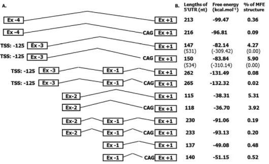
Figure 7. Thermodynamic properties of identified 59UTR isoforms.The comparison of the 59UTR length and RNA secondary structure free energy and percentage of minimal free energy (MFE) for all identifiedATP8B159UTR isoforms, schematically depicted on the left. Prediction for the most frequent isoforms initiating at Ex23 was calculated using TSS at position2125 from 39end of Ex23. Data in brackets (row 3 and 4) represent data for TSS at position2509. Putative secondary RNA structures predicted using RNAfold web tool (http://rna.tbi.univie.ac.at/cgi-bin/RNAfold.cgi) are summarised in Fig. S2.
doi:10.1371/journal.pone.0051650.g007
Cell Culture and Transfection
HepG2 cell lines were purchased from the ATCC (Teddington, UK). Cells were maintained in Dulbecco’s modified Eagle’s medium (PAA, Farnbourough, UK) supplemented with 5% or 10% fetal calf serum (PAA) and glutamine. rNTCP-HepG2 cells, kindly provided by Ulrich Beuers (Department of Gastroenterol-ogy and HepatolGastroenterol-ogy, Academic Medical Center, University of Amsterdam, Amsterdam, the Netherlands) and Christopher Rust (Department of Medicine 2– Grosshadern, University of Munich, Munich, Germany), were maintained in Dulbecco’s modified Eagle’s medium containing 5% fetal calf serum and 1 mg/ml G418 (Invitrogen). For transfection, cells were seeded in 24-well plates (TPP) in medium containing 5% fetal calf serum or 5% charcoal-stripped bovine calf serum (GibcoBRL). For transient transfection, 1.5ml of FuGene HD (Roche) and 500 ng of plasmid DNA were used per well. Plasmid DNA comprised 450 ng of the appropriate LucATP8B1 promoter construct and 50 ng of the
Renilla pRL-TK internal control plasmid (Promega). For some stimulation experiments, 50 ng of pCI_hFXR and 50 ng of pCI_hRXR contructs were co-transfected with the luciferase constructs. Twenty-four hours after transfection, cells were treated with 0 to 100mM dimethyl sulfoxide, chenodeoxycholic acid, CDCA or taurocholic acid, TCA (Sigma Aldrich), respectively.
Reporter Gene Assay
Cells were harvested 48 hours after transfection (24 hours after stimulation with bile acids) and thenFireflyand Renillaluciferase activities in cell lysates were determined using the Dual luciferase system (Promega) on a Glomax luminometer (Promega). All reporter gene assays were performed in triplicate and results are presented as the average value from at least three independent experiments corrected for the transfection efficiency usingRenilla
luciferase activities. The data from individual experiments were related to the activity of the control expression plasmid pGL3 Basic (Promega).
Supporting Information
Figure S1 In silico analysis of first exon and promoter
region forATB8B1gene compared to the 59RACE results.
Four putative first exons were predicted by ‘‘First EF’’ computer prediction software (horizontal double-arrow lines) [1]. Predicted exons correspond to the chromosomal location of the novel exons 22, 23 and 24 identified in the 59RACE experiments. Three independent computer algorithms localised putative promoters (dashed horizontal double-arrow lines) in a CpG island 70–72 kb upstream of Ex+1.
(TIF)
Figure S2 Putative secondary structures of ATP8B1
59UTR isoforms depicted in Fig. 7 of the main text. Drawings of the minimum free energy (MFE) structures for each
splicing variant ofATP8B159UTR suggest their possible different role in regulation of gene expression. One of the regulatory mechanism, formation of stable secondary structures, was shown to impede the progress of the scanning ribosome [2]. Such scanning is influenced by the size and the position of the secondary structure(s) towards the 59cap of the mRNA species: that is, an alternative transcript with a shorter version of the 59UTR is frequently translated more efficiently than the one with a longer 59 region [3,4,5]. Likewise, a stem-loop structure located a consid-erable distance from the 59cap will require a higher free energy compared to one situated closer to it to affect the access of a pre-initiation complex to the mRNA. [2,6].
(TIF)
Table S1 Individually designed TaqManHMGB probes labelled with Fam and non-fluorescent quencher and primers were generated using Primer ExpressH Soft-ware Version 2.0 (Applied Biosytems), to cover all variants of alternative splicing of the untranslated exons.The abundance of each splicing variant was compared relatively to a non-variable coding region ofATP8B1represented by Ex+1/+2 boundary. Probe/primer set for Ex+1/+3 boundary (No.14) was used to test the biological significance of transcript excluding protein coding Ex+2, found in EST database (GenBank accession: DR005588.1). All probe sets were designed across exon/exon boundaries to eliminate the possibility of genomic contamination. Primers used in various amplifications are indicated by upper index (a, b, c). The amplification efficiency
was tested for each probe/primer set on control templates (obtained by cloning the appropriate cDNA region) using different concentrations of positive and negative controls. As each probe set worked with a slightly different efficiency, the concentration of probes was adjusted for each positive control to reach a cycle threshold (Ct) value difference not greater than 1.
(DOC)
Text S1 Supporting references. (DOC)
Acknowledgments
The authors thank to Jonathan Gilthorpe and Vladimı´r Korˇı´nek for helpful discussion and technical support with reporter gene assay and Marek Cebecauer for critical reading of the manuscript. Supported by EASL Sheila Sherlock Post-doc Fellowship (D.C.) and Institutional grant IKEM MZO 00023001 (M.J.)
Author Contributions
Conceived and designed the experiments: DC SSS JAB RJT. Performed the experiments: DC SSS. Analyzed the data: DC JAB MJ RJT. Contributed reagents/materials/analysis tools: DC SSS JAB. Wrote the paper: DC MJ RJT.
References
1. Bull LN, van Eijk MJ, Pawlikowska L, DeYoung JA, Juijn JA, et al. (1998) A gene encoding a P-type ATPase mutated in two forms of hereditary cholestasis. Nat Genet 18: 219–224.
2. Houwen RH, Baharloo S, Blankenship K, Raeymaekers P, Juyn J, et al. (1994) Genome screening by searching for shared segments: mapping a gene for benign recurrent intrahepatic cholestasis. Nat Genet 8: 380–386.
3. van Mil SW, Klomp LW, Bull LN, Houwen RH (2001) FIC1 disease: a spectrum of intrahepatic cholestatic disorders. Semin Liver Dis 21: 535–544. 4. van Mil SW, van Oort MM, van den Berg IE, Berger R, Houwen RH, et al.
(2004) Fic1 is expressed at apical membranes of different epithelial cells in the digestive tract and is induced in the small intestine during postnatal development of mice. Pediatr Res 56: 981–987.
5. Stapelbroek JM, Peters TA, van Beurden DH, Curfs JH, Joosten A, et al. (2009) ATP8B1 is essential for maintaining normal hearing. Proc Natl Acad Sci U S A 106: 9709–9714.
6. Ujhazy P, Ortiz D, Misra S, Li S, Moseley J, et al. (2001) Familial intrahepatic cholestasis 1: studies of localization and function. Hepatology 34: 768–775. 7. Paulusma CC, Folmer DE, Ho-Mok KS, de Waart DR, Hilarius PM, et al.
(2008) ATP8B1 requires an accessory protein for endoplasmic reticulum exit and plasma membrane lipid flippase activity. Hepatology 47: 268–278.
hepatocytes, but FXR expression and activity are maintained. Gastroenterology 136: 1060–1069.
10. Verhulst PM, van der Velden LM, Oorschot V, van Faassen EE, Klumperman J, et al. (2010) A flippase-independent function of ATP8B1, the protein affected in familial intrahepatic cholestasis type 1, is required for apical protein expression and microvillus formation in polarized epithelial cells. Hepatology 51: 2049–2060.
11. Chen F, Ananthanarayanan M, Emre S, Neimark E, Bull LN, et al. (2004) Progressive familial intrahepatic cholestasis, type 1, is associated with decreased farnesoid X receptor activity. Gastroenterology 126: 756–764.
12. Alvarez L, Jara P, Sanchez-Sabate E, Hierro L, Larrauri J, et al. (2004) Reduced hepatic expression of farnesoid X receptor in hereditary cholestasis associated to mutation in ATP8B1. Hum Mol Genet 13: 2451–2460.
13. Frankenberg T, Miloh T, Chen FY, Ananthanarayanan M, Sun AQ, et al. (2008) The membrane protein ATPase class I type 8B member 1 signals through protein kinase C zeta to activate the farnesoid X receptor. Hepatology 48: 1896– 1905.
14. Demeilliers C, Jacquemin E, Barbu V, Mergey M, Paye F, et al. (2006) Altered hepatobiliary gene expressions in PFIC1: ATP8B1 gene defect is associated with CFTR downregulation. Hepatology 43: 1125–1134.
15. Klomp LW, Vargas JC, van Mil SW, Pawlikowska L, Strautnieks SS, et al. (2004) Characterization of mutations in ATP8B1 associated with hereditary cholestasis. Hepatology 40: 27–38.
16. Smale ST, Baltimore D (1989) The ‘‘initiator’’ as a transcription control element. Cell 57: 103–113.
17. Javahery R, Khachi A, Lo K, Zenzie-Gregory B, Smale ST (1994) DNA sequence requirements for transcriptional initiator activity in mammalian cells. Mol Cell Biol 14: 116–127.
18. Kaufmann J, Smale ST (1994) Direct recognition of initiator elements by a component of the transcription factor IID complex. Genes Dev 8: 821–829. 19. Zhu J, He F, Hu S, Yu J (2008) On the nature of human housekeeping genes.
Trends Genet 24: 481–484.
20. Davuluri RV, Suzuki Y, Sugano S, Plass C, Huang TH (2008) The functional consequences of alternative promoter use in mammalian genomes. Trends Genet 24: 167–177.
21. Martianov I, Ramadass A, Serra Barros A, Chow N, Akoulitchev A (2007) Repression of the human dihydrofolate reductase gene by a non-coding interfering transcript. Nature 445: 666–670.
22. Barker DF, Husain A, Neale JR, Martini BD, Zhang X, et al. (2006) Functional properties of an alternative, tissue-specific promoter for human arylamine N-acetyltransferase 1. Pharmacogenet Genomics 16: 515–525.
23. Fusco F, Mercadante V, Miano MG, Ursini MV (2006) Multiple regulatory regions and tissue-specific transcription initiation mediate the expression of NEMO/IKKgamma gene. Gene 383: 99–107.
24. Husain A, Zhang X, Doll MA, States JC, Barker DF, et al. (2007) Functional analysis of the human N-acetyltransferase 1 major promoter: quantitation of tissue expression and identification of critical sequence elements. Drug Metab Dispos 35: 1649–1656.
25. Shibahara S, Takeda K, Yasumoto K, Udono T, Watanabe K, et al. (2001) Microphthalmia-associated transcription factor (MITF): multiplicity in structure, function, and regulation. J Investig Dermatol Symp Proc 6: 99–104. 26. Mignone F, Gissi C, Liuni S, Pesole G (2002) Untranslated regions of mRNAs.
Genome Biol 3: REVIEWS0004.
27. Sorek R, Lev-Maor G, Reznik M, Dagan T, Belinky F, et al. (2004) Minimal conditions for exonization of intronic sequences: 59splice site formation in alu exons. Mol Cell 14: 221–231.
28. Sobczak K, Krzyzosiak WJ (2002) Structural determinants of BRCA1 translational regulation. J Biol Chem 277: 17349–17358.
29. Landry JR, Medstrand P, Mager DL (2001) Repetitive elements in the 59
untranslated region of a human zinc-finger gene modulate transcription and translation efficiency. Genomics 76: 110–116.
30. Diba F, Watson CS, Gametchu B (2001) 59UTR sequences of the glucocorticoid receptor 1A transcript encode a peptide associated with translational regulation of the glucocorticoid receptor. J Cell Biochem 81: 149–161.
31. Lammich S, Buell D, Zilow S, Ludwig AK, Nuscher B, et al. (2010) Expression of the anti-amyloidogenic secretase ADAM10 is suppressed by its 59 -untranslated region. J Biol Chem 285: 15753–15760.
32. Hood HM, Neafsey DE, Galagan J, Sachs MS (2009) Evolutionary roles of upstream open reading frames in mediating gene regulation in fungi. Annu Rev Microbiol 63: 385–409.
33. Zhao C, Datta S, Mandal P, Xu S, Hamilton T (2010) Stress-sensitive regulation of IFRD1 mRNA decay is mediated by an upstream open reading frame. J Biol Chem 285: 8552–8562.
34. Zhang Y, Zhao T, Li W, Vore M (2010) The 59-untranslated region of multidrug resistance associated protein 2 (MRP2; ABCC2) regulates down-stream open reading frame expression through translational regulation. Mol Pharmacol 77: 237–246.
35. Arrick BA, Grendell RL, Griffin LA (1994) Enhanced translational efficiency of a novel transforming growth factor beta 3 mRNA in human breast cancer cells. Mol Cell Biol 14: 619–628.
36. Pickering BM, Willis AE (2005) The implications of structured 59untranslated regions on translation and disease. Semin Cell Dev Biol 16: 39–47.
37. Hughes TA (2006) Regulation of gene expression by alternative untranslated regions. Trends Genet 22: 119–122.
38. Zollner G, Fickert P, Zenz R, Fuchsbichler A, Stumptner C, et al. (2001) Hepatobiliary transporter expression in percutaneous liver biopsies of patients with cholestatic liver diseases. Hepatology 33: 633–646.