Authors
Claudine Maria Jorge de Oliveira1
Roberto Silva Costa2 Osvaldo Merege Vieira Neto3
Rosana Aparecida Spadoti Dantas4
Miguel Moysés Neto5 Elen Almeida Romão6 Gyl Eanes Barros-Silva7 Eduardo Barbosa Coelho8 Márcio Dantas8
1Serviços Médicos de Aneste-sia – SMA
2Department of Pathology of the FMRP - USP
3Division of Nephrology of the Department of Internal Medicine of the HCFMRP - USP 4Department of General Nursing; Nursing School of Ribeirão Preto – USP 5Division of Nephrology of the Department of Internal Medicine of the HCFMRP - USP 6Division of Nephrology of the Department of Internal Medicine of the HCFMRP - USP / FAEPA
7Department of Pathology of the FMRP-USP
8Department of Internal Medicine of the FMRP-USP
Submitted: 08/12/2010 Accepted: 09/07/2010
Corresponding author:
Dr. Márcio Dantas
Divisão de Nefrologia, Depar-tamento de Clínica Médica. HCFMRP-Faculdade de Medi-cina de Ribeirão Preto-USP Av. Bandeirantes, 3900. Ribei-rão Preto – São Paulo - Brasil CEP: 14048-900
E-mail: mdantas@fmrp. usp.br
Tel: 55 (16) 3602-2543; Fax: 55 (16) 3633-6695
This study was conducted at the Medical School of Ribei-rão Preto of the Universidade de São Paulo.
Financial support:
Fundação de Apoio ao Ensi-no, Pesquisa e Assistência do HCFMRP-USP.
A
BSTRACTIntroduction: The elderly population
is growing and aging in better clinical conditions than in the past. However, the distribution and course of kidney diseases in elderly patients are not well known partially due to reluctance to in-dicate renal biopsies in those patients.
Objective: To evaluate the distribution, clinical features, and outcomes of ne-phropathies diagnosed by biopsy in the elderly. Patients and Methods: Seventy-one patients (47 males, 24 females) aged 60 years or older (67.3 ± 6.5 years), undergoing biopsy from January 1990 to December 2006, were evaluated. They were grouped according to their clinical syndromes. Results: Nephrotic syndrome was observed in 35 patients (49.3%), mainly associated with mem-branous nephropathy (17 patients), followed by amyloidosis and focal seg-mental glomerulosclerosis (seven pa-tients each). Acute kidney injury (AKI) was diagnosed in 19 patients, and the main histopathological diagnoses were acute tubular necrosis (six patients) and cast nephropathy (three patients). Of those 19 patients, only two had a favo-rable course, while the others died early or progressed toward advanced chronic kidney disease. Twelve patients under-going biopsy because of asymptomatic hematuria or proteinuria had different diagnoses, but most of them already had significant chronic nephropathy. In five patients with nephritic syndrome, the biopsies also showed several diag-noses. Conclusions: Nephrotic syndro-me was the major indication for renal biopsy, and membranous nephropathy was the most frequent diagnosis. Among patients with AKI and asymptomatic
Renal diseases in the elderly underwent to percutaneous
biopsy of native kidneys
hematuria or proteinuria, different diag-noses were found with high levels of ad-vanced chronic nephropathy.
Keywords: needle biopsy, aged,
nephro-tic syndrome, kidney diseases.
[J Bras Nefrol 2010;32(4): 379-385]©Elsevier Editora Ltda.
I
NTRODUCTIONSimilarly to several other organs, human kidneys also undergo structural and func-tional changes due to aging, such as a re-duction in weight and volume, an increase in the number of glomeruli with global sclerosis, and a reduction in plasma flow and glomerular filtration rate.1-3 However,
such changes rarely cause per se proteinu-ria or any other clinical manifestation. If, on the one hand, clinical and laboratory renal manifestations proper of aging are scarce, on the other, kidney diseases in the elderly have greater clinical repercussions as compared to those in younger individu-als, because the functional renal reserve of the former is smaller.
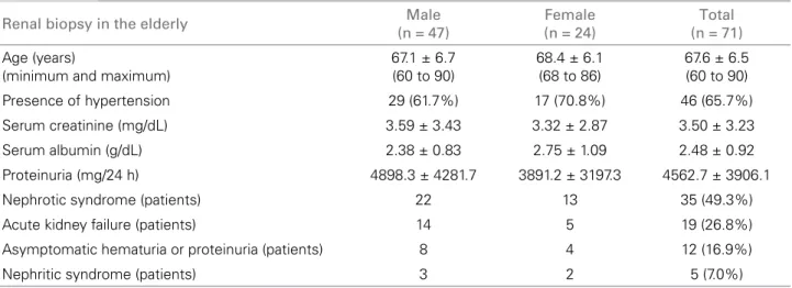
Table 1 CLINICALDATAOF 71 ELDERLYPATIENTSONTHEOCCASIONOFPERCUTANEOUSRENALBIOPSY
increase in life expectancy has led to an increase in the elderly population, whose aging occurs in better clini-cal conditions than in the past. These aspects lead to the need of diagnosis and treatment of nephropathies also in the elderly, yielding several publications encou-raging the indications for renal biopsies. Even in the very elderly, over the age of 80 years, no formal con-traindication to renal biopsy exists, particularly in the diagnoses of nephrotic syndrome and AKI, as long as safety criteria are met.4 In Brazil, the Paulista Registry
of Glomerulopathies has reported that the frequency of biopsies in patients over the age of 60 years accounts for 8.0% of the notified cases, and, in those over the age of 80 years, for 0.4%.5 Another Brazilian study has
reported that 2.2% of the 9,617 biopsies in native kid-neys were performed in elderly patients.6
This study aimed at assessing the distribution, clini-cal characteristics, and evolution of the nephropathies diagnosed by use of renal biopsy in the elderly.
P
ATIENTSANDMETHODSThis retrospective and descriptive study assessed the medical records and all reports of percutaneous biop-sy of topic kidneys of patients aged 60 years or more. The biopsies were performed between January 1990 and December 2006 at the Hospital das Clínicas of the Medical School of Ribeirão Preto of the Universidade de São Paulo (HCFMRP-USP). The study sample com-prised 71 patients (47 males and 24 females), and their mean age was 67.3 ± 6.5 years at the time of biopsy (Table 1). Patients whose biopsies were of transplanted kidneys or performed to diagnose localized kidney di-seases, such as tumors, were excluded from the study. The study was approved by the Committee on Ethics and Research in Human Beings of the HCFMRP-USP.
From the medical record, the following clinical data were assessed: age at the time of biopsy; clini-cal presentation of the kidney disease (nephrotic syn-drome, nephritic synsyn-drome, asymptomatic hematuria or proteinuria, and AKI with no defined cause); du-ration of clinical history; other systemic altedu-rations related; medications used; and laboratory findings (urinary sediment, serum levels of creatinine, urea, glycemia, electrolytes, albumin, protein electrophore-sis, 24-hour proteinuria, serologies, and other com-plementary tests, such as myelography and biopsy of other tissues when relevant).
Clinical presentation was classified as follows: ne-phrotic syndrome (edema, proteinuria ≥ 3.5 g/24 h, and hypoalbuminemia); nephritic syndrome (edema, hematuria, proteinuria < 3.5 g/24 h, and an acute reduction in renal function associated with hyper-tension of recent onset); asymptomatic hematuria or proteinuria (concomitant or non-concomitant glo-merular hematuria and proteinuria); and AKI (sup-posedly recent elevation in serum creatinine, with no defined cause and no conclusive signs of advanced chronic kidney disease).
The histopathological diagnoses were obtained from the renal biopsy reports. According to the routine of the Renal Pathology Laboratory of the HCFMRP-USP, the fragments of tissue obtained through per-cutaneous renal biopsy were analyzed by use of the following: common light microscopy (stained with hematoxylin and eosin, Masson’s trichrome, impreg-nation with methenamine silver stain, and congo red stain, when necessary); immunofluorescence micros-copy [(anti-IgA, -IgG, -IgM, -C3, -C1q, -fibrinogen, -kappa and -lambda polyclonal antibodies conjuga-ted with fluorescein isothiocyanate (Dako, Glostrup, Denmark)]; and, when indicated, electron microscopy.
Renal biopsy in the elderly Male
(n = 47)
Female (n = 24)
Total (n = 71) Age (years)
(minimum and maximum)
67.1 ± 6.7 (60 to 90)
68.4 ± 6.1 (68 to 86)
67.6 ± 6.5 (60 to 90)
Presence of hypertension 29 (61.7%) 17 (70.8%) 46 (65.7%)
Serum creatinine (mg/dL) 3.59 ± 3.43 3.32 ± 2.87 3.50 ± 3.23
Serum albumin (g/dL) 2.38 ± 0.83 2.75 ± 1.09 2.48 ± 0.92
Proteinuria (mg/24 h) 4898.3 ± 4281.7 3891.2 ± 3197.3 4562.7 ± 3906.1
Nephrotic syndrome (patients) 22 13 35 (49.3%)
Acute kidney failure (patients) 14 5 19 (26.8%)
Asymptomatic hematuria or proteinuria (patients) 8 4 12 (16.9%)
R
ESULTSData referring to age, duration of clinical history until biopsy, presence of hypertension, serum crea-tinine and albumin, and 24-hour proteinuria on the occasion of renal biopsy are shown in Table 1. No statistically significant difference between them was observed when compared by gender.
The indications of renal biopsy were as follows: ne-phrotic syndrome, 35 (49.3%) patients; initial diagnostic hypothesis of AKI with no apparent cause, 19 (26.8%); asymptomatic glomerular hematuria or proteinuria, 12 (16.9%); and nephritic syndrome, five (7.0%).
HISTOPATHOLOGICAL DIAGNOSES IN PATIENTS WITH NEPHROTIC
SYNDROME BY THE TIME OF RENAL BIOPSY
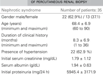
Table 2 shows the clinical data of 35 elderly patients with nephrotic syndrome undergoing renal biop-sy. Hypertension was found in 22 patients (62.9%). Serum creatinine and 24-hour proteinuria on the oc-casion of renal biopsy were 1.79 ± 1.12 mg/dL and 5945.4 ± 3171.9 mg/24 h, respectively, and serum al-bumin was 1.94 ± 0.63 g/dL.
drugs, total remission was observed in two, partial in one, and progression to stages 4 and 5 occurred in two, in addition to one death. Two patients were not followed up in our service.
Amyloidosis was diagnosed in seven patients, fi-ve of whom had no defined underlying disease, but one had associated leprosy, and another had POEMS syndrome (polyneuropathy, organomegaly, endocri-nopathy, M protein, and skin abnormalities). Focal segmental glomerulosclerosis was also diagnosed in seven patients. Although all of them were characte-rized as primary, in one patient, nephrotic syndrome occurred four weeks after a bothrops accident. For that patient, glomerulopathy was treated with pred-nisone, and total remission occurred in six weeks. Primary minimal change disease was diagnosed in two patients, one of whom maintained total remis-sion after 72 months and the other persisted with ne-phrotic proteinuria after 30 months of treatment with prednisone, and also showed a progressive elevation in serum creatinine.
In the patient with leprosy (lepromatous form), the histopathological diagnosis of the glomerulopa-thy was not conclusive. However, the introduction of polychemotherapy resulted in total remission of ne-phrotic syndrome. In the last patient with nene-phrotic syndrome, the histopathological diagnosis could not be established because the biopsy did not reach the kidney. However, the patient presented spontaneous partial remission of nephrotic syndrome and maintai-ned renal function stable after 36 months.
Of the 35 patients with nephrotic syndrome, six were lost to follow-up at our hospital due to referral to their original services right after biopsy. Other se-ven patients died between two and ten months after biopsy, five of whom due to infections.
HISTOPATHOLOGICAL DIAGNOSES IN PATIENTS SUSPECT OF
HA-VING AKI BY THE TIME OF RENAL BIOPSY
Nineteen patients underwent biopsy due to the initial clinical hypothesis of AKI with no defined cause, and their clinical data are shown in Table 3. Mean age was 68.1 ± 6.8 years, and 14 patients were males and five females. Hypertension was present in ten patients (52.6%). The duration of clinical history was 43.8 ± 80.9 days, and initial serum creatinine was 7.47 ± 3.41 mg/dL. The histopathological diagnosis was acute tu-bular necrosis (ATN) in six patients, and its course is discussed below. Three other patients were diagnosed with cast nephropathy due to multiple myeloma, all of whom initiated dialysis, but none of them reversed The most frequent diagnosis was membranous
ne-phropathy (17 patients) as follows: primary, in 15 pa-tients; associated with gastric adenocarcinoma (death after two months due to sepsis in the post-operative period), in one patient; and associated with marginal cell splenic lymphoma (despite treatment with sple-nectomy, after three years, the proteinuria and renal function worsened), one patient. Of the primary mem-branous nephropathies with no specific treatment, to-tal remission occurred in three patients, and progres-sion to stages 4 and 5 chronic kidney disease occurred in four. In patients treated with immunosuppressive
Table 2 CLINICALDATAOFELDERLYPATIENTSWITH
NEPHROTICSYNDROMEONTHEOCCASION OFPERCUTANEOUSRENALBIOPSY
Nephrotic syndrome Number of patients: 35 Gender male/female 22 (62.9%) / 13 (37.1)
Age (years)
(minimum and maximum)
68.4 ± 6.9 (60 to 90)
Duration of clinical history (months)
(minimum and maximum)
8.3 ± 6.9 (1 to 36)
Presence of hypertension 22 (62.9 %)
Initial serum creatinine (mg/dL) 1.79 ± 1.12
Serum albumin (g/dL) 1.94 ± 0.63
Table 3 CLINICALDATAOFELDERLYPATIENTS UNDERGOINGBIOPSYDUETOTHEINITIAL CLINICALDIAGNOSISOFACUTEKIDNEY FAILURE
Table 4 CLINICALDATAOF 12 ELDERLYPATIENTS UNDERGOINGBIOPSYDUETO
ASYMPTOMATICHEMATURIAORPROTEINURIA
kidney failure, dying after 30 days, six months, and 18 months. In another patient, the biopsy established the diagnosis of acute post-infectious glomerulone-phritis associated with diffuse and intense chronic tubulointerstitial nephritis, already requiring dialysis, but the patient never recovered renal function. Renal biopsy diagnosed sarcoidosis in another patient, and he was referred to his original service. Other two pa-tients were diagnosed with pauci-immune crescentic glomerulonephritis, both with diffuse and intense chronic tubulointerstitial lesion and no sign of syste-mic disease. One of them died with acute myocardial infarction after one month, and the other due to hos-pital pneumonia after two months.
The distribution of six causes of ATN was as follo-ws: one patient had obstructive uropathy due to inva-sive neoplasia of the rectum, and died after four weeks; one patient with multiple myeloma and fast progres-sion to the stage 5 of chronic kidney disease had ATN after the use of iodinated contrast medium; one patient with marked ATN associated with bothrops accident did not recover renal function, remaining on dialy-sis; two patients had ATN of undefined etiology, one improved renal function partially but persisted with chronic kidney disease stage 4 after 84 months, while the other patient was referred to his original service; the last patient had ATN of sudden onset, initially of unknown cause, and, after recovery of renal function, showed nephrotic proteinuria with hypoalbuminemia. Revision of the renal biopsy specimen showed nor-mal glomeruli on light microscopy and immunofluo-rescence, but extensive fusion of the foot processes of the visceral epithelial cells on electron microscopy. The diagnosis of minimal change disease was established and the cause of ATN was attributed to probable is-chemia due to reduced oncotic pressure secondary to
hypoalbuminemia. Treatments with prednisone iso-lated or associated with cyclophosphamide were not effective. Remission of proteinuria occurred only after use of mycophenolate mofetil.
In other eight patients, the histopathological findin-gs showed glomerulosclerosis and diffuse and intense chronic tubulointerstitial fibrosis as follows: one had advanced chronic nephropathy due to diabetes melli-tus (death after two months due to pneumonia); one had chronic uncharacteristic glomerulonephritis (dea-th due to uncharacteristic systemic vasculitis); one had multiple myeloma (dialysis was initiated); and four had advanced and unspecific chronic nephropathy, being referred to their original services. One patient with temporal arteritis, whose histopathological diagnosis could not be made because the biopsy did not get renal tissue, was also referred to his original service.
HISTOPATHOLOGICAL DIAGNOSIS IN PATIENTS WITH
ASYMPTO-MATIC HEMATURIA OR PROTEINURIA BY THE TIME OF BIOPSY
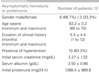
Table 4 shows the clinical data of 12 elderly patients who underwent biopsy because of the initial clinical finding of asymptomatic hematuria or proteinuria. The duration of their clinical history was 5.5 ± 4.4 months and initial serum creatinine and proteinuria were 2.27 ± 1.32 mg/dL and 1388.4 ± 989.8 g/24 h, respectively. Only two patients showed clinical improvement. The first was a woman with class IV lupus nephritis (WHO classification), treated with immunosuppressive drugs, who, after 10 years, had a serum creatinine of 0.9 mg/ dL, and neither proteinuria nor hematuria. The other patient with clinical improvement had post-infectious glomerulonephritis, and, after 48 months, maintained microscopic hematuria, non-nephrotic proteinuria, and serum creatinine of 1.4 mg/dL.
Acute kidney failure Number of patients: 19 Gender male/female 14 (73.7%) / 5 (26.3%)
Age (years)
(minimum and maximum)
68.1 ± 6.8 (60 to 83)
Duration of clinical history (days)
(minimum and maximum)
43.8 ± 80.9 (3 to 360)
Presence of hypertension 10 (52.6%)
Initial serum creatinine (mg/dL) 7.47 ± 3.41
Serum albumin (g/dL) 3.08 ± 0.83
Asymptomatic hematuria
or proteinuria Number of patients: 12 Gender male/female 8 (66.7%) / 3 (33.3%)
Age (years)
(minimum and maximum)
63.2 ± 3.2 (60 to 70)
Duration of clinical history (months)
(minimum and maximum)
5.5 ± 4.4 (1 to 12)
Presence of hypertension 10 (83.3%)
Initial serum creatinine (mg/dL) 2.27 ± 1.32
Serum albumin (g/dL) 3.50 ± 0.66
Two patients were diagnosed with advanced dia-betic nephropathy, and, after 12 months maintained a slow and progressive elevation of serum creatinine. Two other patients were diagnosed with focal seg-mental glomerulosclerosis, one of whom progressed slowly to stage 5 chronic kidney disease after 36 mon-ths, while the other was referred to his original service. Another patient was diagnosed with pauci-immune focal segmental glomerulonephritis and progressed to stage 5 chronic kidney disease after four years. Three other patients with stage 3 chronic kidney disease had unspecific chronic glomerulonephritis and progres-sed as follows: one patient had leprosy and died after two months due to sepsis due to cellulitis of the lower limbs; the other maintained proteinuria close to 2000 mg/24 hours and slow elevation in serum creatinine after four years; the third patient was referred to his original service. The biopsy of two other patients with stage 4 chronic kidney disease showed only unspecific diffuse chronic nephropathy.
HISTOPATHOLOGICAL DIAGNOSES IN PATIENTS WITH NEPHRITIC
SYNDROME OR SUSPICION OF RAPIDLY PROGRESSIVE
GLOMERU-LONEPHRITIS BY THE TIME OF RENAL BIOPSY
Table 5 shows the clinical data of elderly patients un-dergoing biopsy clinically diagnosed as nephritic syn-drome (four patients) or suspected of having rapidly progressive glomerulonephritis (one patient). Three patients were males and two females, and the duration of clinical history was 47.0 ± 54.1 days. Hypertension was observed in four of the five patients. On the occa-sion of biopsy, age was 69.8 ± 4.9 years, proteinuria was 2116.6 ± 1562.2 mg/24 h, serum creatinine was 4.04 ± 3.77 mg/dL, and serum albumin was 2.66 ± 0.66 g/dL.
The histopathological diagnosis of two patients was membranoproliferative glomerulonephritis, and both had negative serologies for hepatitis B and C vi-ruses. In one of them, a reduction in proteinuria and serum creatinine was observed, but, after 24 months, creatinine persisted as 2.2 mg/dL and proteinuria as 560 mg/24 hours. The other patient maintained a slow elevation in serum creatinine (1.8 mg/dL) and persistent proteinuria (3914 mg/24) after six years. Another patient had post-infectious glomerulone-phritis and maintained total remission with serum creatinine of 0.6 mg/dL after two years of follow--up. Another patient was diagnosed with Henoch-Schönlein purpura, and already had glomeruloscle-rosis and advanced tubulointerstitial fibglomeruloscle-rosis, being referred for dialysis. The fifth patient had a clinical diagnosis of rapidly progressive glomerulonephritis, and, on the occasion of biopsy, serum creatinine was 10.3 mg/dL due to essential mixed cryoglobulinemia, but the biopsy did not reach the kidney. The patient was treated with prednisone and cyclophosphamide, and, after 11 months, creatinine level dropped to 2.2 mg/dL and proteinuria persisted as 690 mg/24 hours.
D
ISCUSSIONIn the present study, the histopathological diagnoses were classified and analyzed based on the hypothe-sis of the clinical syndrome on the occasion of renal biopsy, because each clinical syndrome usually cor-responds to specific diseases. The diagnostic hypothe-ses of those syndromes could include a component of advanced chronic kidney disease or could even not be confirmed. This explains why several hypotheses, mainly those of acute kidney failure (AKF), were not confirmed by biopsy, which identified advanced chro-nic kidney disease. However, in face of an undefined diagnosis and a possible component of AKI, which is, thus, reversible, or to perform a complete investiga-tion of systemic diseases, biopsy was indicated.
The major indication of renal biopsy in the present sample was nephrotic syndrome (almost 50% of the patients). However, the frequency of biopsy indica-tion varies in different services. Similarly to our re-sults, in two other studies7,8, nephrotic syndrome was
also the major cause of renal biopsy (64% and 44%, respectively). Another study, however, classified the patients as elderly (66 to 79 years) or very elderly (≥
80 years). In the first age range, the major indications for biopsy were AKF (41%) and nephrotic syndrome (33%), while, in the very elderly group, the frequen-cies of biopsy indications were 33% for nephrotic
Table 5 CLINICALDATAOFFIVEELDERLYPATIENTS UNDERGOINGBIOPSYDUETOTHEINITIAL DIAGNOSISOFNEPHRITICSYNDROME
(FOURPATIENTS) ORRAPIDLYPROGRESSIVE GLOMERULONEPHRITIS (ONEPATIENT)
Gender male/female 3 (60%) / 2 (40%)
Age (years)
(minimum and maximum)
69.8 ± 68.0 (65 to 75)
Duration of clinical history (days) (minimum and maximum)
47.0 ± 54.1 (3 to 120)
Presence of hypertension 4 (80%)
Initial serum creatinine (mg/dL) 4.04 ± 3.77
Serum albumin (g/dL) 2.66 ± 0.66
syndrome, 23% for AKF, and 20% for acute nephritic syndrome.9 Even in the very elderly, the indications
for biopsy have also varied, because, as previously re-ported, the major indications for biopsy were AKF (46.4%), progressive chronic kidney disease (23.8%), nephrotic syndrome (13.2%), and association of AKF and nephrotic syndrome (9.4%).10
In the present study, considering the patients with nephrotic syndrome, renal biopsy most frequently es-tablished the diagnosis of membranous nephropathy (almost 50% of the patients), followed by amyloidosis and focal segmental glomerulosclerosis and other glo-merulopathies. In another study with elderly patients and nephrotic syndrome, primary membranous ne-phropathy was also the most frequent finding (40.8% of the patients), followed by minimal change disease (25%) and others; however, no patient with secon-dary membranous nephropathy has been reported.11
Other studies7,12 have also reported primary
membra-nous nephropathy as the most frequent cause of ne-phrotic syndrome in the elderly, with rates of 44.3% and 28%, respectively. However, other authors ha-ve reported that focal segmental glomerulosclerosis (22.2%) and minimal change disease (18.5%) were the most common, while membranous nephropathy corresponded to 14.8%.8 Changes in the frequencies
of glomerulopathies can also occur as age increases, because an increase in the percentage of patients with minimal change disease has been described over the age of 80 years associated with a reduction in the percentage of patients with membranous nephropa-thy.9 However, that finding is not constant, because,
in another study with patients over the age of 80 ye-ars, membranous nephropathy was the most frequent glomerulopathy, followed by amyloidosis (18%) and minimal change disease (16%).10 It is worth noting
that, in this study sample, two out of the 17 patients with membranous nephropathy, two out of the five patients with amyloidosis, and one unspecific glome-rulopathy were well characterized as secondary. The greater frequency of secondary glomerulopathies in the elderly population has long been known.11,13
The diagnostic hypothesis of AKF was the second cause of biopsy indication in this study. As previou-sly mentioned, it is worth noting the high rate of advanced chronic kidney disease as the final diag-nosis in opposition to the initial hypothesis of acu-te disease. Such finding may be partially due to the presentation with severe functional deficiency and no sign of chronic disease, requiring the elimination of the AKI hypothesis. In this context there is also a role for the presence of diseases that rapidly generate
renal fibrosis, such as cast nephropathy or crescentic glomerulonephritis,. However, even some patients with ATN have not improved, and advanced age and lower renal blood flow rates may have contributed to that, for instance.
The initial diagnosis of ATN and later association with minimal change disease is not uncommon among elderly patients. One study with very elderly patients has shown that of eight patients with minimal change disease, five had AKI associated10. Another study has
reported that association in nine patients.14 The
rea-sons for the high frequency in that population have not been discussed, but the greater susceptibility of the kidneys of elderly patients to that additional com-plication may exist.
Among patients undergoing biopsy because of asymptomatic hematuria or proteinuria, a great di-versity of diagnosis was observed, and, again, it was found an elevated rate of stages 4 and 5 chronic kid-ney disease. In fact, several of those patients alrea-dy had reduced levels of glomerular filtration rate, suggesting the presence of installed chronic kidney disease. In a previously published study, progressive chronic kidney injury in the elderly was the cause of biopsy in 23.8% of the patients10 and the diagnoses
were also extremely varied. The knowledge about the distribution of kidney diseases manifesting only as asymptomatic hematuria or proteinuria is even lower because renal biopsy is less indicated in such situa-tions, because of the supposition that the disease has a benign behavior and no defined treatment. In another study, the indication of biopsy due to isolated hema-turia or proteinuria has occurred in only two patients, diagnosed as having membranous nephropathy and amyloidosis, respectively10. In an additional study, the
authors emphasized that no patient with that clinical manifestation was included.8
Only five patients (7%) with nephritic syndrome or rapidly progressive glomerulonephritis were pre-sent in the prepre-sent sample of renal biopsies in the elderly. That finding is worth noting because such clinical presentations are usually more common as an indication of biopsy in the elderly, with reported rates of 16%,8 when rapidly progressive
glomerulo-nephritis is not included, and of 16%9 and 14.6%,
when that syndrome is included.7 In the very elderly
patients, the incidence of those clinical presentations can increase to 26%.9 The five diagnoses of acute
the pauci-immune pattern, in addition to a variable frequency of usual diagnoses, such as IgA nephropa-thy, type I membranoproliferative glomerulonephri-tis, and post-infectious glomerulonephritis.7,9,10,13,15
This study did not aim at assessing the impact of the biopsy result on the subsequent medical mana-gement. However, the histopathological diagnosis was considered to contribute to a better therapeutic guidance and definition of prognosis. We agree wi-th wi-the current tendency identified in several publi-cations4,8-10,14 that renal biopsy can be indicated in
nephropathies in the elderly as long as safety crite-ria are met.
C
ONCLUSIONSIn conclusion, the present study shows that nephro-tic syndrome is the major indication of renal biop-sy in the elderly in our country. In that population, membranous nephropathy is the main morphological diagnosis, followed by amyloidosis and focal segmen-tal glomerulosclerosis. Among patients undergoing biopsy due to the diagnostic hypothesis of AKI, ATN was the main morphological diagnosis. Although that group is supposed to have AKI, a high rate of advan-ced chronic tubulointerstitial fibrosis was diagnosed, and there was no recovery of renal function in the follow-up of those patients. The diagnoses of patients undergoing biopsy due to asymptomatic hematuria or proteinuria varied, and the histological assessment al-so evidenced a high rate of asal-sociated diffuse chronic tubulointerstitial fibrosis.
A
CKNOWLEDGEMENTSWe thank CNPq for the Scientific Initiation grant provided to Claudine Maria Jorge de Oliveira, and the Productivity in Research grants provided to Roberto Silva Costa, Rosana Aparecida Spadoti Dantas, Eduardo Barbosa Coelho, and Márcio Dantas. We also thank the Fundação de Apoio ao Ensino, Pesquisa e Assistência of the HCFMRP-USP for financial support.
R
EFERENCES1 Bruijn JA, Cotran RS. The aging kidney: patho-logic alterations. In: Martinez-Maldonado M, ed. Hypertension and renal disease in the elderly. Boston: Blackwell Scientific Publications, 1992; pp. 1-9. 2 Davison AM. Renal disease in the elderly. Contrib
Nephrol 1998; 124:126-37 [discussion pp. 137-45]. 3 Lindeman RD. Renal hemodynamics and glomerular
filtration and their relationship to aging. In: Martinez-Maldonado M, ed. Hypertension and Renal Disease in the Elderly. Boston: Blackwell Scientific Publications, 1992; pp. 10-25.
4 Jefferson JA, Alpers CE. Diagnosis: should renal biop-sies be performed in the very elderly? Nat Rev Nephrol 2009; 5:561-2.
5 Malafronte P, Mastroianni-Kirsztajn G, Betonico GN et al. Paulista Registry of glomerulonephritis: 5-year data report. Nephrol Dial Transplant 2006; 21:3098-105. 6 Polito MG, de Moura LA, Kirsztajn GM. An overview
on frequency of renal biopsy diagnosis in Brazil: clinical and pathological patterns based on 9,617 native kid-ney biopsies. Nephrol Dial Transplant 2010; 25:490-6. 7 Shin JH, Pyo HJ, Kwon YJ et al. Renal biopsy in elderly
patients: clinicopathological correlation in 117 Korean patients. Clin Nephrol 2001; 56:19-26.
8 Uezono S, Hara S, Sato Y et al. Renal biopsy in elderly patients: a clinicopathological analysis. Ren Fail 2006; 28:549-55.
9 Nair R, Bell JM, Walker PD. Renal biopsy in patients aged 80 years and older. Am J Kidney Dis 2004; 44:618-26.
10 Moutzouris DA, Herlitz L, Appel GB et al. Renal biop-sy in the very elderly. Clin J Am Soc Nephrol 2009; 4:1073-82.
11 Zech P, Colon S, Pointet P, Deteix P, Labeeuw M, Leitienne P. The nephrotic syndrome in adults aged over 60: etiology, evolution and treatment of 76 ca-ses. Clin Nephrol 1982; 17:232-6.
12 Rivera F, Lopez-Gomez JM, Perez-Garcia R. Clinicopathologic correlations of renal pathology in Spain. Kidney Int 2004; 66:898-904.
13 Glassock RJ. Glomerular disease in the elderly popula-tion. Geriatr Nephrol Urol 1998; 8:149-54.
14 Haas M, Spargo BH, Wit EJ, Meehan SM. Etiologies and outcome of acute renal insufficiency in older adults: a renal biopsy study of 259 cases. Am J Kidney Dis 2000; 35:433-47.