Proteinuria during Follow-Up Period and
Long-Term Renal Survival of Childhood IgA
Nephropathy
Koichi Kamei1*, Ryoko Harada2, Riku Hamada2, Tomoyuki Sakai3, Yuko Hamasaki4, Hiroshi Hataya2, Shuichi Ito5, Kenji Ishikura1,2☯, Masataka Honda2☯
1Division of Nephrology and Rheumatology, National Center for Child Health and Development, Tokyo, Japan,2Department of Nephrology, Tokyo Metropolitan Children’s Medical Center, Fuchu, Tokyo, Japan, 3Department of Pediatrics, Shiga University of Medical Science, Otsu, Shiga, Japan,4Department of Pediatric Nephrology, Toho University Faculty of Medicine, Tokyo, Japan,5Department of Pediatrics, Yokohama City University, Yokohama, Kanagawa, Japan
☯These authors contributed equally to this work.
*kamei-k@ncchd.go.jp
Abstract
Background
Proteinuria is the most important risk factor for IgA nephropathy progression. The purpose of this study is to evaluate the long-term outcome and risk factors for poor prognosis in child-hood IgA nephropathy.
Methods
Patients who were diagnosed with IgA nephropathy between 1972 and 1992 at the Tokyo Metropolitan Kiyose Children’s Hospital were included. We analyzed risk factors for pro-gression to end-stage kidney disease (ESKD) and chronic renal insufficiency (CRI) using Kaplan-Meier method and multivariate analyses of Cox proportional hazard model.
Results
One hundred patients were included and the median observation period was 11.8 years. Twelve and 17 patients progressed to ESKD and CRI, respectively. The survival probabili-ties were 90.0% at 10 years and 79.8% at 20 years for ESKD, and 86.1% at 10 years and 72.3% at 20 years for CRI. Notably, patients with heavy proteinuria with hypoalbuminemia during follow-up period showed extremely poor prognosis. In this group, the survival rate at 10 years from ESKD and CRI was 40.6% and 20.8%, respectively. By multivariate analysis, proteinuria at diagnosis and proteinuria during follow-up period were risk factors for ESKD,
whereas glomeruli showing mesangial proliferation50% and proteinuria during follow-up
period were risk factors for CRI. Patients without heavy proteinuria during follow-up period did not develop CRI and 63% of patients with mild proteinuria during follow-up period showed no proteinuria at the last observation.
a11111
OPEN ACCESS
Citation:Kamei K, Harada R, Hamada R, Sakai T, Hamasaki Y, Hataya H, et al. (2016) Proteinuria during Follow-Up Period and Long-Term Renal Survival of Childhood IgA Nephropathy. PLoS ONE 11(3): e0150885. doi:10.1371/journal.pone.0150885
Editor:Harald Mischak, University of Glasgow, UNITED KINGDOM
Received:November 9, 2015
Accepted:February 19, 2016
Published:March 15, 2016
Copyright:© 2016 Kamei et al. This is an open access article distributed under the terms of the
Creative Commons Attribution License, which permits unrestricted use, distribution, and reproduction in any medium, provided the original author and source are credited.
Data Availability Statement:Data are available on figshare (10.6084/m9.figshare.3079531).
Funding:The authors have no support or funding to report.
Conclusions
The degree of proteinuria during follow-up period is the strongest risk factor for ESKD and CRI.
Introduction
IgA nephropathy is the leading cause of chronic glomerulonephritis worldwide today. Long-term follow-up studies revealed that 20–50% of adult patients progressed to end-stage kidney disease (ESKD) [1]. Heavy proteinuria, hypertension, decreased renal function at diagnosis have been identified as clinical risk factors while diffuse mesangial proliferation, crescents, seg-mental sclerosis, global sclerosis and interstitial fibrosis, and tubular atrophy have been reported as pathological risk factors for poor prognosis [1–7]. Among pediatric patients with IgA nephropathy in Japan, 11% have been reported to reach ESKD within 15 years [8], although there have been few reports regarding long-term prognosis of childhood IgA nephropathy. Risk factors for ESKD in childhood IgA nephropathy have also been reported [9–11]. As the incidence of pediatric IgA nephropathy patients who show hypertension or decreased renal function at onset is relatively rare in comparison with adults, proteinuria is the most important risk factor for progression of the disease in childhood.
Interestingly, there are reports that showed patients with heavy proteinuria or nephrotic syndrome at onset improved promptly with excellent outcome [12,13]. On the other hand, in some cases with mild or no proteinuria at onset, the patients suffered from heavy proteinuria several years later and gradually progressed to chronic renal insufficiency (CRI) defined as esti-mated glomerular filtration rate (eGFR)<60 mL/min/1.73m2[14,15]. Therefore, at onset of
the disease it is difficult to predict the outcome. At present, there are several reports of protein-uria during follow-up period being a stronger predictor than proteinprotein-uria at onset in adult patients [16–19]. Recently, a report highlighted the relationship between proteinuria at two years after onset and the last observation in childhood IgA nephropathy [20]. However, there is no report regarding the relationship between proteinuria during the follow-up period and renal prognosis in childhood IgA nephropathy.
The purpose of this study is to examine the long-term outcome of patients with childhood IgA nephropathy and to evaluate the risk factors for ESKD and CRI, with a focus on protein-uria at onset and during follow-up period.
Materials and Methods
Study design and population
We obtained medical records containing information on gender, age at onset, age at diagno-sis (first renal biopsy), initial presentation (screening, gross hematuria or edema), clinical parameters at diagnosis (serum creatinine level, serum albumin level, 24-h urinary protein excretion, blood pressure), pathological findings (rates of glomeruli showing mesangial prolif-eration, crescents and global sclerosis), treatment after diagnosis, degree of proteinuria during follow-up period and clinical features at the last observation.
We calculated eGFR at diagnosis using the Schwartz formula [21] for patients whose serum creatinine was measured by the Jaffe method before 1989. eGFR was calculated using enzy-matic creatinine-based equation of Japanese children [22] for those whose serum creatinine was measured by an enzymatic method which was introduced in our hospital in 1989. Decreased renal function at diagnosis was defined as eGFR less than 90 mL/min/1.73m2. Serum albumin levels were measured by bromocresol green (BCG) method. Hypertension at diagnosis was defined as systolic or diastolic pressure that exceeded the 95thpercentile on the basis of age-matched standard values [23]. Urinary protein concentrations were measured by Pyrogallol Red Molybdate methods. Patients were grouped into four categories according to the degree of proteinuria defined by 24-h urinary protein excretion at diagnosis: group 1, no proteinuria (<0.2 g/day/1.73 m2); group 2, mild proteinuria (0.2–1.0 g/day/1.73 m2); group 3,
heavy proteinuria without hypoalbuminemia (>1.0 g/day/1.73 m2with serum albumin level 3.0 g/dL); and group 4, heavy proteinuria with hypoalbuminemia (>1.0 g/day/1.73 m2with
serum albumin level<3.0 g/dL). The degree of proteinuria during follow-up period was
defined as the highest level of proteinuria during the period of more than one year after diagno-sis and data which perdiagno-sisted at least more than six months were selected. We also categorized degrees of proteinuria during follow-up period into four groups (group 1–4). Urinary protein to creatinine ratio may be substituted by 24h-urinary protein excretion. For pathological find-ings, the rates of glomeruli (%) showing mesangial proliferation, crescents and global sclerosis were evaluated. Clinical features at the last observation were defined by five categories: no pro-teinuria (urinary protein to creatinine ratio<0.2) with normal renal function (eGFR60 mL/ min/1.73m2); mild proteinuria (0.2–1.0 of urinary protein to creatinine ratio) with normal renal function; heavy proteinuria (urinary protein to creatinine ratio>1.0) with normal renal
function; CRI (eGFR<60 mL/min/1.73m2) without ESKD; and ESKD (initiation of dialysis or
transplantation). Observation period was defined as the time between diagnosis and the last visit. However, observation period of patients who developed ESKD was defined as the time until initiation of renal replacement therapy.
Statistical analyses
The primary outcome was the progression to ESRD and CRI, which was analyzed with the Kaplan-Meier method. Risk factors for ESRD and CRI were calculated by univariate and multi-variate analyses using Cox proportional hazard model. Clinical and pathological parameters (degree of proteinuria, renal function and presence of hypertension at diagnosis, rate of glo-meruli showing mesangial proliferation, steroid treatment and degree of proteinuria during fol-low-up period) were selected for univariate analyses. Multivariate analyses were performed using statistically significant risk factors obtained by univariate analyses. Statistical significance was established atP<0.05. The data were analyzed with JMP version 11.0 (SAS Institute Japan
Ltd., Tokyo, Japan).
Ethics
Declaration of Helsinki. Records of patients were anonymized for personal information could not to be identified.
Results
Characteristics of patient
Characteristics of 100 patients are shown inTable 1. Their clinical features were stratified by degree of proteinuria at diagnosis. Twenty-five patients (25%) received steroid treatment which includes methylprednisolone pulse therapy or combination treatment (prednisolone, immunosuppressive agents, dipyridamole and warfarin) [24–26]. Steroid treatment was intro-duced two patients (6%) in group 2, 15 patients (33%) in group 3 and eight patients (42%) in group 4. Nonsteroidal agents include antiplatelet agents, anticoagulant agents and Sairei-to (Chinese-herb). No patients were treated with angiotensin-converting enzyme inhibitors, nor angiotensin receptor blockers as the initial treatment. Median follow-up period (period between diagnosis and last observation) was 11.8 years.
Fig 1shows the Kaplan-Meier analysis for the time to ESKD and CRI. Twelve patients (12%) reached ESKD and the median duration until ESKD was 6.2 years (0.8–24.8 years). The renal survival probabilities were 93.8% at 5 years, 90.0% at 10 years, 90.0% at 15 years and 79.8% at 20 years. Seventeen patients (17%) reached CRI and the survival probabilities were 91.9% at 5 years, 86.1% at 10 years, 79.5% at 15 years and 72.3% at 20 years.
Renal survival stratified by proteinuria
We compared the prognosis (ESKD and CRI) using Kaplan-Meier analysis stratified by pro-teinuria at onset and during follow-up period. The survival rates from ESKD were 73.7% at 5 years, 61.4% at 10 years and 61.4% at 15 years in group 4 patients at diagnosis, while they were 60.9% at 5 years, 40.6% at 10 years and 40.6% at 15 years in group 4 patients during follow-up period (Fig 2). The survival rates from CRI were 68.4% at 5 years, 57.4% at 10 years and 28.7% at 15 years in group 4 patients at diagnosis, while they were 41.7% at 5 years, 20.8% at 10.0 years in group 4 patients during follow-up period (Fig 3). No patients in groups 1 and 2 during follow-up period developed CRI.
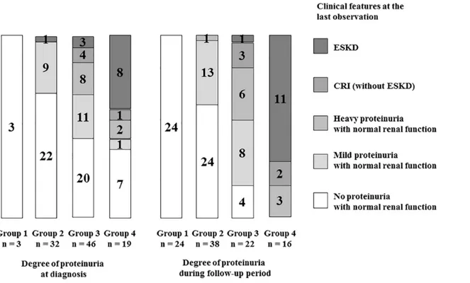
Fig 4shows the relationship between the degree of proteinuria (at diagnosis/during follow-up period) and clinical features at the last observation. Eleven of 16 patients (69%) of grofollow-up 4 during follow-up period developed ESKD at the last observation, while 8 of 19 patients (42%) of group 4 at diagnosis developed ESKD. Almost half patients of group 4 at diagnosis showed no or mild proteinuria at the last observation, while none of group 4 during follow-up period showed such beneficial outcome at the last observation. One patient with mild proteinuria at diagnosis showed heavy proteinuria during follow-up period and developed ESKD at the last observation.Fig 5shows the clinical courses of all patients. All patients with poor prognosis (ESKD or CRI) were in group 3 or 4 during follow-up period, whereas no patients in group 1 or 2 during the same period had poor prognosis. Twenty-four of 38 patients (63%) with mild proteinuria (group 2) during follow-up period showed no proteinuria at the last observation.
Risk factors for ESKD and CRI
diagnosis, glomeruli showing mesangial proliferation50%, steroid treatment and group 4 during follow-up period were calculated as risk factors by univariate analysis. In these factors, glomeruli showing mesangial proliferation50% and group 4 during follow-up period were calculated as statistically significant risk factors for CRI by multivariate analysis (Table 3).
Discussion
To date, few reports on the long-term prognosis of patients with childhood-onset IgA nephropathy are available [8,27,28]. The median observation period of our study was 11.8 years, which is much longer than previous reports. Two-thirds of all patients were followed for more than 10 years. Moreover, several patients who developed ESKD completed the
Table 1. Characteristics of patients.
All patients (n = 100)
Degree of proteinuria at diagnosis
Group 1 (n = 3)
Group 2 (n = 32)
Group 3 (n = 46)
Group 4 (n = 19)
Male gender 50 (50%) 3 (100%) -44% 24 (52%) 9 (47%)
Age at onset (y) 10.1±3.0 8.1±1.9 10.4±3.5 10.4±2.8 9.4±2.8 Age at diagnosis (y) 11.6±3.2 9.3±2.0 12.5±3.7 11.7±2.9 10.0±2.7 Initial presentation
Screening 70 (70%) 0 (0%) 20 (63%) 39 (85%) 11 (58%)
Gross hematuria 21 (21%) 3 (100%) 10 (31%) 6 (13%) 2 (11%)
Edema 9 (9%) 0 (0%) 2 (6%) 1 (2%) 6 (32%)
eGFR at diagnosis (ml/min/1.73 m2) 128.4
±35.7 148.3±66.1 131.7±36.5 128.3±36.6 119.4±26.0
≧90 89 (89%) 3 (100%) 28 (88%) 40 (87%) 18 (95%)
60–90 11 (11%) 0 (0%) 4 (13%) 6 (13%) 1 (5%)
30–60 0 (0%) 0 (0%) 0 (0%) 0 (0%) 0 (0%)
15–30 0 (0%) 0 (0%) 0 (0%) 0 (0%) 0 (0%)
<15 0 (0%) 0 (0%) 0 (0%) 0 (0%) 0 (0%)
Serum albumin level at diagnosis (g/dl) 3.7±0.8 3.9±0.5 4.3±0.4 3.9±0.4 2.3±0.4 24-h urinary protein excretion at diagnosis (g/1.73
m2)
3.0±2.9 0.1±0.1 0.6±0.2 2.9±1.7 7.4±2.3
Hypertension at diagnosis 20 (20%) 2 (67%) 6 (19%) 7 (15%) 5 (26%) Pathologicalfindings at diagnosis
Glomeruli with mesangial proliferation (%) 51.3±32.0 32.3±30.4 30.1±25.4 66.5±29.3 55.2±27.6 Glomeruli with crescents (%) 4.3±9.5 0.0±0.0 2.8±5.3 5.6±11.3 4.7±11.4 Glomeruli with global sclerosis (%) 4.0±9.3 0.0±0.0 1.9±6.2 3.8±5.8 10.4±18.4 Treatment
Steroid 25 (25%) 0 (0%) 2 (6%) 15 (33%) 8 (42%)
Nonsteroidal agents 59 (59%) 3 (100%) 20 (63%) 26 (57%) 10 (53%)
No treatment 16 (16%) 0 (0%) 10 (31%) 5 (11%) 1 (5%)
Observation period (y) 11.8 (0.8–24.8) 16.7 (3.6–16.7) 12.8 (4.9–24.8) 7.0 (2.2–19.8) 10.6 (0.8–19.4)
Clinical features at the last observation
No proteinuria with normal renal function 52 (52%) 3 (100%) 22 (69%) 20 (43%) 7 (37%) Mild proteinuria with normal renal function 21 (21%) 0 (0%) 9 (28%) 11 (24%) 1 (5%) Heavy proteinuria with normal renal function 10 (10%) 0 (0%) 0 (0%) 8 (17%) 2 (11%)
Chronic renal insufficiency 5 (5%) 0 (0%) 0 (0%) 4 (9%) 1 (5%)
End-stage kidney disease 12 (12%) 0 (0%) 1 (3%) 3 (7%) 8 (42%)
Data are expressed as means±SD or median (min-max) or number of patients (%).
Fig 1. Renal survival curve of all patients.a) Kaplan-Meier plot showing time to ESKD. b) Kaplan-Meier plot showing time to CR.
doi:10.1371/journal.pone.0150885.g001
Fig 2. Survival curve showing time to ESKD.a) Kaplan-Meier plot showing time to ESKD stratified by proteinuria at diagnosis. b) Kaplan-Meier plot showing time to ESKD stratified by proteinuria during follow-up period. group 1, no proteinuria (<0.2 g/day/1.73 m2); group 2, mild proteinuria (0.2
–1.0 g/day/ 1.73 m2); group 3, heavy proteinuria without hypoalbuminemia (>1.0 g/day/1.73 m2with serum albumin level3.0 g/dL); group 4, heavy proteinuria with hypoalbuminemia (>1.0 g/day/1.73 m2with serum albumin level<3.0 g/dL).
observation period within 10 years due to the initiation of renal replacement therapy. Our study is also the first report on the relationship between proteinuria during the follow-up period and renal prognosis in childhood IgA nephropathy.
Han et al. reported that five of 24 adult patients (21%) with nephrotic onset IgA nephropa-thy underwent spontaneous remission within six months after onset [12]. Kim et al. summa-rized 100 adult patients with nephrotic onset IgA nephropathy and reported that complete remission, partial remission and no response occurred in 48%, 32% and 20% of patients, respectively, while 24% of them underwent spontaneous remission [13]. In our experience of proteinuria with hypoalbuminemia at diagnosis, 37% showed no proteinuria and 42% devel-oped ESKD at the last observation, which is similar to previous reports. We speculate that some cases of IgA nephropathy of nephrotic onset have a self-limiting character, similar to acute glomerulonephritis and purpura nephritis. On the other hand, nephrotic syndrome or heavy proteinuria during follow-up period may indicate failure of treatment, thus explaining the poor prognosis.
There are also reports of disease activity worsening after several years in patients who showed no or slight proteinuria at diagnosis. Szeto et al. reported that 33% of 72 adult patients with mild proteinuria (0.4 g/day or less) at onset developed proteinuria of 1 g/day or more dur-ing a median follow-up of seven years [14]. Shen et al. reported that 29% of 135 adult patients with isolated microscopic hematuria of IgA nephropathy developed proteinuria, and 20% developed renal insufficiency during a median follow-up of 92 months [15]. In view of these reports, one cannot be optimistic about the prognosis even if proteinuria is mild at onset.
Fig 3. Survival curve showing time to CRI.a) Kaplan-Meier plot showing time to CRI stratified by proteinuria at diagnosis. b) Kaplan-Meier plot showing time to CRI stratified by proteinuria during follow-up period. group 1, no proteinuria (<0.2 g/day/1.73 m2); group 2, mild proteinuria (0.2
–1.0 g/day/1.73 m2); group 3, heavy proteinuria without hypoalbuminemia (>1.0 g/day/1.73 m2with serum albumin level3.0 g/dL); group 4, heavy proteinuria with
hypoalbuminemia (>1.0 g/day/1.73 m2with serum albumin level<3.0 g/dL).
In this study we proved that the degree of proteinuria during follow-up period strongly cor-related with the final prognosis. Although a certain degree of proteinuria at onset is a signifi-cant risk factor, it is not an absolute factor for prognosis. It remains difficult to predict the outcome of each patient based on the clinical and pathological parameters at onset. A similar result has been reported in adult patients [16–19]. In particular, heavy proteinuria with hypoal-buminemia during follow-up period indicated a poor prognosis, with two-thirds of those patients developed ESKD, and others developed CRI or heavy proteinuria at the last observa-tion. None of them showed no or mild proteinuria at the last observaobserva-tion. On the other hand, even if patients showed heavy proteinuria during follow-up period, they are less prone to develop ESKD within a few years without hypoalbuminemia, based on our observations. Patients with mild proteinuria during follow-up period showed excellent prognosis and none of them showed CRI. Moreover, two-thirds of them showed no proteinuria at the last observa-tion. For patients with mild proteinuria, we propose that strong immunosuppression should be avoided. As a goal of the treatment of IgA nephropathy, we should aim to reduce 24-h protein-uria to less than 1 g/1.73 m2(or 1 g/g of urinary protein to creatinine ratio) to reduce the possi-bility of progression to CRI.
There are several limitations to our study. First, we selected the highest level of proteinuria, not time-average proteinuria [16,17], in the follow-up period. It was difficult to calculate time-average proteinuria as data of urinary protein concentrations were often lacking, and only dip-stick tests were performed in some patients who showed no or slight proteinuria. Second, all of the pathological findings were obtained at diagnosis, not at the period when the disease activity became high during the follow-up period. Third, as serum creatinine measurements were dif-ferent before and after 1989, there might be slight errors in calculated data of eGFR. Forth, the
Fig 4. Relationship between degree of proteinuria (at diagnosis/during follow-up period) and clinical features at the last observation.Group 1, no proteinuria (<0.2 g/day/1.73 m2); group 2, mild proteinuria (0.2–1.0 g/day/1.73 m2); group 3, heavy proteinuria without hypoalbuminemia (>1.0 g/day/1.73 m2
with serum albumin level3.0 g/dL); group 4, heavy proteinuria with hypoalbuminemia (>1.0 g/day/1.73 m2with serum albumin level<3.0 g/dL).
sample size was relatively small. For validation of our results, a larger sample cohort is required. Finally, as the period of diagnosis was in the relatively distant past, indication of treatment, such as steroid, is not clear.
In conclusion, the degree of proteinuria during follow-up period is the strongest risk factor for IgA nephropathy progression in children. In particular, proteinuria during follow-up period shows absolutely poor prognosis. Patients who have no heavy proteinuria during
Table 2. Predicted risk factors for end-stage kidney disease.
Univariate Multivariate
HR (95%CI) P value HR (95%CI) P value
Group 4 at diagnosis 13.6 (3.9–62.4) <0.0001 5.4 (1.1–44.8) 0.04
eGFR<90 ml/min/1.73 m2 at diagnosis 0.8 (0.04–4.0) 0.79
Hypertension at diagnosis 0.4 (0.02–2.1) 0.32
Glomeruli with mesangial proliferation≧50% at diagnosis 8.9 (1.6–165.8) 0.008 1.4 (0.1–30.1) 0.79
Steroid treatment 2.6 (0.7–8.7) 0.13
Group 4 during follow-up period (a) <0.0001 (a) <0.0001
HR, hazard ratio; 95%CI, 95% confidence interval
aHazard ratio cannot be obtained since all events were in group 4 during follow-up period.
doi:10.1371/journal.pone.0150885.t002
Fig 5. Clinical course of all patients.Group 1, no proteinuria (<0.2 g/day/1.73 m2); group 2, mild proteinuria (0.2
–1.0 g/day/1.73 m2); group 3, heavy proteinuria without hypoalbuminemia (>1.0 g/day/1.73 m2with serum albumin level3.0 g/dL); group 4, heavy proteinuria with hypoalbuminemia (>1.0 g/
day/1.73 m2with serum albumin level<3.0 g/dL).
follow-up period hardly develop renal insufficiency. Pediatric nephrologists should plan the treatment strategy with the long-term outcome in mind.
Acknowledgments
We would like to thank Dr. Julian Tang from the Department of Education for Clinical Research, National Center for Child Health and Development, for proofreading, editing and rewriting part of this manuscript. We also like to thank Dr. Norishige Yoshikawa from the Center for Clinical Research and Development, National Center for Child Health and Develop-ment, for evaluating the pathological findings.
Author Contributions
Conceived and designed the experiments: KI MH. Performed the experiments: KK. Analyzed the data: KK. Contributed reagents/materials/analysis tools: KK. Wrote the paper: KK. Medical attendants of patients: R. Harada R. Hamada TS YH HH SI.
References
1. Coppo R, D'Amico G. Factors predicting progression of IgA nephropathies. J Nephrol 2005; 18: 503– 12 PMID:16299675
2. D'Amico G. Natural history of idiopathic IgA nephropathy and factors predictive of disease outcome. Semin Nephrol 2004; 24: 179–96 PMID:15156525
3. Berthoux F, Mohey H, Laurent B, Mariat C, Afiani A, Thibaudin L. Predicting the risk for dialysis or death in IgA nephropathy. J Am Soc Nephrol 2011; 22: 752–61 doi:10.1681/ASN.2010040355PMID: 21258035
4. Wakai K, Kawamura T, Endoh M, Kojima M, Tomino Y, Tamakoshi A, et al. A scoring system to predict renal outcome in IgA nephropathy: from a nationwide prospective study. Nephrol Dial Transplant 2006; 21: 2800–8 PMID:16822793
5. Li PK, Ho KK, Szeto CC, Yu L, Lai FM. Prognostic indicators of IgA nephropathy in the Chinese— clini-cal and pathologiclini-cal perspectives. Nephrol Dial Transplant 2002; 17: 64–9 PMID:11773464 6. To KF, Choi PC, Szeto CC, Li PK, Tang NL, Leung CB, et al. Outcome of IgA nephropathy in adults
graded by chronic histological lesions. Am J Kidney Dis 2000; 35: 392–400 PMID:10692264 7. Haas M. Histologic subclassification of IgA nephropathy: a clinicopathologic study of 244 cases. Am J
Kidney Dis 1997; 29: 829–42 PMID:9186068
8. Yoshikawa N, Tanaka R, Iijima K. Pathophysiology and treatment of IgA nephropathy in children. Pediatr Nephrol 2001; 16: 446–57 PMID:11405121
9. Yoshikawa N, Ito H, Nakamura H. Prognostic indicators in childhood IgA nephropathy. Nephron 1992; 60: 60–7 PMID:1738416
Table 3. Predicted risk factors for chronic renal insufficiency.
Univariate Multivariate
HR (95%CI) P value HR (95%CI) P value
Group 4 at diagnosis 6.5 (2.5–17.6) 0.0002 2.4 (0.6–9.5) 0.2
eGFR<90 ml/min/1.73 m2 at diagnosis 2.1 (0.5–6.4) 0.29
Hypertension at diagnosis 0.5 (0.08–1.8) 0.32
Glomeruli with mesangial proliferation≧50% at diagnosis 13.7 (2.7–249.8) 0.0004 8.1 (1.3–165.8) 0.03
Steroid treatment 4.6 (1.7–13.1) 0.003 0.6 (0.1–2.3) 0.43
Group 4 during follow-up period 49.9 (15.5–222.1) <0.0001 78.0 (13.8–730.5) <0.0001
HR, hazard ratio; 95%CI, 95% confidence interval
10. Hogg RJ, Silva FG, Wyatt RJ, Reisch JS, Argyle JC, Savino DA. Prognostic indicators in children with IgA nephropathy—report of the Southwest Pediatric Nephrology Study Group. Pediatr Nephrol 1994; 8: 15–20 PMID:8142218
11. Edström Halling S, Söderberg MP, Berg UB. Predictors of outcome in paediatric IgA nephropathy with regard to clinical and histopathological variables (Oxford classification). Nephrol Dial Transplant 2012; 27: 715–22 doi:10.1093/ndt/gfr339PMID:21750154
12. Han SH, Kang EW, Park JK, Kie JH, Han DS, Kang SW. Spontaneous remission of nephrotic syndrome in patients with IgA nephropathy. Nephrol Dial Transplant 2011; 26: 1570–5 doi:10.1093/ndt/gfq559 PMID:20841490
13. Kim JK, Kim JH, Lee SC, Kang EW, Chang TI, Moon SJ, et al. Clinical features and outcomes of IgA nephropathy with nephrotic syndrome. Clin J Am Soc Nephrol 2012; 7: 427–36 doi:10.2215/CJN. 04820511PMID:22223610
14. Szeto CC, Lai FM, To KF, Wong TY, Chow KM, Choi PC, et al. The natural history of immunoglobulin A nephropathy among patients with hematuria and minimal proteinuria. Am J Med 2001; 110: 434–7 PMID:11331053
15. Shen P, He L, Li Y, Wang Y, Chan M. Natural history and prognostic factors of IgA nephropathy pre-sented with isolated microscopic hematuria in Chinese patients. Nephron Clin Pract 2007; 106: c157– 61 PMID:17596724
16. Le W, Liang S, Hu Y, Deng K, Bao H, Zeng C, et al. Long-term renal survival and related risk factors in patients with IgA nephropathy: results from a cohort of 1155 cases in a Chinese adult population. Nephrol Dial Transplant 2012; 27: 1479–85 doi:10.1093/ndt/gfr527PMID:21965586
17. Reich HN, Troyanov S, Scholey JW, Cattran DC; Toronto Glomerulonephritis Registry. Remission of proteinuria improves prognosis in IgA nephropathy. J Am Soc Nephrol 2007; 18: 3177–83 PMID: 17978307
18. Bartosik LP, Lajoie G, Sugar L, Cattran DC. Predicting progression in IgA nephropathy. Am J Kidney Dis 2001; 38: 728–35 PMID:11576875
19. Donadio JV, Bergstralh EJ, Grande JP, Rademcher DM. Proteinuria patterns and their association with subsequent end-stage renal disease in IgA nephropathy. Nephrol Dial Transplant 2002; 17: 1197–203 PMID:12105241
20. Matsushita S, Ishikura K, Okamoto S, Okuda Y, Nagaoka Y, Harada R, et al. Long-term morbidity of IgA nephropathy in children evaluated with newly proposed remission criteria in Japan. Clin Exp Nephrol [Epub ahead of print]
21. Schwartz GJ, Haycock GB, Edelmann CM Jr, Spitzer A. A simple estimate of glomerular filtration rate in children derived from body length and plasma creatinine. Pediatrics 1976; 58: 259–63 PMID:951142 22. Uemura O, Nagai T, Ishikura K, Ito S, Hataya H, Gotoh Y, et al. Creatinine-based equation to estimate the glomerular filtration rate in Japanese children and adolescents with chronic kidney disease. Clin Exp Nephrol 2014; 18: 626–33 doi:10.1007/s10157-013-0856-yPMID:24013764
23. National High Blood Pressure Education Program Working Group on High Blood Pressure in Children and Adolescents. The fourth report on the diagnosis, evaluation, and treatment of high blood pressure in children and adolescents. Pediatrics 2004; 114: 555–76 PMID:15286277
24. Yoshikawa N, Ito H, Sakai T, Takekoshi Y, Honda M, Awazu M, et al. A controlled trial of combined ther-apy for newly diagnosed severe childhood IgA nephropathy. J Am Soc Nephrol 1999; 10: 101–9 PMID: 9890315
25. Yoshikawa N, Honda M, Iijima K, Awazu M, Hattori S, Nakanishi K, et al. Steroid treatment for severe childhood IgA nephropathy: a randomized, controlled trial. Clin J Am Soc Nephrol 2006; 1: 511–7 PMID:17699253
26. Kamei K, Nakanishi K, Ito S, Saito M, Sako M, Ishikura K, et al. Long-term results of a randomized con-trolled trial in childhood IgA nephropathy. Clin J Am Soc Nephrol 2011; 6: 1301–7 doi:10.2215/CJN. 08630910PMID:21493743
27. Ronkainen J, Ala-Houhala M, Autio-Harmainen H, Jahnukainen T, Koskimies O, Merenmies J, et al. Long-term outcome 19 years after childhood IgA nephritis: a retrospective cohort study. Pediatr Nephrol 2006; 21: 1266–73 PMID:16838184