Idiopathic pulmonary hemorrhage: morphology
and differential diagnosis. Case report
Hemorragia pulmonar idiopática: morfologia e diagnóstico diferencial. Relato de caso
Eduardo Cambruzzi1; Karla Lais Pêgas2; Túlio Vedana3
First submission on 08/03/13; last submission on 04/05/13; accepted for publication on 04/05/13; published on 20/06/13
1. Post-doctorate in Cardiovascular Pathology by Instituto de Cardiologia do Rio Grande do Sul; associate professor at Universidade Luterana do Brasil (ULBRA); pathologist. 2. Master’s in Pathology by Fundação da Universidade Federal de Ciências da Saúde de Porto Alegre; pathologist at Santa Casa de Porto Alegre.
3. Graduate medical student at ULBRA.
ABStrACt
Idiopathic pulmonary hemorrhage (IPH) is a rare cause of alveolar hemorrhage (AH) with unknown etiology that primarily affects children. The process has a variable clinical progression, and its diagnosis is established after excluding all causes of AH. Herein, the authors report a case of IPH in an adult male patient referring cough and hemoptysis. The conventional radiography computed tomography imaging identiied zones of consolidation and areas of ground-glass attenuation in the lower lobes and lingula. Forced spirometry, bronchoscopy, and laboratorial tests yielded normal results. Several alveolar hemosiderin-laden macrophages were identiied on bronchoalveolar lavage and lung biopsy. Thus, the histopathological indings associated with clinical data were compatible with IPH.
Key words:hemosiderosis; lung diseases; alveolar macrophages; pathology; hemorrhage.
introDuCtion
Idiopathic pulmonary hemorrhage (IPH) is a rare cause of diffuse alveolar hemorrhage whose etiology is unknown. The process, which affects primarily children, is characterized by intra-alveolar accumulation of red blood cells and hemosiderin-laden macrophages. The patients present recurrent episodes of pulmonary hemorrhage, which may occasionally determine ventilatory restriction. The clinical presentation is extremely variable, and the most common symptoms include iron deiciency anemia, diffuse pulmonary iniltrates, and hemoptysis(3, 8, 10, 11, 25).
IPH is a diagnosis of exclusion that is reached when all other causes of pulmonary hemorrhage have been ruled out. Herein, the authors report a case of IPH in a male patient referring cough and hemoptysis, and review morphological and clinical indings of this uncommon process.
CASE rEPort
Male patient, 45 years old, hairdresser, came to the hospital service referring cough and hemoptysis for twelve months. The
sliding hiatal hernia were identiied on upper digestive endoscopy. Doppler echocardiography examination showed the presence of altered relaxation of the left ventricle with preserved systolic performance. CT scans imaging of the abdomen revealed normal indings. The relevant laboratory tests are described in Table 1.
The patient underwent biopsy (segmentectomy) of the lingula and lower basal segments of the left lung. The surgical specimen consisted of two cuneiform portions of cortical pulmonary parenchyma, which together weighed 23.0 g and the largest measures were 6.4 × 2.0 × 1.9 cm. On gross examination, red-brownish areas of the lung parenchyma were identiied. Microscopy demonstrated numerous vacuolated macrophages
containing intracytoplasmic brown granular pigment
(Figure 3), which occupied several alveolar spaces and alveolar ducts and some respiratory bronchioles (Figure 4). Alveolar
septa did not show evidence of inlammation and/or ibrosis. Arteries, veins and capillaries also had normal appearance, without evidence of vasculitis (Figure 5). On Prussian blue stains, the intracytoplasmic granules of the alveolar macrophages proved to be hemosiderin (Figure 6). The histopathological indings related to the clinical data were then compatible with IPH. After a 60-day follow-up, including daily use of prednisone 40 mg, the patient reported a marked reduction in the frequency and intensity of hemoptysis.
tABLE 1 – IPH: clinical data
Result Reference values
Erythrocytes 5.26 million/ul 4.5-6.5 million/ul
Hemoglobin 13.9 g/dl 13.5-18.0 d/dl
Hematocrit 42.7% 40.0%-54.0%
Mean corpuscular volume 81.2 fL 76.0-96.0 l
RDW 13.9 11.5-16.0
Leukocytes 5,640/ul 5,000-10,000/ul
Lymphocytes 1,912/ul 1,000-4,000/ul
Neutrophils 3,091/ul 1,500-7,000/ul
Platelets 249,000/ul 130,000-400,000/ul
APTT 34 s 33-43 s
PT 13.1 s 13.0 s
Anti-HIV Nonreactive Nonreactive
Anti-HCV Nonreactive Nonreactive
HBsAg Nonreactive Nonreactive
HBeAg Nonreactive Nonreactive
Strongyloides stercoralis (BAL) Negative Negative
Cryptococcus sp. (BAL) Negative Negative
Streptococcus pneumoniae antibody Nonreactive Nonreactive
Rheumatoid factor < 11 UI/ml < 40 UI/ml
Perinuclear antineutrophil cytoplasmic antibody Nonreactive Nonreactive
Anti-GBM Nonreactive Nonreactive
Antinuclear antibody Nonreactive Nonreactive
Antinucleolar antibody Nonreactive Nonreactive
Anticytoplasmic antibody Nonreactive Nonreactive
Antimitotic apparatus antibody Nonreactive Nonreactive
Antimetaphase plate antibody Nonreactive Nonreactive
Creatinine 0.93 mg/dl 0.4-1.2 mg/dl
Urea 38 mg/dl 10-50 mg/dl
Sodium 137 mEq/l 135-145 mEq/l
Potassium 3.8 mEq/l 3.5-5.0 mEq/l
Calcium 8.9 mg/dl 8.2-10.3 mg/dl
Uroculture Negative for bacterial growth Negative for bacterial growth
CO2 26 mEq/l 21-32 mE/l
TSH 1.92 uUI/ml 0.27-4.20 uUI/ml
Glycemia 91 mg/dl 70-99 mg/dl
Total bilirubin 0.59 mg/dl Up to 1.20 mg/dl
Indirect bilirubin 0.44 mg/dl Up to 0.80 mg/dl
Direct bilirubin 0.15 mg/dl Up to 0.40 mg/dl
Alkaline phosphatase 78 U/l 40- 129 U/l
Albumin 4.8 g/dl 3.5- m5.5 g/dl
Total proteins 7.2 g/dl 6.0- 8.0 g/dl
Gamma glutamyl transferase 40 U/l 8- 61 U/l
Lactate dehydrogenase 379 U/l 240-480 U/l
figurE 5 – IPH: coarse granules of brown hemosiderin are contained within the cytoplasm of numerous macrophages that ill various alveolar spaces (HE 200×) IPH: idiopathic pulmonary hemorrhage; HE: hematoxylin and eosin.
figurE 6 – Granules of hemosiderin can be highlighted on Prussian blue stains (100×)
figurE 2 – Computed tomography through lung bases show zones of consolidation and areas of ground-glass attenuation
figurE 3 – IPH: hemosiderin-laden macrophages illing numerous alveolar spaces, without evidence of pathological changes in the wall of large blood vessels (HE 40×) IPH: idiopathic pulmonary hemorrhage; HE: hematoxylin and eosin.
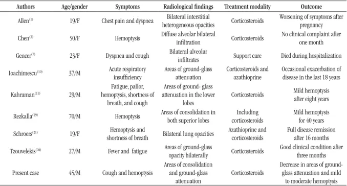
figurE 4 – IPH: hemosiderin-laden macrophages illing alveolar spaces, without evidence of vasculitis in small blood vessels and in alveolar septa (HE 100×) IPH: idiopathic pulmonary hemorrhage; HE: hematoxylin and eosin. figurE 1 – IPH: posteroanterior chest radiographs demonstrate bilateral areas
DiSCuSSion
IPH usually affects children, but in 20 percent of the patients the process is found in adults. There is no gender-based prevalence in children. The clinical presentation is most often associated with dyspnea, cough, hemoptysis, substernal chest pain, and fatigue due to iron-deiciency anemia. The episodes of pulmonary hemorrhage are recurrent and intermittent. Respiratory distress progressing to respiratory failure is uncommon. Chest examination is non-speciic (crackles and consolidations). Radiographic indings of IPH consist of patchy or diffuse bilateral areas of airspace consolidation, mostly affecting the perihilar regions and lower lung zones. Zones of ground-glass attenuation may be visualized on CT scans. On gross examination, the compromised lungs show red, reddish brown, gray, or black, ill-deined areas. IPH is characterized by a diffuse acute and/or chronic hemorrhage with intra-alveolar accumulation of both red blood cells and hemosiderin-laden macrophages. Strong reaction to siderosis is observed on Prussian blue stain. Neutrophilic capillaritis may also be seen, but it tends to be mild. Long-term IPH may lead to interstitial ibrosis. By deinition, IPH does not present renal involvement, immune complexes, and antineutrophil cytoplasmic antibody (ANCAS)(1, 3, 4, 8, 10, 11, 17, 18, 21).
IPH diagnosis is made on the basis of clinical, radiologic, laboratorial and histopathological indings. Final diagnosis can be established only when any evidence of pulmonary vasculitis, nonspeciic granulomatous inlammation or diseases associated with deposition of immunoglobulins have been excluded. Diffuse alveolar hemorrhage (DAH) is characterized by extensive intralveolar hemorrhage, which can be acute or chronic. The process is associated with a great variety of clinical syndromes, and most patients complain of hemoptysis. Microscopy reveals that DAH commonly presents an accumulation of intra-alveolar red blood cells and hemosiderin-laden macrophages. Speciic classiication of DAH syndromes requires correlation with the clinical history, laboratory results, and lung biopsy indings. The differential diagnosis of DAH may be classiied immunologically by the immunoluorescence or electron microscopic indings or histologically by the data on lung biopsy. Alveolar hemorrhage (AH) may be often associated with an organizing pneumonia consisting of intra-alveolar plugs of loose, organizing ibroblastic connective tissue reminiscent of the bronchiolitis obliterans organizing pneumonia (BOOP) pattern(5, 7, 9, 15, 25, 26). Wegener’granulomatosis
(WG) is a systemic granulomatous inlammatory process related to the development of vasculitis on the upper and lower respiratory tract and kidney. The major pathologic features of WG are vasculitis, parenchymal necrosis, granulomatous inlammation (mixed inlammatory iniltrate), and alveolar hemorrhage
in some cases. In active generalized WG, positive staining for C-ANCA in neutrophil cytoplasm on immunoluorescence analysis is found in 84 to 99 percent of patients. Vasculitis of WG may affect arteries, veins, or capillaries in the form of capillaritis(6, 9, 13, 19, 24, 25). Goodpasture’s syndrome (GS) is
characterized by the combination of pulmonary hemorrhage with glomerulonephritis, commonly the rapidly progressive (crescentic) type. It is mediated by circulating cytotoxic antibody (generally immunoglobulin class G [IgG]) that reacts with pulmonary and glomerular basement membrane. Evidence of this anti-basement membrane antibody in the serum, kidney or lung biopsies is required for the diagnosis of GS(25). Bernis
et al.(2) reported a case of GS remission in a female hairdresser
who developed a severe anemia due to pulmonary hemorrhage followed by anti-glomerular basement membrane antibody (GBM) glomerulonephritis with normal renal function. These authors suggested that the use of products related to permanent waving could be linked with the pulmonary disease. In the present case, GS was excluded since anti-GBM and renal disease were not detected. Moreover, the histopathological indings of the sample did not demonstrate the presence of pulmonary vascular lesions or inlammatory process affecting periseptal alveolar capillaries. The authors of the present report also suggest the hypothesis,
similarly to Bernis et al., that this pulmonary hemorrhage may stem from an occupational hazard. Churg-Strauss syndrome (CHS) is characterized by the presence of asthma, peripheral blood eosinophilia, neuropathy, radiographic pulmonary iniltrates, paranasal sinus abnormalities, and vasculitis. On CHS, the vascular inlammatory iniltrates are composed of chronic inlammatory cells, eosinophils, epithelioid cells, multinucleated giant cells, and/ or neutrophils. Presence of diffuse pulmonary hemorrhage and capillaritis may be seen(24, 25). Microscopic polyangiitis (MP) is a
necrotizing vasculitis with few or no immune deposits that involves small vessels, showing necrotizing arteritis of small and medium sized arteries in some occasional cases. The most common lung biopsy indings in MP are pulmonary hemorrhage and neutrophilic capillaritis. On high magniication, thickening of the alveolar walls with many neutrophils on the septal interstitium can be found. The neutrophils may show karyorrhexis and ill in the surrounding alveolar spaces(7, 9, 14, 15, 25). In congestive pulmonary vasculopathy,
the compromised lungs show extensive and severe ibrosis of the elastic arteries. The surrounding lung tissue shows hemosiderosis(25).
to iron is rarely associated with clinically signiicant disease unless other dusts are also present, mainly silica. Microscopy reveals numerous iron-illed macrophages involving respiratory bronchioles and alveolar ducts(20, 22, 27). Airway disease related to
mineral dust and macrophages associated with ibrosis of alveolar ducts can also be identiied. The presence of silica or asbestos (mixed pneumoconiosis) modiies the pathologic indings according to the degree of this deposition. Presence of nodular ibrosis and/or centriacinar emphysema may be found in some cases(16, 23, 25, 26). The
differential diagnosis of IPH also includes cases of vasculitis related to polyarteritis nodosa, Takayasu’s arteritis, Behçet’s syndrome, sarcoidosis, pulmonary infection, and septic emboli(4, 5, 7, 9, 11, 15, 17, 25).
In the present report, the authors reported a case of IPH in an adult
male patient referring cough and hemoptysis, after exhaustive clinical, laboratorial and radiologic investigation to exclude other possible causes of AH. Table 2 describes similar cases of IPH found in the international literature and comparable to the present report in relation to undetermined etiology, clinical course, and treatment.
The most frequent cause of death in IPH is related to acute respiratory failure secondary to massive hemorrhage. Chronic respiratory failure and cor pulmonale are related to the development of severe ibrosis. Some patients develop repeated episodes of hemoptysis, or persistent dyspnea and anemia(5, 11, 21, 26).
The use of corticosteroids must be considered, but the response is
variable(1, 3, 8, 12). A good prognosis is expected in this case, although
the possibility of short episodes of recurrence must be considered.
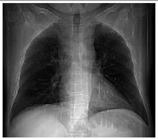
tABLE 2 – Summary of some published cases of IPH
Authors Age/gender Symptoms Radiological findings Treatment modality Outcome
Allen(1) 19/F Chest pain and dyspnea Bilateral interstitial
heterogeneous opacities Corticosteroids
Worsening of symptoms after pregnancy
Chen(2) 50/F Hemoptysis Diffuse alveolar bilateral
iniltration Corticosteroids
No clinical complaint after one month
Gencer(7) 23/F Dyspnea and cough Bilateral alveolar
iniltrates Support care Died during hospitalization
Ioachimescu(10) 37/M Acute respiratory
insuficiency
Areas of ground-glass
attenuation
Corticosteroids and azathioprine
Occasional exacerbation of disease in the last 18 years
Kahraman(11) 29/M
Fatigue, pallor, hemoptysis, shortness of
breath, and cough
Areas of ground- glass attenuation in the lower
lobes
Corticosteroids Mild hemoptysis after eight years
Rezkalla(19) 70/M Hemoptysis Areas of consolidation in
both superior lobes
Including corticosteroids
Mild hemoptysis for 40 years
Schroers(21) 19/F Hemoptysis and
shortness of breath Bilateral lung opacities
Azathioprine and corticosteroids
Full disease remission after 16 months
Tzouvelekis(26) 27/M Fever and fatigue Areas of ground-glass
opacity bilaterally Corticosteroids
Good clinical condition after three months
Present case 45/M Cough and hemoptysis
Areas of consolidation and ground-glass
attenuation
Corticosteroids
Decrease in areas of
ground-glass attenuation and mild
to moderate hemoptysis IPH: idiopathic pulmonary hemorrhage; F: female; M: male.
rESuMo
A hemorragia pulmonar idiopática (HPI) é uma causa rara de hemorragia alveolar (HA) que afeta principalmente crianças, com etiologia desconhecida. O processo tem evolução clínica variável e o diagnóstico é estabelecido depois que todas as causas de HA forem excluídas. Neste artigo, os autores relatam um caso de HPI em paciente do sexo masculino, adulto, referindo tosse e hemoptise. O exame radiológico convencional e a tomograia computadorizada identiicaram zonas de consolidação e áreas de vidro fosco nos lobos inferiores e língula. A espirometria forçada, a broncoscopia e os testes laboratoriais revelaram resultados normais. Numerosos macrófagos alveolares contendo hemossiderina foram identiicados no lavado broncoalveolar e na biópsia pulmonar. Os achados histopatológicos relacionados com os dados clínicos foram então compatíveis com HPI.
rEfErEnCES
1. ALLEN, T. K. et al. Management of a parturient with an acute
exacerbation of idiopathic pulmonary haemosiderosis and posterior
spinal instrumentation. Br J Anaesth, v. 100, n. 2, p. 235-9, 2008.
2. BERNIS, P. et al. Remission of Goodpasture’s syndrome after withdrawal
of an unusual toxic. Clin Nephrol, v. 23, n. 6, p. 312-7, 1985.
3. CHEN, C. H. et al. Idiopathic pulmonary hemosiderosis: favorable
response to corticosteroids. J Chin Med Assoc, v. 71, n. 8, p. 421-4, 2008.
4. COHEN, S. Idiopathic pulmonary hemosiderosis. Am J Med Sci, v. 317,
p. 67-74, 1999.
5. DE PROST, N. et al. Diffuse alveolar hemorrhage in immunocompetent
patients: etiologies and prognosis revisited. Respir Med, v. 106, n. 7, p. 1021-32, 2012.
6. ESPOSITO, S. et al. Wegener’s granulomatosis presenting with
life-threatening lung hemorrhage in a 7-year-old child. Rheumatol Int,
v. 30, n. 12, p. 1665-8, 2010.
7. FISHBEIN, G. A.; FISHBEIN, M. C. Lung vasculitis and alveolar
hemorrhage: pathology. Semin Respir Crit Care Med, v. 32, n. 3, p.
254-63, 2011.
8. GENCER, M. et al. Two sisters with idiopathic pulmonary hemosiderosis.
Can Resp J, v. 14, n. 8, p. 490-3, 2007.
9. GROSS, W. L. et al. New perspectives in pulmonary angiitis. From
pulmonary angiitis and granulomatosis to ANCA associated vasculitis.
Sarcoidosis Vasc Diffuse Lung Dis, v. 17, p. 33-52, 2000.
10. IBRAHEM, R. et al. Case report of idiopathic pulmonary haemosiderosis
in a child with recurrent chest infections. J Radiol Case Rep, v. 5, n. 9,
p. 30-5, 2011.
11. IOACHIMESCU, O. C. et al. Idiopathic pulmonary haemosiderosis
revisited. Eur Respir J, v. 24, p. 162-70, 2004.
12. KAHRAMAN, H. et al. Eight years follow-up of a case with idiopathic
pulmonary hemosiderosis after corticosteroid therapy. N Am J Med Sci,
v. 4, n. 1, p. 49-51, 2012.
13. LANGFORD, C. A.; HOFFMAN, G. S. Wegener’s granulomatosis. Thorax,
v. 54, p. 629-37, 1999.
14. LAUQUE, D. et al. Microscopic polyangiitis with alveolar hemorrhage.
A study of 29 cases and review of the literature. Groupe d’Etudes et
de Recherde sur les Maladies Orphelines Pulmonaries. Medicine
(Blatimore), v. 79, p. 222-33, 2000.
15. LIE, J. T. Illustrated histopathologic classiication criteria for selected vasculitis syndromes. American College of Rheumatology Subcommitee
on Classiication of Vasculitis. Arthritis Rheum, v. 33, p. 1074-87,
1990.
16. MCCORMCK, L. M. et al. Pulmonary ibrosis secondary to siderosis
causing symptomatic respiratory disease: a case report. J Med Case
Reports, v. 2, p. 257, 2008.
17. MIWA, S. et al. Prognosis in adult patients with idiopathic pulmonary
hemosiderosis. Intern Med, v. 50, n. 17, p. 1803-8, 2011.
18. POGGI, V. et al. Idiopathic pulmonary hemosiderosis: a rare cause
of iron-deiciency anemia in childhood. J Pediatr Hematol Oncol, v. 33,
n. 4, p. 160-2, 2001.
19. QIAN, Q. et al. Hemorrhagic colitis as a presenting feature of Wegener
Granulomatosis. J Gastrointestin Liver Dis, v. 19, n. 4, p. 445-7, 2010.
20. REZKALLA, M. A.; SIMMONS, J. L. Idiopathic pulmonary hemosiderosis and alveolar hemorrhage syndrome: case report and review of the literature. S D J Med, v. 48, n. 3, p. 79-85, 1995.
21. SANKARARAMAN, S.et al. Clinical case of the month. Idiopathic
pulmonary hemosiderosis presenting as a rare cause of iron deiciency
anemia in a toddler – diagnostic challenge. J La State Med Soc, v. 164,
n. 5, p. 293-6, 2012.
22. SCHOROERS, R. et al. A female soccer player with recurrent
haemoptysis and iron deiciency aneamia: idiopathic pulmonary
haemosiderosis (IPH) – case report and literature review. BMJ Case Rep,
doi: 10.1136/bcr.06.2009.1969, 2010.
23. SFERLAZZA S. J.; BECKETT W. S. The respiratory health of welders.
Am Rev Respir Dis, v. 143, p. 1134-48, 1991.
24. SPECKS, U.; DEREMEE, R. A. Granulomatous vasculitis. Wegener’s
granulomatosis and Churg-Strauss syndrome. Rheum Dis Clin North
Am, v. 16, p. 377-97, 1990.
25. TRAVIS, W. D. et al. Pulmonary vasculitis. In: TRAVIS, W. D. et al.
(Eds.). Non-neoplastic disorders of the lower respiratory tract – AFIP
atlas of nontumor pathology. Bethesa: ARP, 2002, p. 233-58.
26. TRAVIS, W. D. et al. A clinicopathologic study of 34 cases of diffuse
pulmonary hemorrhage with lung biopsy conirmation. Am J Surg
Pathol, v. 14, p. 1122-5, 1990.
27. TZOUVELEKIS, A. et al. Idiopathic pulmonary hemosiderosis in adults:
a case report and review of the literature. Case Reports in Medicine,
v. 2012, p. 1-5, 2012.
MAiLing ADDrESS
Eduardo Cambruzzi