0021-7557/$ - see front matter © 2013 Sociedade Brasileira de Pediatria. Published by Elsevier Editora Ltda. All rights reserved. http://dx.doi.org/10.1016/j.jped.2013.02.010
www.jped.com.br
☆Please, cite this article as: Ibagy A, Silva DB, Seiben J, Winneshoffer AP, Costa TE, Dacoregio JS, et al. Acute lymphoblastic leukemia in infants: 20 years of experience. J Pediatr (Rio J). 2013;89:64-69.
* Corresponding author.
E-mail: amanda_ibagy@hotmail.com (A. Ibagy).
ORIGINAL ARTICLE
Acute lymphoblastic leukemia in infants: 20 years of experience
☆Amanda Ibagy
a,*, Denise B. Silva
b, Jackline Seiben
c, Ana P.F.F. Winneshoffer
d,
Tatiana E.J.B. Costa
d, Juliana S. Dacoregio
d, Imaruí Costa
d, and Daniel Faraco
da MD. Pediatric Oncologist. Universidade Federal de Santa Catarina (UFSC), Florianópolis, SC, Brazil
b MSc. Pediatric Hematologist. Hospital Infantil Joana de Gusmão, Florianópolis, SC, Brazil c Medical Student, UFSC, Florianópolis, SC, Brazil
d MD. Pediatric Oncologist, Hospital Infantil Joana de Gusmão, Florianópolis, SC, Brazil
Received 8 May 2012; accepted 8 August 2012
KEYWORDS
Infant; Leukemia, classiication; Precursor cell lymphoblastic leukemia-lymphoma
Abstract
Objective: To analyze patients younger than 2 years with acute lymphoblastic leukemia,
treated in the period between 1990 and 2010 in a state reference center.
Methods: This was a clinical-epidemiological, cross-sectional, observational, and descriptive study. It included patients younger than 2 years with acute lymphoblastic
leukemia, treated in the period of 1990 to 2010 in a pediatric oncology unit of a state
reference center, totaling 41 cases.
Results: All patients were white ethnicity, and 60.9% were females. Regarding age, 24.38% were younger than 6 months, 17.07% were between 6 months and 1 year, and 58.53% were older than 1 year. The age of 6 months was statistically significant for the
outcome of death. Predominant signs and symptoms were fever, bruising, and petechiae.
A leukocyte count > 100,000 was found in 34.14% of cases, hemoglobin count < 11 in 95.13%, and platelet count < 100,000 in 75.61. Infiltration of central nervous system was present in 12.91% of patients. According to the lineage, B-cell lineage predominated (73%), but the T-cell line was statistically significant for death. 39% of patients had disease recurrence. In relation to vital status, 70.73% of the patients died; septic shock
was the main cause.
Conclusions: Acute lymphoblastic leukemia in infants has a high mortality rate, especially in children under 1 year and those with T-cell derived lineage.
PALAVRAS-CHAVE Crianças;
Leucemia,
classiicação;
Leucemia-linfoma linfoblástica precursora de células
Leucemia linfoblástica aguda em lactentes: 20 anos de experiência
Resumo
Objetivo: Analisar pacientes com menos de dois anos de idade com leucemia
linfoblásti-ca aguda atendidos no período de 1990 a 2010, em um centro de referência estadual. Métodos: Estudo clínico, epidemiológico, transversal, descritivo e observacional. Pacientes incluídos tinham menos de dois anos de idade, com leucemia linfoblástica
aguda, tratados no período de 1990 a 2010 na unidade de oncologia pediátrica de um centro de referência estadual, totalizando 41 casos.
Resultados: Todos os pacientes eram Caucasianos e 60,9% eram do sexo feminino. Com relação à idade, 24,38% tinham menos de seis meses, 17,07% tinham entre seis meses e um ano e 58,53% mais do que um ano de idade. A idade de seis meses foi estatisticamente
significante para o desfecho de óbito. Os sinais e sintomas predominantes foram febre,
hematomas e petéquias. Uma contagem de leucócitos superior a 100.000 foi observada em 34,14% dos casos; hemoglobina inferior a 11 em 95,13% e contagem de plaquetas inferior a 100.000, em 75,61% dos casos. Infiltração do sistema nervoso central estava presente em 12,91% dos pacientes. Em relação à linhagem, a linhagem B predominou (73%), mas a linhagem de células T foi estatisticamente significativa para o óbito. Trinta e nove por cento dos pacientes tiveram recorrência da doença. Em relação ao estado vital, 70,73% dos pacientes morreram, sendo choque séptico a principal causa.
Conclusões: leucemia linfoblástica aguda em crianças tem uma alta taxa de mortalidade, principalmente em crianças menores de um ano e linhagem derivada de células T. © 2013 Sociedade Brasileira de Pediatria. Publicado por Elsevier Editora Ltda. Todos os direitos reservados.
Introduction
Approximately 10% of all cancers affecting children under
15 years correspond to those diagnosed in the first year of life.1 Leukemia is the second most common cancer in
children under 1 year of age, and acute lymphoblastic
leukemia (ALL) the most frequently observed type.2
Some possible risk factors are genetic syndromes
(Down, Noonan, trisomy 9), high birth weight (> 3.5 kg),
previous abortion, maternal behavior (use of antihistamine, metronidazole, dipyrone, estrogen, alcohol consumption, use of marijuana and hallucinogenic drugs, radiation, and exposure to insecticides and pesticides).3,4
Leukemia in children under 1 year of age has distinct epidemiological, clinical, and biological characteristics, and is associated with unfavorable factors such as hyperleukocytosis and central nervous system (CNS) infiltration.5,6 In this age range, there is a predominance of gene rearrangements that present as 11q23 translocation,
leading to the coexistence of the lymphoid and myeloid phenotypes, known as mixed lineage leukemia (MLL) translocation. The presence of this type of translocation is associated with worse prognosis.6-8
Regarding the immunophenotype, these children’s blast
cells have a very young precursor B-cell (CD34 + / CD 19+)
with CD 10 negativity.9
To treat leukemia in this age range, specific protocols are used, which have been developed by international cooperative groups, as childhood leukemias do not usually respond to traditional treatments, showing resistance to drugs such as corticosteroids and asparaginase.10,11 With the
use of therapy intensification, survival has increased, but
so has toxicity; in the CCG-1953 protocol of the Children’s Cancer Group, 29% of deaths occur during induction.12
The use of allogeneic bone marrow transplantation in these patients is still controversial, showing no difference in survival when compared to chemotherapy alone.13
In spite of all the therapeutic approaches, overall survival
remains poor. In the CCG-1953 protocol of the Children’s
Cancer Group, overall survival at five years ranged between
22% and 30%, while for the Interfant-99 protocol, it was 53.8%.10,12 More recently, a group from Taiwan reported a survival rate of 18%.14
This study aimed to assess patients younger than 2 years of age with ALL, treated in the pediatric oncology unit of
the Hospital Infantil Joana de Gusmão (HIJG) from 1990 to
2010.
Methods
This was a clinical-epidemiological, cross-sectional, observational, and descriptive study. The research protocol was approved by the Research Ethics Committee of the HIJG. The study included patients younger than 2 years diagnosed with ALL, treated at HIJG between January,
1990 and December, 2010. Exclusion criteria were loss at
follow-up, transfer to another department for treatment, and insufficient data in the medical record.
of national (GBTLII-85, GBTLII-93, and GBTLII-99) and international cooperative groups (ALL III-85, Interfant-99,
and ALL-IC BFM 2002).
The variables analyzed were age at diagnosis (stratified
age range as ≤ 6 months; 6 months < age < one year; and ≥
one year); gender; birth weight (greater and less than 3,500 grams); ethnicity according to the Brazilian Institute of Geography and Statistics (Instituto Brasileiro de Geografia e Estatística – IBGE) (white, black, mixed-race, Asian, and Native Brazilian); origin according to Santa Catarina sub regions, as established by IBGE (Greater Florianópolis,
Northern Santa Catarina, Western Santa Catarina, Uplands,
Southern Santa Catarina, Itajaí Valley); parental contact with pesticides; signs and symptoms at diagnosis (fever, bone pain, lymphadenopathy, hepatosplenomegaly, skin and mucosal membrane bleeding); laboratory assessment at diagnosis (leukocyte, hemoglobin, and platelet counts); CNS involvement at diagnosis; presence and type of genetic alterations precursor phenotype (B or T derived); vital status (alive or deceased); death (immediate cause, in remission or not of neoplastic disease, treatment period); relapse (medullary, extra-medullary or combined); time of follow-up.15
Data were collected from the medical records and
statistics service (serviço de arquivo médico e estatística
- SAME) and from the hospital cancer registry of the HIJG.
Statistical analysis was performed using the GraphPad Prism 5® software. The results were analyzed using Pearson’s chi-squared test with 95% level of significance, thereby
analyzing the relationship between two variables.
Results
The total number of patients evaluated was 41, all whites,
of which 60.97% were females. The mean age at diagnosis
was 12.5 months, with a median of 13 months. Regarding
the age range at diagnosis, 24.39% were younger than 6 months, 17.07% were older than 6 months and younger than 1 year, and 58.53% were older than 1 year of age.
The mean birth weight was 3,367 g, with a median of 3,400 g.
History of parental contact with pesticides was described
in 19.51% of cases. A total of 39% of cases denied history of contact with pesticides, and in 41.5% of the cases the data
were not available. Signs and symptoms at the diagnosis are summarized in Table 1.
As for the geographic origin, considering the sub regions of the state of Santa Catarina according to the IBGE,
34.14% (n = 14) of the patients came from the Itajaí Valley, 24.39% (n = 10) from Greater Florianópolis, 17.07% (n = 7) from Southern Santa Catarina, 9.75% (n = 4) from Western Santa Catarina, 7.32% (n = 3) from Northern Santa Catarina, and 7.32% (n = 3) from the Uplands. The overall mean
follow-up time was 2 years; in surviving patients, this time was 4.6 years.
CNS infiltration at diagnosis was present in 12.19% of
patients. Three patients did not undergo CSF analysis, as they died before the exam. CNS infiltration, when compared with vital status, was not statistically significant
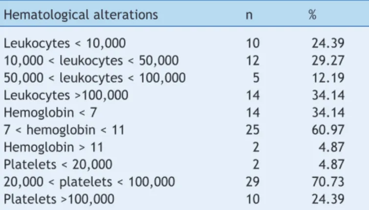
(p = 0.54). Hematological alterations at diagnosis are
detailed in Table 2.
Cytogenetic assessment was performed in 48.78% of cases (n = 20). Of these, 17.07% had genetic alterations, including 11q23:3, chromosome trissomy 8:1, terminal
deletion of the long arm of chromosome 7:1 and t (15, 17).
Data were not available in 51.22% of the cases.
In 73.17% of cases the precursor phenotype was derived from B-lineage, and in14.63%, from T-lineage cells.
Regarding the vital status, 29.27% of the patients were
alive (two patients undergoing treatment). The outcome
of death occurred in 70.73% of patients (two patients off
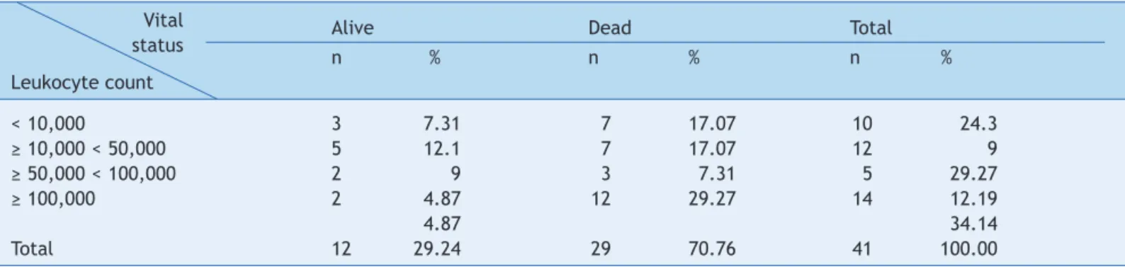
treatment and in clinical remission). Leukocyte count at diagnosis and its comparison with vital status are described in Table 3. Age at diagnosis and its association with vital status is described in Table 4.
Of the patients who died (n = 29), only five of them were
in clinical remission, and three deaths occurred during treatment induction. The main cause of death was septic
shock (41% of deaths). Disease relapse occurred in 39.02% of cases (n = 16); 12 patients presented medullary relapse,
three presented CNS relapse, and one patient presented combined relapse (medullary and CNS). Only two patients underwent allogeneic bone marrow transplant; of these, one is alive and in remission.
Table 1 Infants with acute lymphoblastic leukemia, according to signs and symptoms at diagnosis, as number (n) and percentage (%).
Signs and symptoms n %
Fever 25 60.97
Bone pain 2 4.88
Lymphadenomegaly 12 29.26
Ecchymosis/petechiae 22 53.66
Mucosal bleeding 4 9.76
Hepatomegaly 24 58.53
Splenomegaly 16 39.02
Table 2 Infants with acute lymphoblastic leukemia, according to hematological alterations at diagnosis, as number (n) and percentage (%).
Hematological alterations n %
Leukocytes < 10,000 10 24.39
10,000 < leukocytes < 50,000 12 29.27 50,000 < leukocytes < 100,000 5 12.19
Leukocytes >100,000 14 34.14
Hemoglobin < 7 14 34.14
7 < hemoglobin < 11 25 60.97
Hemoglobin > 11 2 4.87
Platelets < 20,000 2 4.87
20,000 < platelets < 100,000 29 70.73
When comparing the precursor phenotype with vital
status, 40% of patients with B-lineage-derived cells survived and 60% died. All patients with T-lineage-derived cells died. Using the chi-squared test to compare the lines with vital status, a p-value = 0.0006 was obtained, and thus,
T-lineage was statistically significant for death. There was
no statistical significance when comparing birth weight (< and > 3,500 g) with vital status (p = 0.31).
Discussion
Leukemia in children younger than one year is more common in girls (1.17:1), a result confirmed by this study. In the SEER study, the white ethnicity was more affected, but no difference was observed regarding response to treatment when compared to the black ethnicity. In the present research, all patients were whites, consistent with the demographic characteristics of the state of Santa Catarina.1,2
Most patients came from the nearby regions, such as Itajaí valley and the state capital city, Florianópolis, as HIJG is a referral center for cancer treatment in the region.
According to Naumburg, exposure to pesticides is not a proven risk factor for the development of leukemia; however, Slate et al. found an association between leukemia
in infants and gestational exposure to petroleum-derived products, such as benzene. Maternal exposure to pesticides
was verified in 19% of the patients in this study.3,4
Leukemia in children younger than 1 year usually presents as hyperleukocytosis at diagnosis. Mann et al. and Tomizawa et al., evaluating this age range, showed that most patients presented more than 100,000 leukocytes at diagnosis, which is similar to the results found in the
present study, where 34% had a leukocyte count > 100,000
cells.11,16
Pui et al. reported that CNS infiltration is more common in leukemia diagnosed in children younger than 1 year.6 In the Japanese group, 20% of patients showed CNS infiltration; the same was observed for 9% of patients in the Interfant-99 group10,11 Similar to that observed in the Interfant-99 group, CNS infiltration in the present study
was not statistically significant for death.10 However,
in the study by Rives et al., evaluating T-cell-derived leukemia, CNS infiltration at diagnosis was significant for death.17
Most patients in the present study had B-cell precursor phenotype. According to Ribeiro et al., T-cell-derived leukemias are more aggressive, a result corroborated by this study, where this lineage was statistically significant
for death (p = 0.0006).18 Rives et al. achieved overall survival at five years of 74% in T-cell-derived leukemia, whereas Hunger et al. observed an overall survival of 90% Table 3 Infants with acute lymphoblastic leukemia, according to leukocyte count at diagnosis and vital status as number (n) and percentage (%).
Alive Dead Total
n % n % n %
Leukocyte count
< 10,000 3 7.31 7 17.07 10 24.3
≥ 10,000 < 50,000 5 12.1 7 17.07 12 9
≥ 50,000 < 100,000 2 9 3 7.31 5 29.27
≥ 100,000 2 4.87 12 29.27 14 12.19
4.87 34.14
Total 12 29.24 29 70.76 41 100.00
p = 0.09 (chi-squared test). Vital status
Table 4 Infants with acute lymphoblastic leukemia, according to age at diagnosis and vital status as number (n) and percentage (%).
Alive Dead Total
n % n % n %
Age at diagnosis
< 6 months 1 2.43 9 21.95 10 24.3*
6 months < age < 1 year 1 2.43 6 14.63 7 17.07
> 1 year 10 24.39 14 34.14 24 58.53
Total 12 29.24 29 70.76 41 100
in B-cell derived leukemia, considering all age ranges of childhood.17,19
Overall five-year survival rate can reach up to 90%
in children older than 1 year, while in those younger than 1 year, the survival rate decreases dramatically.
The Interfant-99 group had a survival rate of 47%; the
Associazione Italiana Ematologia Pediatric Oncology (AIEOP
91-95), 45%; the Berlin-Frankfurt-Munster (BFM), 43%; the Children’s Cancer Group (CCG) 1953, 42%; and the Taiwan group, 18%.5,10,14,20
This study found an overall survival of 29%. As in the
aforementioned studies, of the patients who died, most were not in remission. The intensive use of chemotherapy in an attempt to remit the disease can cause long periods of severe neutropenia, exposing patients to diverse types of infection. In this study, the main cause of death was septic shock, probably due to the prolonged period of neutropenia. Therefore, intensification of supportive care, prevention, and prompt treatment of infections becomes an extremely important factor in the management of these patients.
The presence of genetic alterations as the MLL rearrangement is associated with a worse prognosis, and is
more frequently observed in younger infants; according to
Bueno, it may originate in the intrauterine period8. Genetic
evaluation was not possible in all patients in this study; however, age younger than 6 months showed statistical significance for death, suggesting a possible correlation with the presence of genetic alterations.8
Considering the poor prognosis associated with ALL in children younger than 1 year, it is important to develop new therapeutic strategies. In this context, the literature describes new treatments, such as trans-retinoic acid, vitamin D3, histone deacetylase, and DNA-methyltransferase-inhibitors, which could improve clinical outcomes in patients younger than one year with leukemia.11,16 However, more studies are needed to
demonstrate the efficacy of these agents.
Conlicts of interest
The authors have no conflicts of interest to declare.
References
1. Gurney JG, Smith MA, Ross JA. Cancer incidence and survival among children and adolescents. US SEER Program 1975-1995. National Cancer Institute, 1999 [accessed 12 May 2012]. Available from: http://www.mindfully.org/Health/Cancer-Infants-SEER75-95.htm
2. Smith MA, Gloeckler Ries LA, Gurrney JG, Ross JA. Leukemia SEER Pediatric Monograph. Vol 1999. Bethesda, Md: National Cancer Institute; 1999 [accessed 17 May 2012]. Available from: http://www.seer.cancer.gov/publications/childhood/ leukemia.pdf
3. Naumburg E. Perinatal risk factors for childhood leukemia. Acta Universitatis Upsaliensis. Comprehensive Summaries of Uppsala Dissertations from the Faculty of Medicine 1111. Uppsala: Eklundshofs Graiska; 2002. p. 44.
4. Slater ME, Linabery AM, Spector LG, Johnson KJ, Hilden JM, Heerema NA, et al. Maternal exposure to household chemicals and risk of infant leukemia: a report from the Children’s Oncology Group. Cancer Causes Control. 2011; 22:1197-204.
5. Hilden JM, Dinndorf PA, Meerbaum SO, Sather H, Villaluna D, Heerema NA, et al. Analysis of prognostic factors of acute lymphoblastic leukemia in infants: report on CCG 1953 from the Children’s Oncology Group. Blood. 2006;108:441-51. 6. Pui CH, Gaynon PS, Boyett JM, Chessells JM, Baruchel A, Kamps
W, et al. Outcome of treatment in childhood acute lymphoblastic leukaemia with rearrangements of the 11q23 chromosomal region. Lancet. 2002;359:1909-15.
7. Sam TN, Kersey JH, Linabery AM, Johnson KJ, Heerema NA, Hilden JM, et al. MLL gene rearrangements in infant leukemia vary with age at diagnosis and selected demographic factors: a Children’s Oncology Group (COG) study. Pediatr Blood Cancer. 2012;58:836-9.
8. Bueno C, Montes R, Catalina P, Rodríguez R, Menendez P. Insights into the cellular origin and etiology of the infant pro-B acute lymphoblastic leukemia with MLL-AF4 rearrangement. Leukemia. 2011;25:400-10.
9. Zweidler-McKay PA, Hilden JM. The ABCs of infant leukemia. Curr Probl Pediatr Adolesc Health Care. 2008;38:78-94. 10. Pieters R, Schrappe M, De Lorenzo P, Hann I, De Rossi G, Felice
M, et al. A treatment protocol for infants younger than 1 year with acute lymphoblastic leukaemia (Interfant-99): an observational study and a multicentre randomised trial. Lancet. 2007;370:240-50.
11. Tomizawa D, Koh K, Hirayama M, Miyamura T, Hatanaka M, Saikawa Y, et al. Outcome of recurrent or refractory acute lymphoblastic leukemia in infants with MLL gene rearrangements: a report from the Japan Infant Leukemia Study Group. Pediatr Blood Cancer. 2009;52:808-13.
12. Hilden JM, Dinndorf PA, Meerbaum SO, Sather H, Villaluna D, Heerema NA, et al. Analysis of prognostic factors of acute lymphoblastic leukemia in infants: report on CCG 1953 from the Children’s Oncology Group. Blood. 2006;108: 441-51.
13. Hunger SP, Loh KM, Baker KS, Schultz KR. Controversies of and unique issues in hematopoietic cell transplantation for infant leukemia. Biol Blood Marrow Transplant. 2009;15:79-83. 14. Chen SH, Yang CP, Hung IJ, Jaing TH, Shih LY, Tsai MH. Clinical
features, molecular diagnosis, and treatment outcome of infants with leukemia in Taiwan. Pediatr Blood Cancer. 2010;55:1264-71.
15. Instituto Brasileiro de Geograia e Estatística (IBGE). Mapas estaduais [accessed 29 Apr 2012]. Available from: http://www. i b g e . g o v. b r / h o m e / d i s s e m i n a c a o / o n l i n e / c a t a l o g o 2 / doccarttema.php?tema=Mapeamentoterrit&pagatual=inicio#M apasestaduais
16. Mann G, Attarbaschi A, Schrappe M, De Lorenzo P, Peters C, Hann I, et al. Improved outcome with hematopoietic stem cell transplantation in a poor prognostic subgroup of infants with mixed-lineage-leukemia (MLL)-rearranged acute lymphoblastic leukemia: results from the Interfant-99 Study. Blood. 2010; 116:2644-50.
17. Rives S, Estella J, Camós M, García-Miguel P, Verdeguer A, Couselo JM, et al. T-cell pediatric acute lymphoblastic leukemia: analysis of survival and prognostic factors in 4 consecutive protocols of the Spanish cooperative study group SHOP. Med Clin (Barc). 2012;139:141-9.
Acute lymphoblastic leukemia in infants 69
19. Hunger SP, Lu X, Devidas M, Camitta BM, Gaynon PS, Winick NJ, et al. Improved survival for children and adolescents with acute lymphoblastic leukemia between 1990 and 2005: a report from the children’s oncology group. J Clin Oncol. 2012;30:1663-9.