DOI: 10.5935/2359-4802.20170084
Introduction
Liver cirrhosis is a progressive pathological process characterized by fibrosis and nodular regeneration. A variety of well-known etiologies can cause cirrhosis, such as hepatitis virus infection (hepatitis B and C virus), drugs (including alcohol), autoimmune diseases, genetic diseases and nonalcoholic steatohepatitis. In addition to liver damage, cirrhotic patients have renal, pulmonary, hemodynamic and cardiac dysfunction that increase morbidity and mortality.1
Cirrhotic cardiomyopathy has been used to describe chronic cardiac dysfunction in cirrhotic patients with
no previous structural heart disease.1 It is defined by
the finding of one or more of the following changes: normal or augmented systolic function at rest but poor contractile response to stress; diastolic dysfunction; structural abnormalities in cardiac chambers; and electrophysiological changes.2 These abnormalities occur
to a varied degree in up to 50% of cirrhotic patients.3
Most patients with cirrhotic cardiomyopathy are asymptomatic and, for this reason, complementary exams are important to identify them.1 The electrocardiography
(ECG) is a low-cost and noninvasive method that may help to identify patients with cirrhotic cardiomyopathy.
ORIGINAL ARTICLES
Mailing Address: Pedro Gemal Lanzieri
Departamento de Clínica Médica (MMC), Hospital Universitário Antônio Pedro Rua Marquês do Paraná, 303, 6° andar, Postal Code: 24033-900 Centro, Niterói, RJ – Brazil E-mail: [email protected]
Cirrhotic Patients with Child-Pugh C Have Longer QT Intervals
Pedro Gemal Lanzieri, Ronaldo Altenburg Gismondi, Matheus de Castro Abi-Ramia Chimelli, Raíssa Pereira Cysne, Thais Guaraná, Cláudio Tinoco Mesquita, Luís Otávio Mocarzel
Programa de Pós-graduação em Ciências Cardiovasculares da Universidade Federal Fluminense (UFF), Niterói, RJ – Brazil
Manuscript received November 25, 2016, revised manuscript April 05, 2017, accepted May 10, 2017
Abstract
Background and aims: Cirrhotic cardiomyopathy has been used to describe chronic cardiac dysfunction in cirrhotic patients with no previous structural heart disease. Additionally, QT prolongation is one of the most important cardiac alterations related to cirrhosis. Previous studies suggest that QT prolongation is associated with a higher mortality rate among cirrhotic patients. The aim of this study was to analyze QT intervals according to cirrhosis severity as measured by the Child-Pugh classification.
Materials and methods: In a cross-sectional study, a total of 67 patients with nonalcoholic cirrhosis underwent clinical and electrocardiographic evaluation. Cirrhosis severity was classified according to the Child-Pugh score. The QT interval was measured by a 12-lead electrocardiogram.
Results: The QT intervals were longer in patients in the Child-Pugh C group than those in the Child-Pugh A and B groups (459 ± 33 vs 436 ± 25 and 428 ± 34 ms, respectively, p = 0.004). There was a positive correlation between
the QT interval and the Child-Pugh score in individuals with Child-Pugh scores ≥ 7 (r = 0.50, p < 0.05) and QT intervals ≥ 440 ms (r = 0.46, p < 0.05).
Conclusion: The present study showed longer QT intervals in patients with Child-Pugh C cirrhosis, which reinforced the relationship between the severity of cirrhosis and electrocardiographic findings of cirrhotic cardiomyopathy. Moreover, this finding emerged in patients with no cardiac symptoms, which highlighted the importance of a simple and noninvasive method (ECG) to identify cirrhotic patients with cardiomyopathy. (Int J Cardiovasc Sci. 2017;30(6)496-503)
QT prolongation is one of the most important cardiac alterations related to cirrhosis and is easily determined by ECG. A prolonged QT interval is associated with a higher mortality rate among patients with chronic liver disease.4
In a study on QT prolongation in cirrhotic patients, when QT intervals were over 440 ms, the survival rate was significantly lower than in patients with shorter QT intervals. In the same study, during a median follow-up of 19 months, cirrhotic patients who died had longer QT intervals than those who survived (463 ± 7 versus 435 ± 3 ms, p < 0.001).5
Most previous studies have included patients with cirrhosis of alcoholic etiology.2,6,7 As alcohol may have
direct cardiac toxicity, it is important to study QT intervals in nonalcoholic cirrhosis. The aim of this study was to analyze QT intervals in patients with nonalcoholic cirrhosis according to their cirrhosis severity estimated by the Child-Pugh classification.
Materials and Methods
Patient population
Patients were recruited from our outpatient cirrhosis clinics. The inclusion criterion was a previous diagnosis of cirrhosis confirmed by histopathology, clinical features and/or liver transient elastography. We excluded patients with alcoholic cirrhosis, electrolytic disorders in the last three months, angina, exertional dyspnea, palpitations, syncope and/or previous structural cardiac disease (including atrial fibrillation, myocardial infarction, valvar disease, heart failure, and/or complete left branch block). All of the patients agreed to participate by written informed consent. The hospital ethics committee approved the study (CAAE 3844813.5.0000.5243).
Study design
The present study had a cross-sectional design. At the same visit, patients underwent a clinical evaluation, ECG and Child-Pugh score calculation.8 The Child-Pugh
score was used to divide patients in groups A, B and C. A post hoc analysis was done to compare cirrhosis etiologies between patients receiving and not receiving beta-blocker therapy. Laboratory data were collected from patient’s medical records, and those conducted more than three months before this study were excluded from the analysis. Patients underwent 12-lead ECGs using an Eletrotouch EP-3 (Philips Dixtal, São Paulo, Brazil) at a
paper speed of 25 mm/s. The QT interval was manually measured from the beginning of the Q wave (or R wave when Q was absent) until the end of the T wave in all 12 leads. The end of the T wave was defined as the point of return to the T-P baseline or the nadir of the curve between the T and the U wave when U waves were present. Leads in which the end of the T wave could not be determined, with a low-amplitude T wave, and isoelectric leads were eliminated. Three consecutive sinus beats were measured per lead, and a mean QT interval was calculated. The maximum average QT interval in any of the 12 leads was obtained for each patient. The measurements were made by a single observer blinded to the Child-Pugh classification. Additionally, QT was corrected according to heart rate (RR interval) using both the Bazett and Fridericia formulas (QTc = QT/2√RR and QTc = QT/3√RR, respectively).9,10 Both formulas have been previously used
to study QT prolongation in cirrhotic patients with no significant difference between them.11
Statistical analysis
We used the Kolmogorov-Smirnov test to assess the normality of data. Parameters with normal distribution are presented as mean ± standard deviation (SD). The other parameters are presented as median ± interquartile range (25-75). An ANOVA (parametric) or Kruskal–Wallis (nonparametric) was used to compare the three groups, and the Tukey’s HSD test was used for the subgroup analysis. Correlations were evaluated using Pearson’s correlation coefficient and categorical variables were compared using chi-square test. We used previous studies on QT intervals in similar populations to estimate the sample size. Assuming an alpha error of 5% and a SD of 35 ms, we included 11 patients in each group with 80% power to detect a 45 ms difference in the QT intervals between the groups.12
Statistical significance was determined by α-level of 0.05. We used Statistica 12 (Statsoft, Tulsa, USA) for the database management and statistical analysis.
Results
Table 1 – Baseline demographic, clinical and laboratory characteristics of 67 patients with nonalcoholic cirrhosis by Child-Pugh classification
A (n = 36) B (n = 20) C (n = 11) p value
Age 58 ± 11 58 ± 13 61 ± 9 0.828
Men/women (n) 72% (26) / 28% (10) 50%(10) / 50% (10) 27% (3) / 63% (8) 0.255
Etiology (n)
Viral 78% (28) 70% (14) 55% (6) 0.408
Autoimmune 17% (6) 10% (2) 9% (1) 0.770
Drugs 3% (1) 0 0 0.675
Biliary 0 10% (2) 0 0.100
NASH 3% (1) 5% (1) 9% (1) 0.576
Criptogenic 0 5% (1) 27% (3) 0.016a
Beta blocker (n) 30% (11) 75% (15) 27% (3) 0.228
Spironolactone (n) 8% (3) 45% (9) 54% (6) 0.265
Diuretics (n) 33% (12) 55% (11) 27% (3) 0.237
eGFR (ml/min) 77 ± 24 78 ± 26 87 ± 33 0.590
Total Bilirrubin (µmol/l) 24.7 ± 11.6 41.5 ± 29.7 43.4 ± 7.8 0.002 a
INR 1.19 ± 0.14 1.33 ± 0.17 1.70 ± 0.18 <0.001 a
Serum K (mEq/L) 4.3 ± 0.5 4.1 ± 0.5 4.2 ± 0.3 0.551
Serum Mg (mEq/L) 1.8 ± 0.4 1.8 ± 0.6 1.8 ± 0.2 0.974 Serum Ca (mEq/L) 8.2 ± 0.4 8.4 ± 0.5 8.0 ± 0.3 0.606
Serum Na (mEq/L) 139 ± 2 139 ± 2 134 ± 6 0.001 a
Values are mean ± standard deviation or frequency distribution, unless otherwise specified. NASH, non-alcoholic steatohepatitis; eGFR, estimated glomerular filtration rate; INR, international normalized ratio. a p < 0.05, Student t test (continuous variables) or chi-square test (categorical variables).
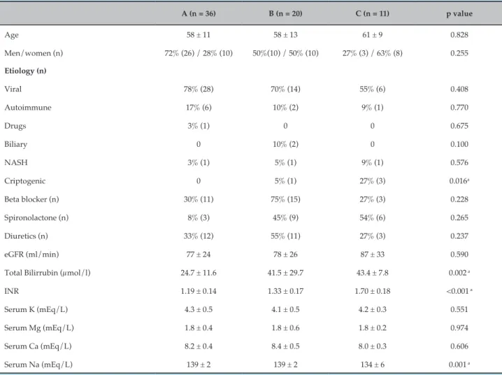
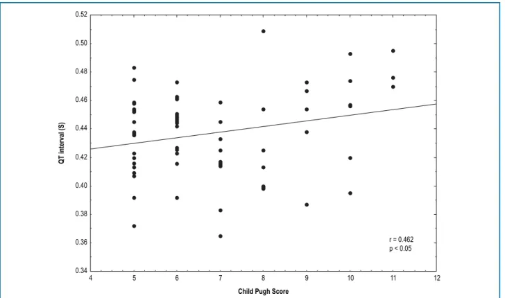
The QT intervals were longer in patients of the Child-Pugh C group (Figure 1). There were no differences between the Bazett and Fridericia QT correction formulas (data not shown). Differences between the groups persisted when we analyzed the subgroups by viral etiology. Other etiologies had too few patients for a statistical analysis. In the subgroup of patients receiving beta-blockers, the QT intervals were also prolonged in the Child-Pugh C group (Figure 2). In addition, there was a positive correlation between the QT interval and the Child-Pugh score in individuals with Child-Pugh scores ≥ 7 (r = 0.50, p < 0.05) (Figure 3). In subjects with QT intervals ≥ 440 ms, the Child-Pugh score and QT interval were also correlated (r = 0.46, p < 0.05) (Figure 4).
Discussion
The present study showed a longer QT interval in Child-Pugh C cirrhotic patients, which reinforced the notion of a relationship between severity of cirrhosis and ECG findings of cirrhotic cardiomyopathy. Other studies have reported similar results, highlighting the importance of a simple and noninvasive method (ECG) to identify cirrhotic patients with cardiomyopathy.1,11,12
Even though the mechanism of the prolongation of QT intervals is not fully elucidated, it is attributed to autonomic changes caused by cirrhosis.14
0.50
0.48
0.46
0.44
0.42
0.40
0.38
A B C
Child Group
QT Interval (S)
* *
* †
Figure 1 – QT intervals in the Child-Pugh A, Child-Pugh B and Child-Pugh C groups. QT intervals are presented as the means + standard deviation. *, p < 0.05 by ANOVA; †, p < 0.05 versus the Child-Pugh A group by Tukey’s HSD test.
0.50
0.48
0.46
0.44
0.42
0.40
0.38
A B C
Child Group
QT Interval (S)
* *
* †
0.52
0.50
0.48
0.46
0.44
0.42
0.40
0.38
0.36
0.34
4 5 6 7 8 9 10 11 12
r = 0.462 p < 0.05
QT interval (S)
Child Pugh Score
Figure 3 – Correlation between QT interval and Child-Pugh score in subjects with Child-Pugh scores ≥ 7.
0.52
0.50
0.48
0.46
0.44
0.42
0.40
0.38
0.36
0.34
6.5 7.0 7.5 8.0 8.5 9.0 9.5 10.0 10.5 11.0 11.5
QT interval (S)
Child-Pugh Score
r = 0.502 p < 0.05
ventricular repolarization. Therefore, QT intervals can be prolonged either because of a delay in the activation or because the complex process of repolarization is prolonged. Repolarization, per se, could be prolonged due to electrolytic abnormalities or previous cardiac disease.7 In our study, we chose to exclude patients
with these conditions so that the QT prolongation could be attributed solely to autonomic dysfunction caused by cirrhosis. In addition, reductions in circulating blood volume and hyperdynamic circulation enhance sympathetic nervous system and renin-angiotensin-aldosterone system activities. The resulting increased cardiac output and reduced systemic vascular resistance may induce myocardial remodeling and left ventricular hypertrophy, causing systolic and diastolic dysfunction and, hence, cardiomyopathy. Sympathetic nervous system hyperactivity also contributes to myocardial dysfunction because of an increase in the levels of inflammatory cytokines such as interleukin-1b, interleukin-6 and tumor necrosis factor alpha.13
Other factors associated with prolonged QT intervals in cirrhosis are disease severity, serum uric acid levels and alcoholic etiology.12 The majority of our patients had
hepatitis virus infection as the cause of cirrhosis, and this subgroup analysis also showed QT prolongation in cirrhotic Child-Pugh C patients. In addition, patients with more severe cirrhosis had a positive correlation between QT interval and Child-Pugh score. As alcohol is directly toxic to myocytes, we chose to exclude patients with alcoholic cirrhosis from this study.13 However, previous
studies on alcoholic cirrhosis observed QT prolongation in patients with Child C cirrhosis.2,14 Regarding electrolytic
alterations, serum potassium, calcium and magnesium levels were similar between the groups. Hyponatremia observed in the Child-Pugh C group was expected in these patients and was not severe.11 We believe that
hyponatremia did not influence QT prolongation in the patients with Child-Pugh C cirrhosis.
In a study with 38 cirrhotic patients (50% male), Mozos et al. showed that corrected QT values were higher in Child-Pugh C cirrhotic patients (Child-Pugh A: 462 ± 25 ms, Child-Pugh B: 493 ± 62 ms and Child-Pugh C: 520 ± 45 ms, p < 0.001). Genovesi et al.12 also observed
that Child-Pugh C patients had higher QT intervals.7
In a study of 409 cirrhotic patients, Bal et al.6 reported
higher corrected QT in the Child-Pugh C group than the Child-Pugh B group (451±43 vs 434 ± 31 ms, p < 0.001).8
Despite similar ages and genders, mean corrected QT in our study was lower than the study by Mozos et al.12 and
similar to the results by Genovesi et al.7 and Bal et al.8
The exclusion of alcoholic etiology and inclusion of patients receiving beta-blockers may explain the observed differences in corrected QT in absolute values. Most of all, these studies strengthened the notion that Child-Pugh C patients should be monitored for QT prolongation.
Beta-blocker use reduces QT intervals as well as portal pressure and hyperdynamic response in cirrhotic patients.13,14 Thus, this medication was indicated in
most patients in the Child-Pugh B and C groups as variceal bleeding prophylaxis.15–17 Hypotension is the
most common reason for beta-blocker withdrawal in Child-Pugh C patients.16,17 Most of our subjects
in the Child-Pugh B and C groups were receiving beta-blockers. In a post hoc analysis of patients receiving beta-blockers, QT was longer in the Child-Pugh C group than in the A and B groups. Most of the previous studies on QT prolongation in cirrhosis have excluded the use of this medication.2,9,10,11,12 In our opinion, it is important
to report this population because most real-world Child-Pugh B and Child-Pugh C cirrhotic patients are beta-blocker users.
A limitation of our study was the small sample size and the cross-sectional design. Thus, a study with a larger cohort is needed to confirm our results and better understand the prognosis of a prolonged QT interval. Such study could also clarify the relationship between cirrhotic cardiomyopathy and the severity of liver disease. In addition, the predominant etiology of cirrhosis in our patients was hepatitis C virus infection. Studies of other common etiologies of cirrhosis, such as nonalcoholic steatohepatitis , are highly desirable.
Conclusion
Patients with Child-Pugh C non-alcoholic cirrhosis show longer QT interval than Child-Pugh A or B patients. This difference persists despite beta-blocker use. Thus, an ECG should be considered in patient with more severe non-alcoholic cirrhosis.
Author contributions
1. Gassanov N, Caglayan E, Semmo N, Massenkeil G, Er F. Cirrhotic cardiomyopathy: a cardiologist’s perspective. World J Gastroenterol.
2014;20(42):15492-8. PMID: 25400434. doi: 10.3748/wjg.v20.i42.15492.
2. Zamirian M, Tavassoli M, Aghasadeghi K. Corrected QT interval and QT dispersion in cirrhotic patients before and after liver transplantation. Arch Iran Med. 2012;15(6):375-7. PMID: 22642249. doi: 012156/AIM.0012.
3. Rahman S, Mallett SV. Cirrhotic cardiomyopathy: implications for the perioperative management of liver transplant patients. World J Hepatol.
2015;7(3):507-20. PMID: 25848474. doi: 10.4254/wjh.v7.i3.507.
4. Kosar F, Ates F, Sahin I, Karincaoglu M, Yldirim B. QT interval analysis in patients with chronic liver disease: a prospective study. Angiology. 2007;58(2):218-24. PMID: 17495272. doi: 10.1177/0003319707300368.
5. Bernardi M, Calandra S, Colantoni A, Trevisani F, Raimondo ML, Sica G, et al. Q-T interval prolongation in cirrhosis: prevalence, relationship with severity, and etiology of the disease and possible pathogenetic factors. Hepatology. 1998;27(1):28-34. PMID: 9425913. doi: 10.1002/ hep.510270106.
6. Genovesi S, Prata Pizzala DM, Pozzi M, Ratti L, Milanese M, Pieruzzi F, et al. QT interval prolongation and decreased heart rate variability in cirrhotic patients: relevance of hepatic venous pressure gradient and serum calcium. Clin Sci (Lond). 2009;116(12):851-9. PMID: 19076059. doi: 10.1042/CS20080325.
7. Bal JS, Thuluvath PJ. Prolongation of QTc interval: relationship with etiology and severity of liver disease, mortality and liver transplantation.
Liver Int. 2003;23(4):243-8. PMID: 12895263. doi:
10.1111/j.1399-3046.2008.00909.
8. Pugh RN, Murray-Lyon IM, Dawson JL, Pietroni MC, Williams R. Transection of the oesophagus for bleeding oesophageal varices. Br J
Surg. 1973;60(8):646-9. PMID: 4541913. doi: 10.1002/bjs.1800600817.
9. Bazett H. An analysis of the time-relations of electrocardiograms. Heart.
1920;7:353-70. doi: 10.1111/j.1542-474X.1997.tb00325.
10. Fridericia L. Die Systolendauer im Elektrokardiogramm bei normalen Menshenund bei Herzkranken. Acta Med Scand. 1920;53:469-86. doi:
10.1111/j.0954-6820.1920.tb18266.
11. Zambruni A, Di Micoli A, Lubisco A, Domenicali M, Trevisani F, Bernardi M. QT interval correction in patients with cirrhosis. J Cardiovasc
Electrophysiol. 2007;18(1):77-82. PMID: 17229304. doi:
10.1111/j.1540-8167.2006.00622.
12. Mozos I, Costea C, Serban C, Susan L. Factors associated with a prolonged QT interval in liver cirrhosis patients. J Electrocardiol. 2011;44(2):105-8.
PMID: 21146831. doi: 10.1016/j.jelectrocard.2010.10.034.
13. Mozos I. Arrhythmia risk in liver cirrhosis. World J Hepatol.
2015;7(4):662-72. PMID: 25866603. doi: 10.4254/wjh.v7.i4.662.
14. Kim YK, Hwang GS, Shin WJ, Bang JY, Cho SK, Han SM. Effect of propranolol on the relationship between QT interval and vagal modulation of heart rate variability in cirrhotic patients awaiting liver transplantation. Transplant Proc. 2011;43(5):1654-9. PMID: 21693252.
doi: 10.1016/j.transproceed.2011.02.017.
15. Bari K, Garcia-Tsao G. Treatment of portal hypertension. World J
Gastroenterol. 2012;18(11):1166-75. PMID: 22468079. doi: 10.3748/wjg.
v18.i11.1166.
16. Mela M, Mancuso A, Burroughs A. Drug treatment for portal hypertension. Ann Hepatol. 2002;1(3):102-20. PMID: 15280809.
17. Garcia-Tsao G, Sanyal AJ, Grace ND, Carey WD; Practice Guidelines Committee of American Association for Study of Liver Diseases, Practice Parameters Committee of American College of Gastroenterology. Prevention and management of gastroesophageal varices and variceal hemorrhage in cirrhosis. Am J Gastroenterol. 2007;102(9):2086-102. PMID: 17879356. doi: 10.1002/hep.21907.
References
Lanzieri PG, Gismondi RA, Chimelli MCA, Cysne RP, Guaraná T, Mesquita CT, Mocarzel LO. Writing of the manuscript: Lanzieri PG, Gismondi RA, Chimelli MCA, Cysne RP, Guaraná T, Mesquita CT, Mocarzel LO. Critical revision of the manuscript for intellectual content: Lanzieri PG, Gismondi RA, Chimelli MCA, Cysne RP, Guaraná T, Mesquita CT, Mocarzel LO.
Potential Conflict of Interest
No potential conflict of interest relevant to this article was reported.
Sources of Funding
There were no external funding sources for this study.
Study Association
This article is part of the thesis of master submitted by Pedro Gemal Lanzieri, from Universidade Federal Fluminense.
Ethics approval and consent to participate