321 Radiol Bras. 2018 Set/Out;51(5):321–327
Idiopathic interstitial pneumonias: review of the latest
American Thoracic Society/European Respiratory Society
classification
Pneumonias intersticiais idiopáticas: revisão da última classificação da American Thoracic Society/ European Respiratory Society
Daniel Simões Oliveira1, José de Arimatéia Araújo Filho1, Antonio Fernando Lins Paiva1, Eduardo Seigo Ikari1, Rodrigo Caruso Chate1, César Higa Nomura1
Oliveira DS, Araújo Filho JA, Paiva AFL, Ikari ES, Chate RC, Nomura CH. Idiopathic interstitial pneumonias: review of the latest American Thoracic Society/European Respiratory Society classification. Radiol Bras. 2018 Set/Out;51(5):321–327.
Abstract
Resumo
The diagnosis of idiopathic interstitial pneumonias (IIPs) involves a multidisciplinary scenario in which the radiologist assumes a
key role. The latest (2013) update of the IIP classification by the American Thoracic Society/European Respiratory Society proposed some important changes to the original classification of 2002. The novelties include the addition of a new disease (idiopathic pleu
-roparenchymal fibroelastosis) and the subdivision of the IIPs into four main groups: chronic fibrosing IIPs (idiopathic pulmonary fibro
-sis and nonspecific interstitial pneumonia); smoking-related IIPs (desquamative interstitial pneumonia and respiratory bronchiolitis-associated interstitial lung disease); acute or subacute IIPs (cryptogenic organizing pneumonia and acute interstitial pneumonia); rare IIPs (lymphoid interstitial pneumonia and idiopathic pleuroparenchymal fibroelastosis); and the so-called “unclassifiable” IIPs.
In this study, we review the main clinical, tomographic, and pathological characteristics of each IIP.
Keywords: Idiopathic interstitial pneumonia; American Thoracic Society; European Respiratory Society.
O diagnóstico das pneumonias intersticiais idiopáticas (PIIs) envolve um cenário multidisciplinar no qual o radiologista assume papel fundamental. A última atualização (2013) da classificação das PIIs pela American Thoracic Society/European Respiratory Society propõe algumas mudanças importantes em relação à classificação original de 2002. Dentre as novidades, destacam-se o acréscimo de uma nova doença (fibroelastose pleuroparenquimatosa idiopática) e a subdivisão das PIIs em quatro grupos princi
-pais: PIIs crônicas fibrosantes (fibrose pulmonar idiopática e pneumonia intersticial não específica); PIIs relacionadas ao tabagismo (pneumonia intersticial descamativa e bronquiolite respiratória com doença intersticial pulmonar); PIIs agudas/subagudas (pneu
-monia em organização e pneu-monia intersticial aguda); PIIs raras (pneu-monia intersticial linfocítica e fibroelastose pleuroparenqui
-matosa idiopática); além das ditas “inclassificáveis”. Foram revisadas, de forma didática neste estudo, as principais características clínicas, tomográficas e patológicas de cada uma das PIIs.
Unitermos: Pneumonias intersticiais idiopáticas; American Thoracic Society; European Respiratory Society.
Study conducted at the Instituto do Coração do Hospital das Clínicas da Facul
-dade de Medicina da Universi-dade de São Paulo (InCor/HC-FMUSP), São Paulo, SP, Brazil.
1. Instituto do Coração do Hospital das Clínicas da Faculdade de Medicina da Universidade de São Paulo (InCor/HC-FMUSP), São Paulo, SP, Brazil.
Correspondence: Dr. Daniel Simões Oliveira. InCor/HC-FMUSP – Radiologia e Diagnóstico por Imagem. Avenida Doutor Enéas de Carvalho Aguiar, 44, Pinheiros.
São Paulo, SP, Brazil, 05403-900. E-mail: danieloliveira8@live.com.
Received July 27, 2016. Accepted after revision January 5, 2017.
scenario in which the radiologist assumes a fundamental role. High-resolution computed tomography of the chest has been the subject of recent publications in the radiol-ogy literature of Brazil(1–7). The integrated approach not-withstanding, the final classification and definitive diag -nosis might not be achieved in all cases. Some cases do not meet the criteria for classification into any of the clas -sic interstitial lung disease categories and are therefore designated “unclassifiable”. Such cases continue to pose a challenge for the entire multidisciplinary team.
The most recent (2013) update of the American Tho-racic Society/European Respiratory Society IIP classifica -tion(8,9) proposes some important changes in relation to the original (2002) classification. Notable among those changes are the subdivision of IIPs into four main groups (chronic fibrosing, smoking-related, acute/subacute, and rare) and the addition of a new disease: idiopathic pleuro-parenchymal fibroelastosis.
INTRODUCTION
Oliveira DS et al. / Review of idiopathic interstitial pneumonias
In this study, we provide a brief clinical description of each of these four IIP groups, as well as presenting their main radiological characteristics (Table 1), including to-mographic findings and distribution patterns, together with the main differential diagnoses. The images presented were selected from the teaching files of our institution, from cases in which the tomographic pattern was typical of each IIP, with pathological confirmation.
CHRONIC FIBROSING IIPs
Idiopathic pulmonary fibrosis (Figure 1)
Among all IIPs(10), idiopathic pulmonary fibrosis is the most common; it is an interstitial lung disease of un-known cause, characterized histologically by the usual interstitial pneumonia pattern(11,12), with dispersed fibro -blastic foci and heterogeneous involvement of the paren -chyma, areas of tissue preservation being interspersed with areas of interstitial inflammation and honeycomb -ing. It occurs mainly in adults over 50 years of age who are smokers or former smokers, typically manifesting as progressive dyspnea and dry cough. In general, it has a poor prognosis, with an estimated survival of less than five years after the diagnosis. Patients usually have fewer acute exacerbations when treated with cyclosporine combined with corticosteroids, and most are considered candidates for lung transplantation(10). In an appropriate clinical set -ting (typical clinical and radiological findings), the diag -nosis of idiopathic pulmonary fibrosis can be established without the need for biopsy(10).
Nonspecific interstitial pneumonia (Figure 2)
Nonspecific interstitial pneumonia is characterized histologically by homogeneous inflammation and expan -sion of the alveolar walls, with or without fibrosis; it can be classified as belonging to one of three subtypes(11): cel-lular (less common and with a better prognosis); fibrotic (with a worse prognosis); or mixed. Nonspecific intersti -tial pneumonia can be idiopathic, although it most often manifests as pulmonary symptoms associated with con-nective tissue diseases (especially scleroderma), hyper -sensitivity pneumonia, drug reactions, or diffuse alveolar damage. It typically occurs in women between 40 and 50 years of age, with symptoms similar to, although usually milder than, those of idiopathic pulmonary fibrosis. Treat -ment is directed to the underlying disease and can include the use of a combination of systemic corticosteroids and cytotoxic drugs, which is successful in most cases(10). Al-though the tomographic findings of idiopathic pulmonary fibrosis and nonspecific interstitial pneumonia are often similar(13), an experienced radiologist knows the main dif -ferences between them (Table 1).
SMOKING-RELATED IIPs
Desquamative interstitial pneumonia (Figure 3)
Desquamative interstitial pneumonia is a rare disease that is strongly associated with smoking; in some cases, it progresses to respiratory failure and advanced pulmonary fibrosis(8). Histopathological findings include homoge -neous thickening of the alveolar septa and intra-alveolar
Table 1—Pattern of distribution, tomographic findings, and main differential diagnoses of each IIP.
IIP category
Chronic fibrosis
Idiopathic pulmonary fibrosis
Nonspecific interstitial pneu -monia
Smoking related
Desquamative interstitial pneumonia
Respiratory bronchiolitis-associated interstitial lung disease
Acute/subacute
Cryptogenic organizing pneu -monia
Acute interstitial pneumonia
Rare
Lymphocytic interstitial pneu-monia
Idiopathic pleuroparenchy-mal fibroelastosis
Pattern of distribution
Peripheral, subpleural, basal
Peripheral, basal, symmetric
Lower fields, predominantly peripheral
Principally upper fields, cen -trilobular
Subpleural, peribronchial
Diffuse or focal
Predominantly in the upper lung fields
Peripheral, upper fields
Tomographic findings
Reticular opacities; honeycombing; minimal ground-glass opacity; architectural distortion
Extensive ground-glass opacity; irregular lin-ear opacities; traction bronchiectasis; sub -pleural preservation
Ground-glass attenuation; cysts; reticular opacities
Bronchial wall thickening; centrilobular nod -ules; ground-glass opacities
Focal ground-glass opacities; consolidations; reversed halo sign
Consolidations; ground-glass opacities; trac-tion bronchiectasis
Thin-walled cysts; centrilobular nodules; ground-glass attenuation; peribronchovascu -lar septal thickening
Pleural thickening; subpleural fibrotic changes
Differential diagnoses
Collagen diseases; hypersensitivity pneumo-nia; pneumoconiosis
Usual interstitial pneumonia; desquamative interstitial pneumonia; cryptogenic organiz -ing pneumonia; hypersensitivity pneumonia
Respiratory bronchiolitis-associated intersti -tial lung disease; nonspecific interstitial pneu -monia; hypersensitivity pneumonia Desquamative interstitial pneumonia; non-specific interstitial pneumonia; hypersensitiv-ity pneumonia
Infection; aspiration; eosinophilic pneumo-nia; vasculitis
Hydrostatic edema; acute respiratory distress syndrome; alveolar hemorrhage
Nonspecific interstitial pneumonia; sarcoid-osis; histiocytosis X; cystic lung diseases
accumulation of pigmented macrophages(11). Men be-tween 30 and 50 years of age constitute the majority of patients; after smoking cessation and corticosteroid ther -apy, there is significant improvement of the symptoms(11).
Respiratory bronchiolitis-associated interstitial lung disease (Figure 4)
Classically characterized by the combination of in-terstitial disease and respiratory bronchiolitis, the typical histological findings of respiratory bronchiolitis-associated interstitial lung disease include the accumulation of pig-mented macrophages in the respiratory bronchioles, alveo -lar ducts, and adjacent alveoli, together with minimal fibro -sis and inflammatory infiltrate(11). The affected patients are
usually men between 30 and 50 years of age who have few or no symptoms. The treatment consists of smoking cessa -tion and corticosteroid therapy(10).
ACUTE AND SUBACUTE IIPs
Cryptogenic organizing pneumonia (Figure 5)
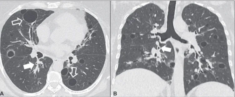
Formerly known as bronchiolitis obliterans organizing pneumonia, organizing pneumonia is characterized histo-logically by buds of granulation tissue within the alveolar ducts and adjacent alveoli, accompanied by chronic in -flammatory infiltrate involving the interstitium and alveo -lar spaces(8). That is a common pattern of response to vari -ous types of lung injury and is most commonly found in conjunction with connective tissue diseases, drug-induced Figure 1. Idiopathic pulmonary fibrosis. A 66-year-old male smoker. Axial high-resolution computed tomography scan of the chest (A) and coronal reformatting (B). In A, fine reticular opacities (closed arrow), with traction bronchiectasis, traction bronchiolectasis, and honeycombing (open arrow). In B, predominantly peripheral and basal pattern of distribution.
Oliveira DS et al. / Review of idiopathic interstitial pneumonias
adverse pulmonary reactions, hypersensitivity pneumonia, infectious processes, and aspiration, among others. When no cause is identified, it is known as cryptogenic organi -zation pneumonia(10). It predominantly affects individuals between 50 and 70 years of age, with no gender prefer -ence; most patients seek treatment in one to three months after the symptom onset, which is often preceded by respi-ratory tract infections(11). Corticosteroid treatment usually produces an excellent response(9).
Acute interstitial pneumonia (Figure 6)
Acute interstitial pneumonia is an extremely severe idiopathic acute interstitial disease, characterized by a
histopathological pattern of diffuse alveolar damage, the exudative phase of which is defined by interstitial and in -tra-alveolar edema, formation of hyaline membranes, and diffuse alveolar infiltration of inflammatory cells. It has a poor prognosis (mortality greater than 50%), mainly affect -ing previously healthy patients between 50 and 60 years of age, with severe sudden-onset dyspnea (occurring within the first three weeks), typically progressing to the need for mechanical ventilation(10). The clinical, radiological, and histological findings are the same as those of acute respi -ratory distress syndrome(11). The treatment is basically supportive, with oxygen supplementation, and the use of corticosteroids is effective in the acute phase(10).
Figure 3. Desquamative interstitial pneumonia. A 65-year-old male smoker. Axial high-resolution computed tomography scan of the chest (A) and coronal reformatting (B). In A, linear reticular opacities (closed arrow), with sparse bilateral ground-glass opacity (open arrow). In B, predominantly peripheral pattern of distribution.
RARE IIPs
Lymphocytic interstitial pneumonia (Figure 7)
In the vast majority of patients, lymphocytic intersti -tial pneumonia is associated with systemic autoimmune or immunodeficiency disorders, including connective tis -sue diseases (mainly Sjögren’s syndrome, autoimmune thyroiditis, and primary biliary cirrhosis). In its idiopathic form, it is considered extremely rare. Histologically, it is characterized by diffuse infiltration of the interstitium by polyclonal lymphocytes, histiocytes, and variable numbers of plasma cells, as well as reactive lymphoid follicles dis -tributed throughout the peribronchovascular regions, ac -companied by intense inflammation(11). It mainly affects
women between 50 and 70 years of age, with nonspecific symptoms of chronic coughing and dyspnea (typically for more than three years). The response to corticosteroid therapy is unpredictable(10).
Idiopathic pleuroparenchymal fibroelastosis (Figure 8)
Idiopathic pleuroparenchymal fibroelastosis is a rare disease affecting the pleura and lungs. It is character -ized histologically by homogeneous subpleural fibrosis and abundant elastic fibers, with variable inflammation and lymphoid aggregates. Currently, there is no consen-sus regarding its diagnostic criteria or regarding the fact that it is a new entity(14). With a little more than 40 cases Figure 5. Cryptogenic organizing pneumonia. A 62-year-old female patient with a one-year history of dyspnea and a two-month history of symptom worsening. Axial high-resolution computed tomography scan of the chest (A) and coronal reformatting (B). In A, ground-glass opacities/sparse bilateral consolidations (open arrows). In B, bilateral subpleural and peribronchial pattern of distribution.
Oliveira DS et al. / Review of idiopathic interstitial pneumonias
described in the literature to date, this entity has been associated with infections, bone marrow transplantation, autoimmune diseases, and genetic predisposition. Its symptoms are nonspecific (dry cough and dyspnea), and it is believed to progress slowly (over the course of 10– 20 years), with few therapeutic options other than lung transplantation(15).
CONCLUSION
The correct diagnosis and classification of IIPs in -volves a broad multidisciplinary debate, with many gaps. Given the similarity of the clinical and pathological find -ings, together with the complexity of the differential di-agnoses and the need for constant follow-up,
high-reso-lution computed tomography plays a fundamental role in the diagnostic assessment of these diseases.
REFERENCES
1. Torres PPTS, Moreira MAR, Silva DGST, et al. High-resolution computed tomography and histopathological findings in hypersen -sitivity pneumonitis: a pictorial essay. Radiol Bras. 2016;49:112–6. 2. Mogami R, Goldenberg T, Marca PGC, et al. Pulmonary infection
caused by Mycobacterium kansasii: findings on computed tomogra -phy of the chest. Radiol Bras. 2016;49:209–13.
3. Queiroz RM, Gomes MP, Valentin MVN. Pulmonary paracoccidioi -domycosis showing reversed halo sign with nodular/coarse contour. Radiol Bras. 2016;49:59–60.
4. Koenigkam-Santos M, Cruvinel DL, Menezes MB, et al. Quantitative computed tomography analysis of the airways in patients with cystic fibrosis using automated software: correlation with spirometry in the evaluation of severity. Radiol Bras. 2016;49:351–7.
Figure 7. Lymphocytic interstitial pneumonia. A 62-year-old female patient with Sjögren’s syndrome. Axial high-resolution computed tomography scan of the chest (A) and coronal reformatting (B). In A, diffuse thickening of the bronchial walls (closed arrows), some ground-glass opacities and thin-walled cysts of vary -ing sizes, with a diffuse, bilateral distribution (open arrows). In B, distribution predominantly in the lower fields.
This is an open-access article distributed under the terms of the Creative Commons Attribution License.
5. Bastos AL, Corrêa RA, Ferreira GA. Tomography patterns of lung disease in systemic sclerosis. Radiol Bras. 2016;49:316–21. 6. Francisco FAF, Rodrigues RS, Barreto MM, et al. Can chest
high-resolution computed tomography findings diagnose pulmonary al -veolar microlithiasis? Radiol Bras. 2015;48:205–10.
7. Alves UD, Lopes AJ, Maioli MCP, et al. Changes seen on computed tomography of the chest in mildly symptomatic adult patients with sickle cell disease. Radiol Bras. 2016;49:214–9.
8. Sverzellati N, Lynch DA, Hansell DM, et al. American Thoracic So -ciety-European Respiratory Society classification of the idiopathic interstitial pneumonias: advances in knowledge since 2002. Radio -graphics. 2015;35:1849–71.
9. Travis WD, Costabel U, Hansell DM, et al. An official American Thoracic Society/European Respiratory Society statement: up -date of the international multidisciplinary classification of the idiopathic interstitial pneumonias. Am J Respir Crit Care Med. 2013;188:733–48.
10. Mueller-Mang C, Grosse C, Schmid K, et al. What every radiologist
should know about idiopathic interstitial pneumonias. Radiograph -ics. 2007;27:595–615.
11. Silva CIS, Jasinowodolinski D, Terra Filho M, et al. Atlas e diag -nóstico diferencial: tomografia computadorizada de alta resolução do tórax – Jorge Kavakama. 1ª ed. Rio de Janeiro, RJ: Revinter; 2008.
12. Lynch DA, Travis WD, Müller NL, et al. Idiopathic interstitial pneumonias: CT features. Radiology. 2005;236:10–21.
13. Silva CIS, Müller NL, Lynch DA, et al. Chronic hypersensitivity pneumonitis: differentiation from idiopathic pulmonary fibrosis and nonspecific interstitial pneumonia by using thin-section CT. Radiology. 2008;246:288–97.
14. Frankel SK, Cool CD, Lynch DA, et al. Idiopathic pleuroparenchy -mal fibroelastosis: description of a novel clinicopathologic entity. Chest. 2004;126:2007–13.