217
1. Full professor, Brazilian Society of Pediatrics – Department of Allergy andImmunology.
2. Associate professor, Brazilian Society of Pediatrics – Department of Allergy and Immunology.
3. Assistant professor, Brazilian Society of Pediatrics – Department of Allergy and Immunology.
4. Specialist in Allergology and Immunopathology, Brazilian Society of Pediatrics – Department of Allergy and Immunology.
PROAL Group - Cristina Abe Jacob, Emanuel C. S. Sarinho, Francisco J. P. Soares, Luiza Karla de Paula Arruda, Márcia C. Mallozi, Maria Marluce Santos Vilela, Neusa F. Wandalsen, Paulo Silva da Silva, Thales Barba, Vera Dantas, Wellington Borges, Wilson Rocha Filho, Judith Arruda.
Financial support: Pharmacia (donation of dosage kits).
Manuscript received Sep 01 2003, accepted for publication Mar 03 2004. Abstract
Objectives: To evaluate the positivity of Phadiatop® in children from several Brazilian pediatric allergology centers and to compare its results with the presence of serum specific IgE to inhalant and food allergens.
Patients and method: Phadiatop® and serum specific IgE levels (RAST) to inhalant and food allergens (UniCAP® - Pharmacia) were measured in 457 children from several pediatric allergy centers and in a non-allergic control group (n = 62), distributed across five age groups.
Results: Phadiatop was positive in 305 atopic children (67.6%) and in 25.8% of controls (p < 0.001). Among atopic children the distribution of positive test varied according to age: 7.9% (24/305) among under 2 year-olds, 15.4% (47/305) in 2 to 3 year-olds, 22% (67/305) in 3 to 4 year-olds, 19.3% (59/305) in 4 to 5 year-olds and 35.4% (108/305) in 5 to 12 year-olds. No concordance between food allergens and Phadiatop® was observed. Analysis of the relationship between positive inhaled allergen RASTs and positive Phadiatop® showed best indices with house dust mites (D. pteronyssinus, D. farinae and Blomia tropicalis).
Conclusions: Phadiatop® is a useful tool for diagnosing domestic mite allergy.
J Pediatr (Rio J). 2004;80(3):217-22: Allergy, food hypersensitivity, IgE, asthma, rinithis.
Phadiatop
®in the diagnosis of respiratory allergy
in children: Allergy Project PROAL
Charles K. Naspitz1, Dirceu Solé1, Maria Cecília Aguiar4
, Maria Letícia Chavarria2,
Nelson Rosário Filho1, Antônio Zuliani2, Eliana C. Toledo3, Bruno A. P. Barreto3,
Leda S. F. Souza2, Grupo PROAL Copyright © 2004 by Sociedade Brasileira de Pediatria
O
RIGINALA
RTICLEIntroduction
Recent studies have documented an increase in the prevalence of allergic diseases in several different parts of the world. In Brazil the prevalence of medical diagnosis of asthma, allergic rhinitis and atopic eczema were recorded for the first time as part of an international study and were
found to be, on average, 21%, 39% and 8% respectively.1-3 The presence of IgE antibodies specific to usual antigens that characterizes allergic diseases is an important parameter on which diagnosis confirmation can be based.4
When assessing an allergic individual, clinical history plays a fundamental role. It allows causative and aggravating factors to be verified, which is important for the future establishment of treatment plans, in addition to the providing information on the natural history of the disease. Recent studies, however, have pointed to false positive rates of up to 22.6% when clinical history is used in isolation to diagnose allergic disease.5-7
In the diagnosis of respiratory allergy Phadiatop® has become one of the most widely used in vitro tests. Phadiatop® is a simple test that is capable of detecting the presence of IgE specific to the inhalant allergens that are most common in the tested environment simultaneously.8 Nevertheless, it must have the allergens with the greatest prevalence of sensitization, among atopic individuals, in the environment it is being used in.8
In Brazil, despite its use as a diagnostic screening test for respiratory allergies, the real correlation between Phadiatop® and positivity to the inhalant allergens most common among the Brazilian allergic population is unknown. This being the case, the objectives of this study were to evaluate the frequency of Phadiatop® in children from different locations in Brazil treated at pediatric allergology centers and to compare it with sensitization to inhalant and food allergens as evaluated by specific IgE assay in serum, in the same locations.
Patients and methods
This was a case-controlled study in which 457 children (177 girls [38.7%] and 280 boys [61.3%]) participated. They were aged between 12 and 144 months, cared for at allergology services located in all five regions of the country. The children were split into five age groups as follows: younger than 2 years, 2 to 3 years, 3 to 4 years, 4 to 5 years and 5 to 12 years. Children were classed as atopic if they exhibited at least one positive immediate hypersensitivity skin test (average wheal diameter greater than or equal to 3 mm) to at least one inhalant or food allergen, tested and selected randomly. The control group was made up of 62 other children originating from investigative centers of the Northeast, Southeast and Southregions who had needed blood tests for other reasons, such as preoperative assessment for elective surgery. All controls had a history free of allergic disease and negative immediate hypersensitivity skin test for the allergens employed at the respective centers.9
Depending on the reason for referral, patients were classed as: wheezing infants (n = 20), food allergy (n = 16), atopic dermatitis (n = 56), and respiratory allergy (n = 348).Babies were defined as wheezing if they were less than two years old and presented recurrent episodes of wheezing and other possible causes had been ruled out (aspiration syndromes, fibrocystic disease, airway malformations among others). Patients with proven asthma and/or rhinitis were defined as having respiratory allergies.
Peripheral blood samples were taken from both allergic and control patients so that IgE serum levels specific to inhalant allergens (Dermatophagoides pteronyssinus,
Dermatophagoides farinae, Blomia tropicalis, cat, dog, fungus, cows epithelium, horse, grasses and cockroach) and food allergens (cows milk, egg, peanut, wheat and seafood panel) could be assayed and inhalant allergens tested with Phadiatop® (UniCAP®-Pharmacia).12,13 Phadiatop® is a selective test composed mainly of a mixture of domestic dust and mites: Dermatophagooides
pteronyssinus and D. farinae and as such its result is qualitative (present or not). Specific IgE levels (RAST) greater than or equal to 0.35 UI/ml (class 1) were defined as positive.10,12 The study was approved by the relevant Ethics Committees and informed consent forms were signed.
Non-parametric tests were employed to analyze variables and coefficients for sensitivity, and specificity, positive and negative predictive values and agreement were calculated.13 In all cases the cut off for null hypothesis rejection was set at 5%.
Results
Divided by sex, the group of allergic children contained 38.6% (174/451) girls and 61.4% (277/451) boys. Comparable figures were observed in the non-allergic controls: 41.9% (26/62) girls and 58.1% (36/62) boys. Distribution by age group is shown in Table 1.
Age group Allergic Controls (years) Total n % Total n %
< 2 (a) 79 24 30.8 12 2 16.7 2-3 (b) 83 47 56.6 13 3 23.1 3-4 (c) 102 67 65.7 9 3 33.3 4-5 (d) 81 59 72.8 13 4 30.7 5-12 (e) 112 108 96.4 15 4 26.7
Total 457 305 67.6 62 16 25.8
Table 1 - Distribution of Phadiatop® positive children by age
group
Chi-square: allergic children > controls. Chi-square test. Allergic significant value: a < b, c, d, e; b, c < d, e; c, d <e; d < e.
rates of RAST for domestic mites and Phadiatop® positivity are shown by primary complaint. We observed the lowest values among the wheezing babies and patients with food allergies. In contrast, among patients with atopic dermatitis sensitization to domestic mites was as elevated as among those with respiratory allergies.
Discussion
The clinical manifestations of allergic diseases are often the same as with other diseases which makes their diagnosis difficult. Therefore, clinical practitioners often feel the need
for a laboratory test that is capable of identifying them in a suitable manner. If the test combines low costs, speed of execution, availability at most laboratories and a high level of sensitivity in identifying its target population, then we have a good instrument to be used in epidemiological studies of allergic disease prevalence. Phadiatop® has been widely used as a screening test for respiratory allergies.5,14,15
In principal Phadiatop® is an in vitro test that allows individuals who are sensitive to multiple inhalant allergens to be identified simultaneously. In order to be a useful and sensitive method Phadiatop® must be constituted of the aeroallergens that are most prevalent in the area of study. In Brazil, a number of different studies of selected allergic populations have also shown elevated prevalence of sensitization to the domestic dust mites: D. pteronyssinus, D. farinae and Blomia tropicalis and, less often: animal epithelia, cockroach allergens and, more seldom, fungi.16-18
Depending on the population being studied, the rate of Phadiatop® positivity can vary. It will be lower among younger children or normal subjects and higher among patients with respiratory allergies.19,20 Smith-Sivertsen et al. performed a comparative study of the prevalence of allergic respiratory disease in two adult populations living in two different countries, Russia and Norway, but living in adjacent regions.21 In addition to a specific questionnaire about symptoms, Phadiatop® was used as a screening test to identify sensitized individuals.21 The authors reported that 20.7% of the Norwegians and 25.7% of the Russians were Phadiatop® positive. Tschopp et al. observed Phadiatop® positivity in 29% of the normal adults they assessed.19 On the other hand, Williams et al. observed a rate of 71.7% positivity among patients with a clinical diagnosis of respiratory allergy.5
In the current study we found that 67.6% of the children with allergic diseases and 25.8% of those without allergies were positive when tested with Phadiatop®. The apparently
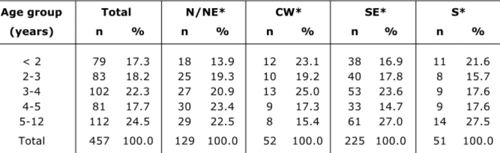
Table 2 - Distribution of allergic patients by age group and region of origin
Chi-square test: no differences between age groups in each region and total. *N/NE = north and northeast; CW = central west; SE = southeast; S = south.
Age group Total N/NE* CW* SE* S* (years) n % n % n % n % n %
< 2 79 17.3 18 13.9 12 23.1 38 16.9 11 21.6 2-3 83 18.2 25 19.3 10 19.2 40 17.8 8 15.7 3-4 102 22.3 27 20.9 13 25.0 53 23.6 9 17.6 4-5 81 17.7 30 23.4 9 17.3 33 14.7 9 17.6 5-12 112 24.5 29 22.5 8 15.4 61 27.0 14 27.5
Total 457 100.0 129 100.0 52 100.0 225 100.0 51 100.0
Allergen Agreement
positive negative geral (%)
D. pteronyssinus 288 133 93.3*
D. farinae 286 141 94.7*
Blomia tropicalis 239 136 83.2*
Cockroach 146 144 64.3
Cat 53 145 43.9
Grasses 45 146 42.2
Cows epithelium 34 135 37.5
Dog 33 144 39.2
Horse 19 146 36.6
Fungus 12 145 34.8
Fish 129 142 60.1
Egg 89 125 47.5
Wheat 79 135 47.5
Cows milk 78 120 43.9
Peanut 59 142 44.6
Soya 50 143 42.8
Corn 44 142 41.2
Table 3 - Results of positive and negative agreement between the RAST and Phadiatop® tests
reduced rate of positivity to Phadiatop® among allergic children is the result of the fact that we included patients with non-respiratory manifestations in the calculation. There was a 40% positivity level among the wheezing babies and those with food allergies were 56% positive. These results are explained by young patients and type of allergic disease respectively.22,23 Nevertheless, positivity to Phadiatop® was elevated among atopic dermatitis patients, i.e. 82.1%. There is controversy over the role played by mites as aggravating or causative agents of atopic dermatitis. Additionally, it is known that around 50% of these patients will progress to allergic respiratory disease.22,23 Patients with atopic dermatitis and serum IgE specific to domestic dust mites exhibit improvements when their environmental exposure to domestic dust mites is controlled.24,25 The rate of positivity among patients with respiratory allergies was 77.3%, which is comparable to those observed by others.5,15,21
Another use of a allergy diagnostic test would be its use as a predictive method for later allergic disease development. A number of different researchers studied the positivity of Phadiatop® as a marker for allergic disease, in particular respiratory ones. Lilja et al. in a follow-up study from birth to five years of age, of children at risk of developing asthma, children of asthmatics parents, were tested with Phadiatop® at six months, at 18 months and at five years.14 Of the children whose exams were positive at six months, 75% had
manifested as asthmatic at 18 months and all of them had done so by five years of age.14 This being the case, the authors found that Phadiatop® had a predictive value of 80% for predicting asthma in at-risk infants. This value is significantly superior than the 53% that would be the case if a positive aeroallergen skin test result had been included in the diagnostic criteria.14
Kotaniemi-Syrjanen et al. studied Phadiatop® as a predictive test for the development of asthma in infants less than two years old that had been hospitalized for acute wheezing. Blood samples were taken at this point and stored until the children were at least five years old.15 They were then followed until school age at which point they were assessed for asthma and/or other allergic manifestations and levels of serum IgE specific to inhalant and food allergens were assayed. Forty percent of the initial population progressed to asthma and Phadiatop® was positive in 18% of the entire group and all of these patients became asthmatic.15 In an attempt to identify risk factors for hospital admission and readmission because of acute exacerbations in children under four years old with asthma, Wever-Hess et al. recorded that sensitization to aeroallergens (positive Phadiatop® test) was associated with an eight times greater risk of recurring exacerbations.26
Doubt remains as to which of the many different screening methods routinely employed is the most appropriate: anamnesis, immediate hypersensitivity skin tests, specific
Test Wheezing Food Atopic Respiratory babies allergy dermatitis allergy (n = 20) (n = 16) (n = 66) (n = 348)
D. pteronyssinus 40.0 62.5 80.4 76.7
D. farinae 35.0 43.8 78.6 73.6
Blomia tropicalis 20.0 18.8 64.3 69.8
Phadiatop® 40.0 56.3 82.1 77.3
Table 5 - Distribution of patients (%) according to primary complaint and positivity for domestic mites and Phadiatop®
RAST positive SS (%) S (%) PPV (%) NPV (%) A (%)
D. pteronyssinus 94.3 91.5 96.1 87.8 93.3
D. farinae 94.6 96.4 98.4 88.9 94.7
Blomia tropicalis 79.4 92.9 96.2 66.8 64.3
Table 4 - Indices for sensitivity (SS), specificity (S), positive and negative predictive values (PPV, NPV) and for agreement (A) between Phadiatop® and RAST for
IgE serum assay or Phadiatop®. Williams et al. studied the value of Phadiatop® as a diagnostic method for sensitization in allergic children and adolescents, treated at specialist services.5 Results were compared with those from immediate hypersensitivity skin tests for aeroallergens, with specific IgE and with clinical history. According to Williams et al. clinical history induced a false positive result in around 23% of cases. The laboratory tests had a good correlation. Phadiatop® was positive in 73.1% of those who had positive skin tests, in 71% of those with positive RAST and in 71.7% of all patients. There was 99% agreement between Phadiatop® positive patients with specific IgE serum tests and 98% agreement with positive immediate hypersensitivity skin tests and 83.2% with positive clinical history. Agreement levels were also elevated among those with negative Phadiatop® tests.5
Tschopp et al. studied the diagnostic value of immediate hypersensitivity skin test, total IgE serum levels and Phadiatop® in a Swiss adult population.26 The prevalence of allergic asthma was 1.8% and that of allergic rhinitis was 16.3%. In the overall population the prevalence of positive Phadiatop® results was 29%, and positivity was 23% for both the skin tests and total IgE serum assays. The sensitivity of Phadiatop® for the diagnosis of allergic asthma was significantly higher than that of the skin tests (72.5% vs. 65.4%) and the same was true of allergic rhinitis (77.1% vs. 68.4%); both were better than IgE serum levels. Nevertheless, specificity and efficiency were better for the skin tests than for Phadiatop®.27
While it was already known that Phadiatop® is constituted from inhalant allergens, no studies had been performed in our country that identified these allergens. This being the case, a comparative study of Phadiatop® positivity and the various tests for serum IgE specific for several different allergens was performed. With the intention of confirming that food allergens were not included in Phadiatop® we included children with food allergies in the research. The highest levels of agreement were found with the domestic dust mites: D. pteronyssinus, D farinae and Blomia tropiclais. Using Phadiatop® as a method for diagnosing sensitization to each of the mites we were able to establish its sensitivity, specificity and positive and negative predictive values (Table 4).
In conclusion, we have proved in this study that Phadiatop® positivity is elevated among children with respiratory allergy, and that domestic dust mites are the main constituents. The high levels of agreement, sensitivity, specificity and positive and negative predictive values indicate that Phadiatop® is a useful test for identifying patients sensitized to domestic dust mites.
Acknowledgements
We would like to thank Mr. Fábio Arcuri (Pharmacia do Brasil) for donating the test kits and to Doctors Paulo G Leser and Roseli Dobner dos Santos at the Laboratório Fleury for performing experiments and to the other members of the PROAL Group for data collection.
References
1. Solé D, Yamada E, Vana AT, Werneck G, Solano LSF, Sologuren MJ, et al. International Study of Asthma and Allergies in Childhood (ISAAC): prevalence of asthma and asthma-related symptoms among Brazilian schoolchildren. J Investig Allergol Clin Immunol. 2001;11:123-8.
2. Strachan DP, Sibbald B, Weiland SK, Ait-Khaled N, Anabwani G, Anderson HR, et al. Worldwide variations in prevalence of symptoms of allergic rhinoconjunctivitis in children: the International Study of Asthma and Allergies in Childhood (ISAAC). Pediatr Allergy Immunol. 1997;8:161-76.
3. Williams H, Robertson C, Stewart A, Ait-Khaled N, Anabwani G, Anderson R, et al. Worldwide variations in prevalence of symptoms of atopic eczema in the International Study of Asthma and Allergies in Childhood. J Allergy Clin Immunol. 1999;103:125-38.
4. EAACI Position paper A revised nomenclature for allergy: EAACI position statement from the EAACI nomenclature task force. Allergy. 2001;56:813-24.
5. Williams PB, Siegel C, Portnoy J. Efficacy of a single diagnostic test for sensitization to common inhalant allergens. Ann Allergy Asthma Immunol. 2001;86:196-202.
6. Williams PB, Dolen WK, Koepe JW, Selner JC. Comparison of skin testing and three in vitro assays for specific IgE in the clinical evaluation of immediate hypersensitivity. Ann Allergy. 1992;68:35-45.
7. Kam KL, Hsieh KH. Comparison of three in vitro assays for serum IgE with skin testing in asthmatic children. Ann Allergy. 1994;73:329-36.
8. Yunginger JW, Ahlsted S, Egglestone PA, Homburger A, Nelson HS, Ownby DR, et al. Quantitative IgE antibody assays in allergic diseases. J Allergy Clin Immunol. 2000;105:1077-84. 9. Tripathi A, Patterson R. Clinical interpretation of skin results.
Immunol Allergy Clin N Amer. 2001;21:291-300.
10. Ownby DR, Anderson JA, Jacob GL. Development and comparative evaluation of a multiple-antigen RAST as a screening test for inhalant allergy. J Allergy Clin Immunol. 1984;73:466-72. 11. Wraith K, Merret J, Roth A, Yman L, Merret TG. Recognition of
food-allergic patients and their allergens by the RAST technique and clinical investigation. Clin Allergy. 1979;9:25-36. 12. Warner JO, ETAC Study Group. Early treatment of the atopic
child. A double-blinded, randomized, placebo-controlled trial with cetririzine in preventing the onset of asthma in children with atopic dermatitis: 18 month-treatment and 18 months-posttreatment follow-up. J Allergy Clin Immunol. 2001;108: 929-37.
13. Fletcher RH, Fletcher SW, Wagner EH. Epidemiologia clínica: elementos essenciais. 3ª ed. Porto Alegre: Artes Médicas; 1996. 281p.
14. Lilja G, Oman H, Johansson SGO. Development of atopic disease during childhood and its prediction by Phadiatop Paediatric. Clin Exp Allergy. 1996;26:1073-9.
15. Kotaniemi-Syrjanen A, Reijonen TM, Romppanem J, Korhonen K, Savolainen K, Korppi M. Allergen-specific immunoglobulin E antibodies in wheezing infants: the risk for asthma in later childhood. Pediatrics. 2003;111:e255-61.
16. Rizzo MC, Solé D, Rizzo A, Holanda MA, Rios JBM, Wandalsen NF, et al. Etiologia da doença atópica em crianças brasileiras, estudo multicêntrico. J Pedaitr (Rio J). 1995;71:31-5.
17. Arruda LK, Rizzo MC, Chapman MD, Fernandez-Caldas E, Baggio D, Platts-Mills TAE, et al. Exposure and sensitization of dust mite allergens among asthmatic children in São Paulo, Brazil. Clin Exp Allergy. 1991;21:433-9.
18. Camelo-Nunes IC, Solé D, Naspitz CK. Fatores de risco e evolução clínica da asma em crianças. J Pediatr (Rio J). 1997;73; 161-70.
19. Wahn U, Lau S, Bergmann R, Kulig M, Forster J, Bergmann K, et al. Indoor allergen exposure is a risk factor for sensibilization during the first three years of life. J Allergy Clin Immunol. 1999;97:763-9.
20. Hattevig G, Kjellman B, Björksten B. Clinical symptoms and IgE responses to common food proteins and inhalants in the first 7 years of life. Clin Allergy. 1987;17:571-8.
21. Smith-Sivertsen T, Tchachtchine V, Lund E. Atopy in Norwegian and Russian adults: a population-based study from the common border area. Allergy. 2003;58:357-62.
23. ETAC® Study Group Allergic factors associated with the
development of asthma and the influence of cetirizine in a double-blind, randomized, placebo controlled-trial: first result of ETAC®. Pediatr Allergy Immunol. 1998;9:116-24.
24. Jones SM. Triggers in atopic dermatitis. Immunol Allergy Clin North Am. 2002;22:55-72.
25. Pajno GB, Peroni DG, Barberio G, Pietrobelli A, Boner AL. Predictive features for persistence of atopic dermatitis in children. Pediatr Allergy Immunol. 2003;14:292-5.
26. Wever-Hess J, Kouwenberg JM, Duiverman EJ, Hermans J, Wever AM. Risk factors for exacerbations and hospital admissions in asthma of early childhood. Pedaitr Pulmonol. 2000;29: 250-6.
Corresponding author: Dirceu Solé
Rua Mirassol, 236/72
CEP 04044-010 - São Paulo, SP, Brazil Tel.: +55 (11) 5579.3778
E-mail: dirceus@nox.net; dirceus@ajato.com.br