223
1. Associate professor, Department of Endocrinology, School of Medicine, Universidade do Estado do Rio de Janeiro (UERJ). Coordinator of the Division of Pediatric Endocrinology, Pedro Ernesto University Hospital, Rio de Janeiro, RJ, Brazil.
2. Department of Endocrinology, School of Medicine, Universidade do Estado do Rio de Janeiro, Pedro Ernesto University Hospital, Rio de Janeiro, RJ, Brazil. 3. Undergraduate student, School of Medicine, Universidade do Estado do Rio de Janeiro, Pedro Ernesto University Hospital, Rio de Janeiro, RJ, Brazil. 4. MD, Department of Endocrinology, School of Medicine, Universidade do Estado do Rio de Janeiro, Pedro Ernesto University Hospital, Rio de Janeiro, RJ,
Brazil.
5. Assistant professor, Department of Endocrinology and Pediatrics, School of Medicine, Universidade do Estado do Rio de Janeiro, Pedro Ernesto University Hospital, Rio de Janeiro, RJ, Brazil.
6. Associate professor, Department of Radiology, School of Medicine, Universidade do Estado do Rio de Janeiro, Pedro Ernesto University Hospital, Rio de Janeiro, RJ.
7. Associate professor and chief of the Department of Endocrinology, School of Medicine, Universidade do Estado do Rio de Janeiro, Pedro Ernesto University Hospital, Rio de Janeiro, RJ, Brazil.
Manuscript received Jul 14 2003, accepted for publication Mar 03 2004. Abstract
Objective: The aim of this study was to analyze the type and frequency of cranial computed tomography and magnetic resonance imaging anomalies in patients with idiopathic growth hormone deficiency (GHD), and also to investigate the possible relationship between neuroradiological images and the presence of isolated GH or multiple pituitary hormone deficiency.
Methods: Magnetic resonance and computed tomography images were obtained for 37 patients with idiopathic growth hormone deficiency (GHD). The patients were divided into two groups: patients with isolated GH (group A) and patients with multiple pituitary hormone deficiencies (group B).
Results: Computed tomography was normal in 25(68%), and abnormal in 12(32%) patients. We observed empty sella in 50%, partially empty sella in 17% and anterior pituitary hypoplasia in 33% patients. MRI studies revealed normal findings in the hypothalamus-pituitary area in 17 (46%) and abnormal in 20 (54%) patients. We didnt observed differences in the frequency of computed tomography alterations when groups A and B were compared (p = 0.55). With magnetic resonance imaging we observed, empty sella in 10%, partially empty sella in 15% and anterior pituitary hypoplasia in 75% patients. Among those patients whose magnetic resonance images were altered, the posterior lobe of the pituitary gland was identified in an abnormal position in 70%, and the hypophyseal stalk was thin or interrupted in 60%. The patients from group B presented a higher frequency of magnetic resonance imaging anomalies (90%) when compared to group A (10%), p = 0.03. There was disagreement between the two methods in 43% of cases, but we didnt observe a difference in the frequency of alterations when computed tomography was compared with magnetic resonance imaging (p = 0.06).
Conclusions: The most frequent defects observed using magnetic resonance imaging are anterior pituitary hypoplasia and ectopic posterior pituitary lobe. The association of glandular hypoplasia with other magnetic resonance imaging abnormalities can suggest the presence of multiple anterior pituitary deficiencies.
J Pediatr (Rio J). 2004;80(3):223-8: Growth hormone deficiency, magnetic resonance images, cranial computed tomography, hypoplastic anterior pituitary, ectopic posterior pituitary.
Neuroradiological investigation
in patients with
idiopathic growth hormone deficiency
Maria Alice N. Bordallo1, Leandro D. Tellerman2, Rodrigo Bosignoli3, Fernando F. R. M. Oliveira3,Fernanda M. Gazolla4, Isabel R. Madeira5,
José Fernando C. Zanier6, Jodélia L. M. Henriques7 Copyright © 2004 by Sociedade Brasileira de Pediatria
Introduction
Since the turn of the century, with the advent of radiography, studies of the sella turcica and the pituitary have been valued. Radiography remained the only method for many decades, until the introduction of computerized tomography (CT), which increased the capacity for recognition and definition of what is normal and of the variations of what is pathological. During the eighties, with the advent of magnetic resonance imaging (MRI) and, later, angiomagnetic resonance imaging, the quality of images was improved further, putting pneumoencephalography and angiography in the shade.
The techniques of CT and MRI have made it possible to improve the definition of structures in the hypothalamic-pituitary region and, consequently, improve detection of anatomic abnormalities indicative of growth hormone deficiency (GHD). In children with reduced stature, the investigation and diagnosis of GHD, is sometimes problematic because a number of different causes may have been involved in the etiopathogenesis of this deficiency. In such cases, images obtained by CT and/or MRI facilitate the detection of anatomic abnormalities, which, in the majority of cases are indicators of permanent growth hormone deficiency.1-5
Magnetic resonance imaging is a non-invasive method which offers greater sensitivity than CT for identification of abnormalities in the hypothalamic-pituitary region 5 and is considered useful, not just in diagnosis, but also when making treatment decisions for patients with GHD.
Previously the majority of GHD cases were diagnosed as idiopathic, but with the advent of MRI it has been possible to observe in such patients a characteristic structural anomaly described as the pituitary stalk interruption syndrome,3,4,6,7 which is more difficult to identify with CT.5 A number of different publications1,3,4,6,8 have tried to correlate the morphological alterations seen with MRI with isolated growth hormone deficiency (IGHD) or as multiple anterior pituitary deficiencies (MPHD). Studies have shown that children with MPHD exhibit a higher frequency of abnormalities on MRI when compared with those that have IGHD.3,9 Pituitary stalk interruption syndrome has been associated with MPHD, and therefore with greater severity of hypopituitarism, while an intact stalk has been observed with greater frequency in patients with IGHD.1,4,8
This study is based on the hypothesis that structural abnormalities in the hypothalamic-pituitary region would be common in our group of patients with GHD, and that abnormal MRI findings could be an indicator of hypopituitarism severity. As ectopic posterior pituitary is frequently observed in patients with GH deficiency,4,6,7 we postulated that if this finding is present in our short stature patients it would allow us to diagnose doubtful cases, thus avoiding unnecessary exhaustive laboratory investigation.
The objective of this study, therefore, is to analyze the frequency and types of abnormalities observed with CT and MRI in patients with apparently idiopathic GH deficiency and also to investigate a possible relationship between the frequency of MRI findings and severity of hypopituitarism.
Methods
During the period between 1996 and 2003, 64 patients were registered at the Pedro Ernesto University Hospital with a suspicion of growth hormone deficiency. All of the patients were submitted to neuroradiological investigation of the hypothalamic-pituitary region by CT as part of the investigation protocol.
In a retrospective, observational study based on a review of medical records 37 patients were assessed who had originally undergone CT and, later, MRI of the sella turcica. The interval between CT and MRI scans was 10.2±4.1 months.
Diagnostic criteria for GHD were: short stature (height below the 3rd percentile on the Tanner and Whitehouse curve), growth of less than 4 cm/year; retarded bone age (> 2 SD below chronological age according to the Greulich and Pyle method); peak GH below 7 ng/ml in prepubescent patients and below 10 ng/ml in pubescent patients measured by two pharmacological tests (insulin and clonidine); the presence of neuroradiological abnormalities such as hypoplastic anterior pituitary, empty sella or pituitary stalk interruption syndrome demonstrated by CT or MRI.
With the intention of relating the frequency of structural anomalies of the hypothalamic-pituitary region with hypopituitarism severity, patients were divided into two groups: group A patients with isolated growth hormone deficiency (IGHD) and group B with multiple pituitary hormone deficiency (MPHD).
Computerized tomography was performed with GE helical equipment, Hi Speed model and MRI was performed with 1.5 Tesla Toshiba equipment. The protocol selected for sella turcica imaging was to use coronal sections before and after application of a venous contrast, spaced 2 mm apart, on both CT and MRI. Magnetic resonance images were obtained in sequence, T1-weighted, including gadolinium-enhanced dynamic scanning. Complementary sagittal sections were also scanned at T1 with MRI and, in some cases sequences were scanned at T2 weighting for better cerebrospinal fluid cavity visualization.
Morphological and structural abnormalities were noted in the hypothalamic-pituitary region, including size and morphology of the anterior pituitary lobe, location of the posterior lobe, characteristics of the pituitary stalk, the hypothalamic region and intraglandular cleft. The pituitary stalk was described as normal, thinned or absent. We defined empty sella as cerebrospinal fluid invagination with no pituitary tissues identified and partially empty sella as the existence of both cerebrospinal fluid and some pituitary tissue within the cavity. Maximum pituitary height was defined as a perpendicular measurement giving the greatest distance between the base and the top of the gland. We defined the anterior pituitary as hypoplastic when its height was below -2 SD, compared with normal controls.10
School of Medicine of the Universidade do Estado do Rio de Janeiro.
Data was analyzed using version 6.04b of Epi-Info. The Chi-squared test, Fishers exact test and the Kruskal-Wallis test were employed.
Results
In this study 37 patients with a diagnosis of an apparently idiopathic GH deficiency initially had CT scans and, later, MRI of the hypothalamic-pituitary region. Patients were split into two groups. Group A included those with an isolated GH deficiency (IGHD) and group B those with multiple pituitary hormone deficiency (MPHD). Clinical data, the number of patients in each group, mean and standard deviation of z score for height and peak GH after clonidine and/or insulin stimulation are all contained in Table 1.
abnormalities in the hypothalamic-pituitary are when viewed by MRI than did the patients in group A (p = 0.01), as can be observed in Table 3.
Table 3 - Statistical analysis of the comparison of the percenta-ge of abnormalities when viewed by MRI and CT in the patients in group A (IGHD*) and Group B (MPHD)
Table 1 - Number of patients in each group, mean and standard deviation of z score for height and peak growth hormone after clonidine
* IGHD = isolated growth hormone deficiency. † MPHD = multiple anterior pituitary deficiencies. ‡ p significant < 0.05.
§ Clonidine or insulin.
Group A Group B p
(IGHD*) (MPHD)
Number of patients 10 (27%) 27 (73%)
z score for height -3,7±0,8 -4,7±1,7 0,1
Peak GH after clonidine § 2,2±1,9 1,4±2,2 0,7
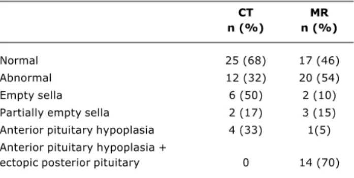
Table 2 - A comparison of the findings from magnetic resonan-ce imaging with those from computerized tomography in patients with idiopathic growth hormone deficiency
CT MR
n (%) n (%)
Normal 25 (68) 17 (46)
Abnormal 12 (32) 20 (54)
Empty sella 6 (50) 2 (10)
Partially empty sella 2 (17) 3 (15)
Anterior pituitary hypoplasia 4 (33) 1(5) Anterior pituitary hypoplasia +
ectopic posterior pituitary 0 14 (70)
* IGHD = isolated growth hormone deficiency. † MPHD = multiple pituitary hormone deficiency. ‡ p significant < 0.05.
Group A Group B p
Normal CT 8 (32%) 17 (68%) 0.55
Abnormal CT 2 (17%) 10 (83%)
Normal MRI 8 (47%) 2 (10%) 0.01
Abnormal MRI 9 (53%) 18 (90%)
Thirty patients were prepubescent and seven had already entered puberty when the study was performed (Tanner II). Peak GH after clonidine or insulin stimulation (ITT) was less than 10 ng/ml for all pubescent patients and less than 7 ng/ml for the prepubescent patients with a single exception who presented a peak GH of 8.5 ng/ml after ITT. Computerized tomography was normal in 25 (68%) patients, and abnormal in 12 (32%) patients.
Magnetic resonance imaging was normal in 17 (46%) patients and abnormal in 20 (54%) patients. We did not observe any significant differences in terms of the frequency of abnormalities in the pituitary area when we compared CT with MRI for patients with GHD (p = 0.06). A comparison of the findings from magnetic resonance imaging with those from computerized tomography in patients with idiopathic GH deficiency (GHD) is shown in Table 2. We did not observe any significant differences in terms of frequency of abnormalities in the pituitary area viewed by CT scan between the patients in groups A and B (p = 0.55). However, patients in group B exhibited a greater percentage of
The posterior pituitary was found in an ectopic position in two (20%) of the group A patients and 12 (44%) of the group B ones. The pituitary stalk was narrowed or absent in two (20%) of the group A patients and 10 (37%) of the patients in group B. We did not observe differences between the percentages of ectopic posterior pituitaries and pituitary stalk abnormalities between the two groups (p = 0.3 and p = 0.6). An association was observed between hypoplasia of the anterior pituitary and ectopic posterior pituitary was observed in 38% of the patients. Ectopic posterior pituitary was only observed with MRI and was the most common finding (70%) of all abnormalities.
The two methods (CT and MRI) disagreed in 16 (43%) cases. In 10 (62.5%) patients initial CT was normal and then MRI performed at a later date found anterior pituitary hypoplasia associated with ectopic posterior pituitary. In four patients CT found empty sella, but MRI did not confirm the diagnosis showing a significant reduction in anterior pituitary volume in two cases and showed normal for the other two patients. One patient presented anterior CT = Computerized tomography.
pituitary hypoplasia on CT while MRI was normal and one patient whose CT was normal had partially empty sella on MRI
Discussion
In patients with restricted height and a suspicion of growth hormone deficiency (GHD) neuroradiological studies of the hypothalamic-pituitary area using CT and/or MRI have made it possible to identify destructive lesions and anatomic structural anomalies which are usually indicators of a permanent growth hormone deficiency. 1-5 In certain situations, as is observed with partial hormone
deficiencies, GHD diagnosis may be problematic and require laboratory investigation that is expensive, complex, and often difficult to interpret. Images obtained by MRI offer greater accuracy compared with CT, and MRI is therefore considered a better method than CT for investigating patients with GHD.5
Recent studies of children with reduced height known to have GHD, resulting from causes other than tumors, demonstrate that the principal abnormality found with MRI is hypoplastic anterior pituitary associated with an interrupted stalk and ectopic posterior pituitary,1-4,6 this last being rarely seen with CT.5 It is, therefore, consensus that when investigating restricted stature, the observation of a hypoplastic anterior pituitary associated with an ectopic posterior pituitary on MRI can be a strong indication of GHD.1,3,4,11,12 Among our patients, 54% presented abnormalities on MRI and, of the abnormalities observed, a hypoplastic anterior pituitary associated with an ectopic posterior pituitary was the most common finding, being present in 70% of our cases. The results found by this
study are comparable with published data that also records a large number of morphological anomalies in MRI of the hypothalamic-pituitary area of patients with GHD, among which an ectopic posterior pituitary was common.4,6,9 One of our patients with short stature and suspected GHD had a normal CT scan, IGF-1 above the minimum for normality (-2 SD), normal growth hormone response to ITT for pubertal stage, making the MRI findings of a hypoplastic anterior pituitary associated with an ectopic posterior pituitary (Figure 1) decisive to GHD diagnosis. Other authors11,12 also value the abnormalities visible on MRI as being important for differential diagnosis of children with restricted stature, since, in the face of difficulties with establishing normal values and with reproducibility of GH stimulation tests, the presence of structural abnormalities in the hypothalamic-pituitary area of such children, reinforces the possibility of a hormone deficiency. In the present study findings of hypoplastic anterior pituitary and ectopic posterior pituitary were also of aid in GHD diagnosis, particularly for our patients in the lowest age group, weighing least and presenting the greatest difficulties in terms of performing pharmacological GH stimulation tests and in whom the large variations in IGF-1 levels inherent in the age group make it impossible to discriminate between normal and pathological states.
One criticism of our study could be that the exams were performed at different points in time, which may have contributed to the discord between CT and MRI results. Changes in sella volume have been described in patients with GHD as the disease progresses. We cannot rule out the possibility that our patients may have exhibited changes in terms of hypophyseal volume during the
observation period as a result of the normal course of hypopituitarism. This hypothesis would explain the disagreement between the two imaging methods that occurred in 46% of our patients, reinforcing the idea that the differences between CT and MRI findings could be the result of the natural progress of the disease and not inherent in the investigation performed. In our sample, 10 (62.5%) of the 16 patients in which CT findings disagreed with MRI exhibited a normal CT scan and it was MRI that found abnormalities (hypoplastic anterior pituitary and ectopic posterior pituitary). One of the motives for such a high level of disagreement between the results obtained by the two methods may have been the fact that an ectopic posterior pituitary is rarely diagnosed by CT and, because MRI gives a greater degree of accuracy in identifying pituitary tissues,5 we find variations in anterior pituitary measurements when CT is compared with MRI. One further advantage of MRI is the fact that images are obtained in multiple planes in contrast with CT in which sagittal reconstructions are created form the images, which can lead to diagnostic confusion, in particular when non-helical tomographs are used and when sections are spaced more widely apart.
In our study, we did not observe any significant differences between the prevalence of abnormalities observed in the hypothalamic-pituitary area when CT (32%) was compared with MRI (54%) although we agree that, comparing the two methods, MRI appears to be superior for the study of the hypothalamic-pituitary area of patients with com GHD because it is more sensitive at evaluating pituitary tissues.5 The fact that our patients with MPHD presented a greater frequency of abnormalities on MRI when compared with patients with IGHD, confirms our hypothesis that the presence of abnormal MRI findings may be an indicator of hypopituitarism severity, although, in contrast with other studies,1,4,8 we did not observe any relationship between absent/narrowed pituitary stalk visible on MRI and panhypopituitarism.
Knowing that GHD is frequently associated with abnormal secretion of other pituitary hormones, we believe that it is important for patients with isolated GHD that exhibit abnormalities on MRI, be periodically subjected to hormone analysis since, during the clinical course of their diseases they may develop multiple pituitary hormone failure. Based on the results of this study we might suspect that some of our patients with an isolated GHD and narrowed or absent pituitary stalk on MRI may also progress to deficiencies of other pituitary hormones that were not detected during the initial assessment. In common with descriptions by other authors,1,3,4,7,8 we did not observe diabetes insipidus among our patients with ectopic posterior pituitaries. Recent studies13,14 h a v e s h o w n t h a t i n d i v i d u a l s w i t h c o n g e n i t a l panhypopituitarism caused by mutations in the PROP-1 gene may present anomalies in the hypothalamic-pituitary area that can be viewed by MRI, suggesting that PROP-1 plays a crucial part in the organogenesis and differentiation of the anterior pituitary.14 It has also been demonstrated that patients with genetic mutations to PROP-1, GHRH-R,
References
1. Arrigo T, De Luca F, Maghnie M, Blandino A, Lombardo F, Messina MF, et al. Relationships between neuroradiological and clinical features in apparently idiopathic hypopituitarism. Europ J Endocrinol. 1998;139:84-8.
2. Barros L, Ribeiro C, Bastos M, Rodrigues D, Moura C, Geraldes E, et al. Craniocerebral imaging in children with short stature. Acta Med Port. 1997;10(5):361-5.
3. Bozzolla M, Adamsbaum C, Biscaldi I, Zecca M, Cisternino M, Genovese E, et al. Role of magnetic resonance imaging in the diagnosis and prognosis of growth hormone deficiency. Clin Endocrinol. 1996;45:21-6.
4. Hamilton J, Blaser S, Danemen D. MR imaging in the diagnosis and prognosis of growth hormone deficiency. Am J Neuroradiol. 1998;19:1609-15.
5. Maghnie M, Triulzi F, Larizza D, Scotti G, Cecchini A, Severi F. Hypotalamic-pituitary dwarfism: comparison between MR imaging and CT findings. Pediatr Radiol. 1990;20(4):229-35. 6. Tillman V, Tang VW, Price DA, Hughes DG, Wright NB, Clayton
PE. Magnetic resonance imaging of the hypotalamic-pituitary axis in the diagnosis of growth hormone deficiency. J Pediatr Endocrinol Metab. 2000;13(9):1577-83.
7. Chen S, LEger J, Garel C, Hassam M, Czernichow P. Growth hormone deficiency with ectopic neurohypophysis: anatomical variations and relationship between the visibility of the pituitary stalk asserted by magnetic resonance imaging and anterior pituitary function. J Clin Endocrinol Metab. 1999;84:2408-13. 8. Maghnie M, Triulzi F, Larizza D, Preti P, Priora C, Scotti G.
Hypothalamic-pituitary dysfunction in growth-hormone-deficient patients with pituitary abnormalities. J Clin Endocrinol Metab. 1991;73:79-83.
9. Truilzi F, Scotti G, di Natale B, Pellini C, Lukezic M, Scognamiglio M. Evidence of a congenital midline brain anomaly in pituitary dwarfs: a magnetic resonance imaging study in 101 patients. Pediatrics. 1994;93:409-16.
10. Argyropoulou M, Perignon F, Brunelle F, Brauner R, Rappaport R. Height of normal pituitary gland as a function of age evaluated by magnetic resonance imaging in children. Pediatr Radiol. 1991;21:247-9.
11. Argyropoulou M, Perignon F, Brauner R, Brunelle F. Magnetic resonance imaging in the diagnosis of growth hormone deficiency. J Pediatr. 1992;120:886-91.
12. Drummond JB, Martins JCT, Soares MMS, Dias EP. Alterações da haste hipofisária e suas implicações clínicas. Arq Bras Endocrinol Metab. 2003;47(4):458-66.
13. Teinturier C, Vallet S, Adamsbaum C, Bendaoud M, Brue T, Bougnères PF. Pseudotumor of pituitary due to Prop-1 deletion. J Pediatr Endocrinol Metab. 2002;15:95-101.
14. Fofanova O, Takamura N, Kinoshita Ei-ichi, Vorontsov A, Vladimirova V, et al. MR imaging of the pituitary gland in children and young adults with congenital combined pituitary hormone deficiency associated with PROP I mutations. Am J Roentgenol. 2000;174:555-9.
and GH-1, present an entire pituitary stalk and normal posterior pituitary location with greater frequency than do patients with GHD and no mutation.15,16 It did not prove possible to carry out genetic studies of our patients, but we believe that GHD patients with an entire pituitary stalk and normal posterior pituitary location merit further investigation into the possibility of genetic mutations.
Corresponding author: Maria Alice Neves Bordallo Rua Praia de Botafogo, 132/501
CEP 22250-040- Rio de Janeiro, RJ, Brazil Tel.: +55 (21) 2551.4986
Fax: +55 (21) 2551.4986/2553.5553 E-mail: [email protected] 15. Osório MGF, Marui S, Jorge AAL, Latronico AC, Lo LSS, Leite CC,
et al. Pituitary magnetic resonance imaging and function in patients with growth hormone deficiency with and without mutations in GHRH, GH-1, or PROP-1 genes. J Clin Endocrinol Metab. 2002;87(11):5076-84.
16. Sung-Su K, Youngho K, Young-Lim S, Gu-Hwan K, Tae-Ue K, Han-Wook Y.Clinical characteristics and molecular analysis of