Abstract
Objective: Investigate the prevalence of hearing impairment in newborns hospitalized at the Intensive and Intermediate Care Unit at the Womens Comprehensive Health Center Neonatology Service (UNICAMP) and associated risk factors.
Methods: 979 newborn babies were assessed between January 2000 and January 2003, through automated auditory brainstem response (AABR) (ALGO 2e color screener). The result was considered normal when the newborn showed response to a 35dBNA signal bilaterally. The prevalence of AABR impairment and the odds ratio were analyzed with a 95% confidence interval using bivariate analysis. To identify the independent risk factors for hearing alterations, multivariate analyses were used with logistic regression.
Results: The prevalence of AABR impairment was 10.2%, of which 5.3% was unilateral and 4.9% bilateral. From the multivariate analyses, the following observations were made: family history of congenital hearing loss (OR = 5.192; p = 0.016), craniofacial deformity (OR = 5.530; p < 0.001), genetic syndromes associated with hearing loss (OR = 4.212; p < 0.001), weight below 1,000 g (OR = 3.230; p < 0.001), asphyxia (OR = 3.532; p < 0.001), hyperbilirubinemia (OR = 4.099; p = 0.002) and use of mechanical ventilation (OR = 1.826; p < 0.031) were the indicators that best characterized the group at risk for hearing impairment.
Conclusions: The prevalence of hearing impairment using AABR is high. Therefore, it is essential for all newborns who present isolated or associated risk factors to undergo hearing screening in situations in which it is not possible to have universal hearing screening.
J Pediatr (Rio J). 2006;82(2):110-4: Newborn, diagnosis, hearing loss.
Hearing screening in a neonatal intensive care unit
Gisele M. L. Lima,1 Sérgio T. M. Marba,2 Maria Francisca C. Santos3
1. Mestre, Faculdade de Ciências Médicas, Universidade Estadual de Campinas (UNICAMP), Campinas, SP, Brasil. Médica neonatologista. 2. Doutor. Professor, Departamento de Pediatria, Faculdade de Ciências
Médicas, UNICAMP, Campinas, SP, Brasil. Diretor, Área de Neonatologia, Centro de Assistência Integral à Saúde da Mulher (CAISM), UNICAMP, Campinas, SP, Brasil.
3. Doutora. Professora, Curso de Fonoaudiologia, Faculdade de Ciências Médicas e Centro de Estudos e Pesquisas em Reabilitação Gabriel Porto, UNICAMP, Campinas, SP, Brasil.
Partial financial support: FAPESP.
Manuscript received Jun 27 2005, accepted for publication Nov 02 2005.
Suggested citation: Lima GM, Marba ST,Santos MF. Hearing screening in a neonatal intensive care unit. J Pediatr (Rio J). 2006;82:110-4. Introduction
Hearing is the means by which the newborn comes into contact with the world of sound and with language structures. It is through oral language that humans are able to make contact with their fellowmen, and develop the ability to share their experiences, thoughts and ideas in the search for new knowledge.1
Bilateral hearing loss presents high incidence, affecting around 3 out of every 1,000 live births, and from 2 to 4 out of every 100 newborns leaving the neonatal intensive care unit (NICU). The initial signs of hearing loss are very subtle and systematic neonatal hearing screening is the most effective means of early detection. Early diagnosis and immediate intervention are decisive factors in the development and prognosis of these children.2-5
The hearing loss risk indicators, as well as the use of objective methods for performing hearing screening and follow-up were established and reviewed by the Joint Committee on Infant Hearing (JCIH).6 In Brazil, The
Brazilian Committee on Hearing Loss in Childhood recommends neonatal hearing screening.7
Screening procedures may be divided into two categories: behavioral and electrophysiologic assessments. Behavioral techniques, due to the relative subjectivity of the assessment and difficulty in detecting mild or unilateral losses, determine a high number of false negative results.8
110
O
RIGINALA
RTICLEElectrophysiologic procedures have greater sensitivity and specificity, and the following may be used: auditory brainstem response (ABR), automated auditory brainstem response (AABR) and evoked oto-acoustic emissions (EOAE).9,10 Various studies have analyzed the cost of
hearing screening in the neonatal period as well as the difference between the methods available.11,12
The objective of the present study was to assess the prevalence of hearing impairment by AABR in newborns admitted to an intensive and intermediate care unit and analyze the associated risk factors.
Methods
An observational cross-sectional study was conducted between January 2000 and January 2003. All the newborns admitted to the ICU and Neonatal Intermediate Care Unit at the Comprehensive Womens Health Assistance Center (School of Medical Sciences, UNICAMP, Brazil), whose admission lasted for over 48 hours, were included in the study. At the time of discharge from the hospital, they were submitted to hearing assessment by means of AABR using the ALGO Model 2e color Natus screener.
The test was performed in a silent room, reserved for this purpose within the unit, by the researcher, with the child in a state of natural sleep in a common crib. The equipment sends approximately 1,000 clicks at 35 dB by means of phones placed over the newborns ears, and after comparing the response obtained with an internal normal response model, automatically sends the objective pass/fail result, with statistical confidence of 99.96%. The result was considered normal when the newborn responded to a 35 dB signal bilaterally and impaired when it did not present response to 35 dB in at least one ear.
After the test, a pre-coded card was filled out, using data from the newborns case history record. The population was characterized as follows: birthweight, gestational age (by the Capurro or New Ballard method), weight for gestational age (Denver Curve and classification according to Battaglia and Lubchenco), sex, family history of congenital hearing loss and consanguinity. The presence of the following neonatal pathologies was investigated: craniofacial malformations, neonatal asphyxia (defined by the presence of three of more of the following characteristics: Apgar score at 5 minutes < 6; umbilical cord blood pH < 7.10; hypoxic-ischemic encephalopathy; systemic involvement), genetic syndromes, congenital infections, peri intraventricular hemorrhage, bacterial meningitis, and hyperbilirubinemia (total bilirubin > 20 mg/dl for all newborns). Specialized procedures: use of phototherapy for more than two days, use of an incubator for more than five days, use of ototoxic medication for more than
five days and mechanical ventilation for more than five days.
The data contained in the pre-coded card were introduced into a data file on a microcomputer in Epi-Info 6.0.
The prevalence of hearing impairment and the odds ratio were analyzed with a confidence interval of 95% using bivariate analysis. To identify the independent risk factors for impaired AABR, a multiple analysis was done with a logistic regression model, including all the variables of the bivariate analysis and a stepwise variable selection process, using the SAS system for Windows, version 8.2 (SAS Institute Inc, 1999-2001, Cary, NC, USA).
The protocol was assessed and approved by the Research Ethics Committee of the MSF/UNICAMP.
Results
Nine hundred and seventy-nine newborns were assessed, 494 (50.4%) being boys and 485 (49.6 %) girls. One hundred (10.2%) of the newborns failed in the hearing screening, 55 (11.3%) being girls and 45 (9.1%) being boys (p = 0.2535). Involvement was bilateral in 48 newborn and unilateral in 52.
The bivariate analysis revealed a statistically significant association between family history of congenital hearing loss and impaired AABR results, as well as for weight lower than 1,000 g, presence of genetic syndrome, asphyxia, presence of craniofacial malformation, occurrence of meningitis, use of ototoxic medication for more than five days and mechanical ventilation for more than five days (Table 1).
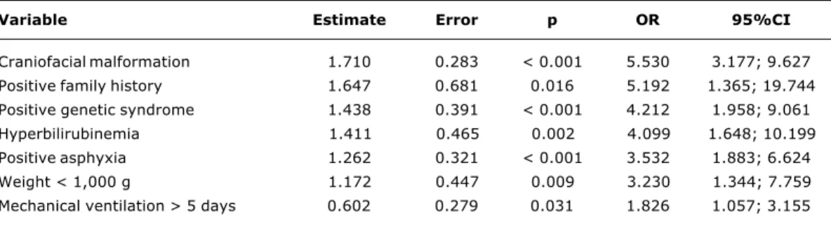
According to the multivariate logistic regression model, the main characteristics of the group of children at greater risk of presenting with impaired hearing screening were as follows: family history of congenital hearing loss; craniofacial malformation; weight < 1,000 g; mechanical ventilation for more than five days; hyperbilirubinemia and genetic syndrome (Table 2).
Discussion
Table 2 - Multiple analysis by logistic regression of the risk factors for impaired hearing screening
Variable Estimate Error p OR 95%CI
Craniofacial malformation 1.710 0.283 < 0.001 5.530 3.177; 9.627
Positive family history 1.647 0.681 0.016 5.192 1.365; 19.744
Positive genetic syndrome 1.438 0.391 < 0.001 4.212 1.958; 9.061
Hyperbilirubinemia 1.411 0.465 0.002 4.099 1.648; 10.199
Positive asphyxia 1.262 0.321 < 0.001 3.532 1.883; 6.624
Weight < 1,000 g 1.172 0.447 0.009 3.230 1.344; 7.759
Mechanical ventilation > 5 days 0.602 0.279 0.031 1.826 1.057; 3.155
CI = confidence interval; OR = odds ratio.
Table 1 - Bivariate analysis of the variables associated with the result of neonatal hearing screening (n = 979)
Variable n (%) Normal Impaired OR (95%CI) p
Neonatal characteristics
Family history 13 (1.3) 9 4 4.03 (1.22-13.33) 0.022
Consanguinity 7 (0.7) 5 2 3.57 (0.68-8.64) 0.131
Weight < 1,000 g 50 (5.1) 39 11 2.51 (1.20-5.22) 0.014
Weight 1,000-2,500 g 454 (46.4) 413 41 2.51 (1.20-5.22) 0.579
Gestational age < 30 weeks 75 (7.7) 63 12 1.57 (0.79-3.13) 0.196
Gestational age 31-34 weeks 267 (27.3) 248 19 0.63 (0.36-1.10) 0.106
Gestational age 35-36 weeks 201 (20.5) 179 22 1.01 (0.59-1.73) 0.957
Small for gestational age 209 (21.3) 188 21 0.98 (0.59-1.63) 0.937
Large for gestational age 47 (4.8) 42 5 1.04 (0.40-2.72) 0.929
Girls 485 (49.6) 430 55 1.27 (0.84-1.93) 0.254
Neonatal pathologies
Craniofacial malformation 104 (10.6) 74 30 4.66 (2.86-7.60) < 0.001
Genetic syndrome 42 (4.3) 28 14 4.95 (2.51-9.75) < 0.001
Hyperbilirubinemia (TB > 20 mg/dl) 42 (4.3) 35 7 1.81 (0.78-4.20) 0.164
PIH 33 (3.4) 29 4 1.22 (0.42-3.55) 0.713
Asphyxia 81 (8.3) 64 17 2.61 (1.46-4.66) 0.001
Congenital infection 103 (10.5) 95 8 0.72 (1.20-5.22) 0.388
Meningitis 20 (2.0) 15 5 3.03 (1.08-8.53) 0.035
Specialized procedures
Ototoxic medication > 5 days 213 (21.7) 181 32 1.81 (1.16-2.85) 0.009
Incubator > 5 days 317 (32.4) 285 32 0.98 (0.63-1.53) 0.932
Phototherapy > 2 days 387 (39.5) 345 42 1.17 (0.77-1.78) 0.465
Mechanical ventilation > 5 days 200 (20.4) 169 31 1.88 (1.20-2.98) 0.006
CI = confidence interval; OR = odds ratio; PIH = peri intraventricular hemorrhage; TB = total bilirubin.
so that the prevalence of failure in the neonatal hearing screening test must be understood as possible hearing loss, and false negatives may be linked with incomplete myelinization in the studied age group. The diagnosis must be confirmed with the conventional ABR, considering the maturation of the Central Nervous System.16
A study conducted in Germany found 5% of children with impaired AABR, 2% bilaterally.17 In another study
conducted in an NICU with the use of two-stage AABR, 8% and 3.1% failed the screening test, and confirmation of the diagnosis by conventional ABR was obtained in 2.5%. The exam performed close to hospital discharge had with greater specificity.18
newborns.19 Another study assessed newborns weighing
under 1,500 g with distortion product otoacoustic emissions and found a prevalence of 6.3% for hearing loss.20
Although the data in the literature show variable results, the prevalence we observed is above the average, which may be related to the characteristics of the present group, comprised of newborns admitted to the intensive and intermediate care unit at a University Hospital that is a regional excellence center and therefore provides care to highly complex cases. Thus, a higher prevalence of hearing loss in the population treated at this facility can be expected.
Family history of congenital hearing loss was identified in the bivariate analysis as a factor significantly associated with hearing impairment. This shows that among the sensorineural deficiencies of known etiology, those of a hereditary origin are highly representative. With the advances in the field of molecular genetics, new genes responsible for sensorineural deafness have been identified, and the mechanisms involving cases of deafness due to non-syndromic causes have been explained in such a way that the influence of this variable may be better understood in the future.18,21,22
Since birthweight is a continuous variable, it was separately analyzed in the following categories: < 1,000 g, from 1,000 to 1,500 g, from 1,501 to 2,500 g and > 2,500 g. Newborns weighing > 2,500 g were considered to be at less risk, and were considered as the reference category, with which the other categories were compared. A significant association was noted in newborns < 1,000 g. Weight from 1,000 to 1,500 g, as well as from 1,501 to 2,500 g were not a significant risk factor. The current literature considers weight < 1,500 g to be a risk factor, although this finding is not consistent and its greater or lesser significance depends on the differences in the populations assessed, as well as the conditions of perinatal care.17,22
The presence of craniofacial malformations was shown to be significantly related to hearing impairment and has been a frequent finding in the literature. In different studies, this variable is responsible for between 11 and 16% of the cases of hearing loss, increasing the risk of these children presenting with impairment by up to 5 times.17,23,24
In the literature, syndromes associated with sensorineural deafness, as well as structural defects with or without chromosomal abnormalities, have been described as a risk factor, which is in agreement with the results found in the present study, in which the presence of the genetic syndrome was significantly associated with greater risk of hearing impairment.22,24
As regards comorbidities, asphyxia was associated with 3.5 times more risk of hearing impairment. Although the brain is more sensitive to anoxia than the auditory
system, severe hypoxic ischemic encephalopathy was an important risk factor for hearing loss. In current literature, these results also varied in accordance with the studied population and mainly with the definition of neonatal asphyxia.16,21 Nevertheless, the impact of asphyxia on
hearing may be minimized or abolished if it is controlled, which may be achieved by training the teams that give assistance to newborns in the delivery room.25
In spite of a declining trend in the occurrence of meningitis in the neonatal period, in the present studied it was identified as being significantly associated with hearing impairment in the bivariate analysis, which is in agreement with the literature. However, in the multiple analysis, in which we attempted to eliminate the joint action of various other factors, this association was not maintained.17,26
As regards hyperbilirubinemia, the data in the literature point it out as an important cause of deafness.27 In the
present study assessment, it was not identified as a risk factor in the bivariate analysis. However, when analyzed in conjunction with the other variables, it acquired great statistical power, becoming an important event in the occurrence of hearing impairment.
These results suggest that it is necessary to implement protocols with strict control of cases of jaundice, including objective measures for assessing the serum level of bilirubin and highly efficient phototherapy, which represent measures to prevent hearing impairment resulting from hyperbilirubinemia.
Although the use of phototherapy has not been the object of study in the literature, this analysis was made because it is a very common procedure in our unit, and involves a noise level above the ideal proposed (mean of 45 dB during the day and 35 dB at night, the maximum limit suggested being 58 dB).29 The number of children
submitted to phototherapy is higher than the number considered as having hyperbilirubinemia, since the indication of phototherapy contemplates other children in addition to those with total bilirubin > 20 mg/dl. We did not observe an association between this variable and hearing impairment.
Correspondence:
Gisele Marafon Lopes de Lima Rua Coronel Quirino, 1343/72
CEP 13025-002 Campinas, SP Brazil Tel./Fax: +55 (19) 3294.9573
E-mail: marafon@supernet.com.br References
1. Lewis DR. As habilidades auditivas do recém-nascido e a triagem auditiva neonatal. In: Andrade CRF. Fonoaudiologia em berçário normal e de risco. São Paulo: Lovise; 1996. p. 149-68. 2. Downs MP, Yoshinaga-Itano C. The efficacy of early identification and intervention for children with hearing impairment. Pediatr Clin North Am. 1999;46:79-87.
3. American Academy of Pediatrics. Task Force on Newborn and Newborn and Infant Hearing. Newborn and infant hearing loss: detection and intervention. Pediatrics. 1999;103:527-30. 4. Kenna MA. Neonatal hearing screening. Pediatr Clin North Am.
2003;50:301-13.
5. Moeller MP. Early intervention and language development in children who are deaf and hard of hearing. Pediatrics. 2000;106:e43.
6. American Academy of Pediatrics. Joint Committee on Infant Hearing. Year 2000 position statement: principles and guidelines for early hearing detection and intervention programs. Pediatrics. 2000;106:798-817.
7. Comitê Brasileiro sobre Perdas Auditivas na Infância. Recomendação 01/99 do Comitê Brasileiro sobre Perdas Auditivas na Infância. Jornal do Conselho Federal de Fonoaudiologia. 2000;3-7.
8. Bassetto MCA. Triagem auditiva em berçário. In: Bassetto MCA, Brock R, Wajnsztejn R. Neonatologia um convite à atuação fonoaudiológica. São Paulo: Lovise; 1995. p. 289-93. 9. Matas CG. Medidas eletrofisiológicas da audição audiometria
de tronco cerebral. In: Carvalho RMM. Fonoaudiologia: informação para a formação. Rio de Janeiro: Guanabara Koogan; 2003. p. 43-57.
10. Carvalho RMM. Emissões otoacústicas: conceitos básicos e aplicações. In: Carvalho RMM. Fonoaudiologia: informação para a formação. Rio de Janeiro: Guanabara Koogan; 2003. p. 22-41. 11. National Center for Hearing Assessment and Management. What does a newborn hearing screening program cost? http:// w w w . i n f a n t h e a r i n g . o r g / r e s o u r c e s / c o s t / i n d e x . h t m l . Access: 23/06/2005.
12. Keren R, Helfand M, Homer C, McPhillips H, Lieu TA. Projected cost-effectiveness of statewide universal newborn hearing screening. Pediatrics. 2002;110:855-64.
13. Oudesluys-Murphy AM, Bhoalsingh R, Van Zanten GA, Van Straaten HLM. Neonatal hearing screening. Eur J Pediatr. 1996;155:429-35.
14. Doyle KJ, Fujikawa S, Rogers P, Newman E. Comparison of newborn hearing screening by transient otoacoustic emissions and automated auditory brainstem response using ALGO-2Ò. Int J Pediatr Otorhinolaryngol. 1998;43:207-11.
15. Van Straaten HL. Automated auditory brainstem response in neonatal hearing screening. Acta Paediatr Suppl. 1999;88:76-9. 16. Northern JL, Downs MP. Audição em crianças. 5ª ed. Rio de
Janeiro: Guanabara Koogan; 2005.
17. Meyer C, Witte J, Hildmann A, Hennecke KH, Schunck KU, Maul K, et al. Neonatal screening for hearing disorders in infant at risk: incidence, risk factors and follow-up. Pediatrics. 1999;104:900-4.
18. van Straaten HLM, Hille ET, Kok JH, Verkerk PH. Dutch NICU Neonatal Hearing Screening Working Group. Implementation of a nation-wide automated auditory brainstem response hearing screening program in neonatal intensive care units. Acta Paediatr. 2003;92:332-8.
19. Chapchap MJ, Segre CM. Universal newborn hearing screening and transient evoked otoacoustic emission: new concepts in Brazil. Scand Audiol Suppl. 2001;53:33-6.
20. Uchôa NT, Procianoy RS, Lavinsky L, Sleifer P. Prevalência de perda auditiva em recém-nascidos de muito baixo peso. J Pediatr (Rio J). 2003;79:123-8.
21. Kitamura K, Takahashi K, Tamagawa Y, Noguchi Y, Kuroishikawa Y, Ishicawa K, et al. Deafness genes. J Med Dent Sci. 2000;47: 1-11.
22. Chu K, Elimian A, Barbera J, Ogburn P, Spitzer A, Quirk JG. Antecedents of newborn hearing loss. Obstet Gynecol. 2003;101:584-8.
23. Watkin PM, Baldwin M, Mcenery G. Neonatal at risk screening and the identification of deafness. Arch Dis Child. 1991;66:1130-5.
24. Homer JJ, Linney SL, Strachan DR. Neonatal hearing screening using the auditory brainstem response. Clin Otolaryngol. 2000;25:66-70.
25. American Academy of Pediatrics, American Heart Association. International guideline for neonatal resuscitation: an excerpt from the guideline 2000 for cardiopulmonary resuscitation and emergency cardiovascular care: international consensus on science. http://www.pediatrics.org/cgi/content/full/106/3/e29. Access: 23/06/2005.
26. Roizen NJ. Non-genetic causes of hearing loss. Ment Retard Dev Disabil Res Rev. 2003;9:120-7.
27. Oysu C, Aslan I, Ulubil A, Baserer N. Incidence of cochlear involvement in hyperbilirubinemic deafness. Ann Otol Rhinol Laryngol. 2002;111:1021-5.
28. Facchini FP. Icterícia neonatal. In: Marba STM, Mezzacappa F, editores. Manual de neonatologia da UNICAMP. Rio de Janeiro: Revinter; 1998. p. 59-64.
29. American Academy of Pediatrics. Noise: a hazard for the fetus and newborn. Pediatrics. 1997;100:724-7.
30. Hess M, Finckh-Kramer U, Bartsch M, Kewitz G, Versmold H, Gross M. Hearing screening in at-risk neonate cohort. Int J Pediatr Otorhinolaryngol. 1998;46:81-9.
Mechanical ventilation for more than five days showed a significant association with hearing impairment. Various aspects have been related to the greater occurrence of deafness in children submitted to assisted ventilation, including the noise level of the appliances, duration of mechanical ventilation, and the pulmonary pathologies involved.