103 Abstract
Objective: To measure and compare the activity of the masseter, temporalis and buccinator muscles in different infant feeding methods.
Method: Cross-sectional study of 60 full-term infants with no intercurrent diseases, aged between two and three months, classified into the following groups: 1) exclusive breastfeeding; 2) breastfeeding plus bottle-feeding; and 3) exclusive breastfeeding plus cup feeding. Surface electromyography was performed during infant feeding. The Krushal-Wallis test was used, complemented by multiple paired comparisons of the groups. A 5% significance level was chosen for the tests.
Results: Statistically higher results were verified in the breastfeeding group in relation to the bottle-feeding one, both in the range of movement and the mean contraction of the masseter. With regard to the temporalis muscle, statistically higher results were found in the breastfeeding group comparatively to the bottle-feeding one. As to the buccinator muscle, statistically higher results were observed in the breastfeeding group in relation to the bottle-feeding one, although in this case, the difference concerned only the range of contraction.
Conclusion: The similarities between the muscle activity in the breastfeeding and in the cup-feeding groups suggests that cup-feeding can be used as an alternative infant feeding method, being better than bottle-feeding, due to the hyperactivity of the buccinator muscle, which could result in changes to the structural growth and development of the stomatognathic system functions.
J Pediatr (Rio J). 2006;82(2):103-9: Electromyography, breastfeeding, facial muscles, sucking, infants.
Surface electromyography of facial muscles
during natural and artificial feeding of infants
Cristiane F. Gomes,1 Ercília M. C. Trezza,2 Emílio C. M. Murade,3 Carlos R. Padovani4
1. Doutora, Faculdade de Medicina de Botucatu, Universidade Estadual Paulista (UNESP), São Paulo, SP, Brasil.
2. Doutora. Professora assistente, Faculdade de Medicina de Botucatu, UNESP, São Paulo, SP, Brasil.
3. Doutor, Faculdade de Ciências Médicas da Santa Casa de São Paulo, São Paulo, SP. Docente, Departamento de Ortopedia e Traumatologia, Universidade de Marília (UNIMAR), Marília, SP, Brasil.
4. Professor titular, Departamento de Bioestatística, Faculdade de Medici-na de Botucatu, UNESP, São Paulo, SP, Brasil.
Part of the doctoral dissertation presented on November 11, 2005 at the School of Medicine of Botucatu, UNESP, São Paulo, SP, Brazil. Support: CAPES.
Manuscript received Sep 26 2005, accepted for publication Dec 14 2005.
Suggested citation: Gomes CF, Trezza EM, Murade EC, Padovani CR. Surface electromyography of facial muscles during natural and artificial feeding of infants. J Pediatr (Rio J). 2006;82:103-9.
Introduction
The stomatognathic system comprises static and dynamic oral structures, i.e., hard parts (dental arches, maxilla, jaw, hyoid bone and skull bones) and active parts (muscles, body spaces, nerves, and blood vessels) that perform neurovegetative functions (sucking, chewing,
swallowing and breathing), speech and facial expression. These structures are interconnected in such a way that if any of them is disordered, all of them will become disorganized and unbalanced.1-3
Craniofacial growth and development rely on genetic and external stimuli, which are provided by breathing, sucking (breastfeeding), swallowing and chewing. Stimuli provided by muscles are the major determinants of growth, as they stretch the bones, resulting in growth or wear, depending on where stretching occurs.2
As to the effects of breastfeeding on the growth of structures and on the development of stomatognathic system functions, it is common knowledge that appropriate facial growth occurs by means of movements made by the infant during milk expression, when the maxillae are encouraged to grow in a well-directed course. Moreover, breastfeeding allows for oral maturation by stimulating muscle tone and the development of the temporomandibular joint, providing enough room for tooth eruption.4-12
O
RIGINALA
RTICLE Copyright © 2006 by Sociedade Brasileira de PediatriaElectromyography consists of the assessment of muscle function by analyzing the electrical signal produced during muscle contraction. An electromyograph is used to detect the electrical variables found on the cells during nerve transmission and muscle contraction, which are converted into electrical signals and, after amplification, are displayed on the oscilloscope screen, for later analysis.
Surface electromyography has been currently used to assess various types of pathologies (respiratory disorders, sleep disorders), in the study of specific muscles in athletes and animals, the speech of children with occlusion disorders, in the assessment of infant feeding methods, among others.
The use of electromyography in research is quite recent and has allowed to determine the activity of oral muscles on the different types of feeding, and to compare their strength and activity.
Given the specificities of surface electromyography in the objective assessment of muscle activity and the paucity of studies showing the differences between the activities of the muscles in charge of sucking during breastfeeding, bottle-feeding, and cup-feeding, the aim of the present study is to assess the activity of the masseter, temporalis and buccinator muscles during exclusive breastfeeding, breastfeeding plus bottle-feeding, and exclusive breastfeeding plus cup-feeding as an alternative feeding method.
Methods
A cross-sectional double-blind design was used, in which cause and effect occur concomitantly, which characterizes the moment analyzed herein.13
Sixty full-term infants aged between 2 and 3 months and with no intercurrent diseases were included in the study. They were divided into three groups: Group 1: 20 infants on exclusive breastfeeding; Group 2: 20 infants on breastfeeding plus bottle-feeding; Group 3: 20 infants on exclusive breastfeeding who could be cup-fed during the electromyography.
A convenience sample was used, since 212 invitations had been sent out and 64 mothers accepted to participate in the study. Four infants were excluded from the study: three for being exclusively breastfed and one for having inconclusive exam results. The following inclusion criteria were used: infants aged between 60 and 90 days at the time of electromyography on either exclusive breastfeeding or mixed feeding; full-term infants, with birth weight greater than 2,500 g; and infants with no intercurrent diseases in the prenatal, perinatal and postnatal periods. The following were not included in the study: twins; those on artificial exclusive breastfeeding, and those who could not be submitted to electromyography.
The differences in the activity of the masseter in breastfed and bottle-fed infants, in the study conducted by Sakashita et al.,14 were used to calculate the sample size of this study. By considering
α
= 5% and ß = 80%, we stipulated 18 infants per group; however, we assessed 20 infants in each group to maintain a safe margin.Sixty electromyographies were performed between February 17 and July 31, 2003. Participants were evaluated in terms of age (days of life at the time of the exam), gender, gestational age at birth, type of delivery, birth weight, head circumference, length, first-minute Apgar score and use of pacifier. In addition, mothers were evaluated in terms of age, schooling, and type of health plan adopted at the time of delivery.
The study was carried out at the Teaching Hospital of Universidade de Marília (UNIMAR) after being approved by the Research Ethics Committee of UNESP School of Medicine of Botucatu and was authorized by the Municipal Department of Health and Sanitation of Marília to gain access to the Human Milk Bank records, regarding the Nascer Cidadão Program, which aims to register and refer newborns at risk to municipal health units in order to reduce infant mortality.
To begin with, mothers were informed about the study objectives and about the importance of their participation and, after their verbal agreement, they signed an informed consent form.
The following devices were used for the exam: a portable two-channel Dantec (model LBM Firmware version 4.03, 1993 by Neuro Diagnostics, Inc.) electromyograph with a 70 Hz filter, 5-ms time constant per division and a sensitivity of 50 µv (microvolts) per division; monopolar, disc, gold-plated skin surface electrodes (Maxxigold, MF), with 9 mm in diameter and 16 mm of distance between the reference and the working electrodes; electrode cream (Maxxifix cream, MF); adhesive tape for electrode fixation (Transpore®, 3M); Thermwhite (Alfamedic Ind. Com. e Serviços Ltda.) paper for printing the results.
Electromyographic activity was graded and quantitatively tabulated by an expert for later statistical analysis and for the comparison between the different groups and muscles analyzed.
The expert carried out an independent analysis, i.e., he was blinded to the feeding method used at the time of electromyography, with no interference from the researcher.
Non-parametric analysis of variance for data with non-normal distribution (Kruskal-Wallis test) was used, complemented by multiple paired comparisons.15
Table 1 - Absolute frequency and percentage distribution of the characteristics of assessed individuals (n = 60)
Characteristics f %
Infant age
60-70 days 3 5.00
71-80 days 10 16.67
81-90 days 47 78.33
Gender
Female 22 36.67
Male 38 63.33
Gestational age
38 weeks 12 20.00
39 weeks 18 30.00
40 weeks 30 50.00
Type of delivery
Vaginal 22 36.67
C-section 38 63.33
Birth weight
2,500-3,000 g 19 31.67
3,001-3,599 g 29 48.33
3,600-4,000 g 8 13.33
+ 4,000 g 4 6.67
Head circumference
33-35.9 cm 36 60.00
36-37.9 cm 18 30.00
Not informed 6 10.00
Length
44-47.9 cm 15 25.00
48-51.9 cm 35 58.33
52-53.9 cm 10 16.67
First-minute Apgar score
7 4 6.67
8 17 28.33
9 35 58.33
Not informed 4 6.67
Maternal age
- 18 years 4 6.67
19-25 years 38 63.33
26-30 years 8 13.33
31-35 years 8 13.33
+ 35 years 2 3.33
Maternal schooling
incomplete elementary education 3 5.00
Completed elementary education 9 15.00
Completed high school education 39 65.00
Incomplete college education 2 3.33
Completed college education 7 11.67
Type of health plan
Unified Health System 23 38.3
Private 14 23.3
Health insurance 16 26.7
Not informed 7 11.7
Use of pacifier
Bottle-feeding 10 16.67
Breastfeeding 0
Cup-feeding 0
A significance level of 5% and a 95%CI were used.
Results
Patients
The statistical analysis did not show any differences between the studied groups and age (medians of 82.50 days for those infants on exclusive breastfeeding, 83.00 days for those on breastfeeding plus bottle-feeding, and 84.00 days for those on exclusive breastfeeding plus cup feeding), since the variable is an inclusion criterion, i.e., only infants between 30 and 90 days participated in the study (Table 1).
Exam results
The assessment of muscles included range of contraction and mean contraction of the masseter, temporalis, and buccinator muscles.
Range of contraction and mean contraction of the masseter
The statistical analysis showed a significant result when the groups were compared as to the type of feeding, allowing us to conclude that the cup-feeding (median of 3.07 µv) and bottle-feeding (median of 1.94 µv) groups differ between each other.
Moreover, the mean contraction of the masseter in the breastfeeding group (median of 80 µv) differs from that of the bottle-feeding group (median of 50 µv) (Table 2).
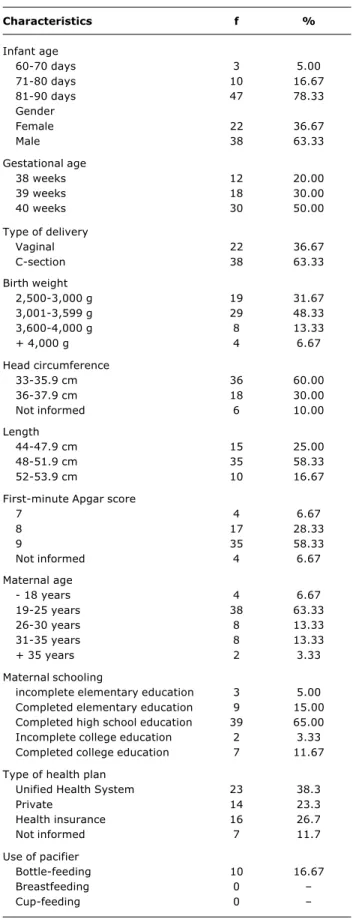
Range of contraction and mean contraction of the temporalis muscle
There are differences between the groups regarding the range of contraction of the temporalis muscle, which corroborate that the breastfeeding (median of 4.02 µv) and cup-feeding (median of 3.84 µv) groups differ from the bottle-feeding group (median of 1.81 µv).
As to the mean contraction of the temporalis muscle, the breastfeeding (median of 111.25 µv) and cup-feeding (median of 96.04 µv) groups differ from the bottle-feeding group (median of 48.03 µv) (Table 3).
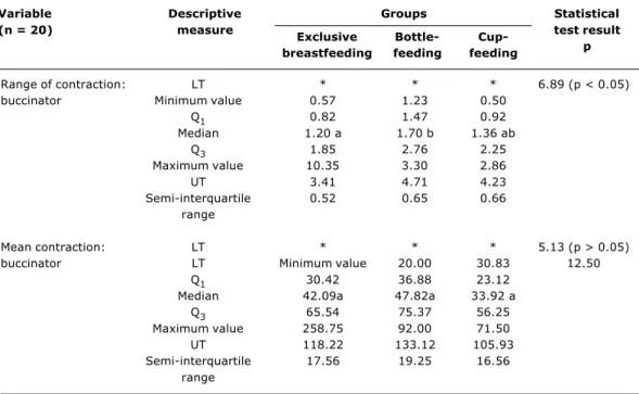
Range of contraction and mean contraction of the buccinator muscle
As shown in Table 4, there are differences between the groups as to the range of contraction of the buccinator muscle, corroborating that the bottle-feeding group (median of 1.70 µv) differs from the breastfeeding group (median of 1.20 µv).
With regard to the mean contraction of the buccinator muscle, no differences were observed between the groups
Table 2 - Median, semi-interquartile range, minimum and maximum values, lower and upper thresholds, first and third quartiles of range of contraction and mean contraction of the masseter (n = 20)
LT = lower threshold; UT = upper threshold. * Negative values: values that cannot be obtained.
† Two medians followed by at least one identical letter do not differ at the 5% significance level.
Range of contraction: LT * * * 6.61 (p < 0.05)
masseter Minimum value 0.84 0.77 1.70
Q1 1.62 1.40 2.12
Median 2.38 ab 1.94 a 3.07 b
Q3 3.14 2.80 4.31
Maximum value 4.48 5.27 9.10
UT 5.42 4.90 7.42
Semi-interquartile 0.76 0.70 1.10
range
Mean contraction: LT * 1.25 * 7.35 (p < 0.05)
masseter Minimum value 31.88 19.17 42.50
Q1 57.60 42.50 53.03
Median 80.00 b 50.00 a 76.59 ab
Q3 99.90 70.0 107.82
Maximum value 298.33 131.67 227.50
UT 163.35 111.50 202.80
Semi-interquartile 21.15 13.75 27.40
range
Variable Descriptive Statistical
(n = 20) measure test result
p Groups
Exclusive Bottle-
Cup-breastfeeding feeding feeding Discussion
With regard to the comparison between the groups as to the type of feeding, several studies show that the masseter is the muscle that most actively participates in
Table 3 - Median, semi-interquartile range, minimum and maximum values, lower and upper thresholds, first and third quartiles of range of contraction and mean contraction of the temporalis muscle (n = 20)
Range of contraction: LT * * 0.81 14.21 (p < 0.01)
temporalis Minimum value 0.00 0.00 2.00
Q1 2.58 1.25 3.21
Median 4.02 b 1.81 a 3.84 b
Q3 4.81 3.18 4.80
Maximum value 15.87 5.35 6.20
UT 8.17 6.09 7.20
Semi-interquartile 1.12 0.97 0.80
range
Mean contraction: LT 38.68 * 20.36 17.68 (p < 0.01)
temporalis Minimum value 0.00 0.00 50.00
Q1 100.0 36.88 80.21
Median 111.25 b 48.03 a 96.04 b
Q3 140.87 83.56 120.10
Maximum value 793.33 133.75 155.00
UT 202.19 203.77 179.95
Semi-interquartile 20.44 23.34 19.95
range
LT = lower threshold; UT = upper threshold. * Negative values: values that cannot be obtained.
Variable Descriptive Statistical
(n = 20) measure test result
p Groups
Exclusive Bottle-
Cup-breastfeeding feeding feeding
Table 4 - Median, semi-interquartile range, minimum and maximum values, lower and upper thresholds, first and third quartiles of range of contraction and mean contraction of the buccinator muscle (n = 20)
Range of contraction: LT * * * 6.89 (p < 0.05)
buccinator Minimum value 0.57 1.23 0.50
Q1 0.82 1.47 0.92
Median 1.20 a 1.70 b 1.36 ab
Q3 1.85 2.76 2.25
Maximum value 10.35 3.30 2.86
UT 3.41 4.71 4.23
Semi-interquartile 0.52 0.65 0.66
range
Mean contraction: LT * * * 5.13 (p > 0.05)
buccinator LT Minimum value 20.00 30.83 12.50
Q1 30.42 36.88 23.12
Median 42.09a 47.82a 33.92 a
Q3 65.54 75.37 56.25
Maximum value 258.75 92.00 71.50
UT 118.22 133.12 105.93
Semi-interquartile 17.56 19.25 16.56
range
LT = lower threshold; UT = upper threshold. * Negative values: values that cannot be obtained.
Variable Descriptive Statistical
(n = 20) measure test result
p Groups
Exclusive Bottle-
Cup-breastfeeding feeding feeding
According to Palmer,16 Madeira17 and Almeida et al.,18 jaw movements favor the appropriate growth and positioning of the jaw for tooth eruption. Therefore, the masseter plays a crucial role in infant feeding, which is accomplished only by breastfeeding and cup-feeding, because bottle-feeding reduces masseter activity, increases buccinator activity, reduces jaw movements, causes the tongue to retract, with possible hypoactivity and hyperactivity, depending on the type of sucking.19
The previously described results are similar to those reported in the literature. In the study by Sakashita et al.,14 the electromyographic results reveal greater masseter activity in breastfeeding than in bottle-feeding, leading to the conclusion that bottle-fed infants may have masticatory dysfunctions, resulting in possible masticatory and swallowing disorders.
Likewise, the study conducted by Tamura et al.20 shows that, in bottle-feeding, the masseter has a remarkably lower activity compared to breastfeeding.
Although most studies corroborate the data obtained herein, Sakashita et al.14 concluded in their study, which compares the sucking pattern of infants being breastfed, bottle-fed with a conventional teat and bottle-fed with a new type of artificial teat, that masseter activity is greater
among those infants who are bottle-fed using the new type of artificial teat than in the breastfeeding group.
With regard to cup-feeding, Kuehl21 asserts that the movements of the tongue and jaw are similar to the movements necessary for successful breastfeeding, suggesting that cup-feeding may be an opportunity for the infant to develop the muscles necessary for these movements.
The data obtained allow stating that if breastfeeding is not possible at certain times, cup-feeding may be indicated, since the range of contraction and mean contraction of the masseter are greater than in bottle-feeding, being an intermediary between this group and the breastfeeding group.
Few studies have assessed the activity of the temporalis muscle, possibly because it is more related to movement than to strength, being responsible, just like the masseter, for elevation of the jawbone due to the combination of its fibers with those of the masseter and medial pterygoid.16-18 The temporalis muscle serves a double purpose: elevation of the jawbone, due to its vertical fibers, and retraction, due to its oblique and horizontal fibers.19,22
References
1. Bianchini EMG. Crescimento e desenvolvimento craniofacial. In: Bianchini EMG. A cefalometria nas alterações miofuncionais orais: diagnóstico e tratamento fonoaudiológico. 2ª ed. São Paulo: Pró-Fono; 1994. p. 5-15.
2. Tanigute CC. Desenvolvimento das funções estomatognáticas. In: Marchesan IQ. Fundamentos em fonoaudiologia:aspectos clínicos da motricidade oral. Rio de Janeiro: Guanabara-Koogan; 1998. p. 1-6.
3. Carvalho GD. Amamentação e o sistema estomatognático. In: Carvalho MR, Tamez RN. Amamentação: bases científicas para a prática profissional. Rio de Janeiro: Guanabara-Koogan; 2002. p. 37-49.
4. Vardes V, Sanchez AP, Labbok M. Manejo clínico da lactação: assistência à nutriz e ao lactente. Rio de Janeiro: Revinter; 1996. p. 1-25.
5. Righard L, Alade MO. Sucking technique and its effect on success of breastfeeding. Birth. 1992;19:185-9.
6. Fernandes FBU. Pensando no bebê: benefícios, técnicas e dificuldades do aleitamento materno [monografia]. Rio de Janeiro: 2000. Centro de Especialização em Fonoaudiologia Clínica; 2000.
7. Matida MY. Amamentação: uma visão fonoaudiológica [monografia]. Londrina: Centro de Especialização em Fonoaudiologia Clínica; 2000.
8. Medeiros EB, Rodrigues MJ. Importância da amamentação natural para o desenvolvimento do sistema estomatognático do bebê. Rev Cons Reg Odontol Pernambuco. 2001;4:79-83. 9. Gava-Simioni LR, Jacinto SR, Gaviao MBD, Puppin Rontani RM.
Amamentação e odontologia.J Bras Odontopediatr Odontol Bebê. 2001;4:125-31.
10. Jacinto-Goncalves SR, Gaviao MBD, Berzin F, Oliveira AS, Semeguini TA. Electromyographic activity of perioral muscles in breastfed and non-breastfed children. J Clin Pediatr Dent. 2004;29:57-62.
11. Woolridge MW. The anatomy of infant sucking. Midwiferi. 1986;2:164-71.
12. Naylor AJ, Danner S, Lang S. Development of oral motor function. In: Naylor AJ, Morrow AL. Developmental readiness of normal full term infants to progress from exclusive breastfeeding to the introduction of complementary foods: reviews of the relevant literature concerning infant gastrointestinal, immunologic, oral motor and maternal reproductive and lactational development. San Diego: Wellstart International; 2001. p. 21-6.
13. Campana AO. Pesquisa clínica estudos observacionais. In: Campana AO. Introdução à investigação científica. Botucatu: UNESP; 1995. p. 87-100.
14. Sakashita R, Kamegai T, Inoue N. Masseter muscle activity in bottle feeding with the chewing type bottle teat: evidence from electromyographs. Early Hum Dev. 1996;45:83-92.
15. Norman GR, Streiner DL. Bioestatistics: the bare essentials. St. Louis: Mosby-Year Book; 1994.
16. Palmer JM. Oral cavity. In: Palmer JM. Anatomy for speech and hearing. 14th ed. Baltimore: Williams & Wilkins; 1993. p. 53-90. 17. Madeira MC. Músculos da face. In: Madeira MC. Anatomia da face:bases anátomo-funcionais para a prática odontológica. 3ª ed. São Paulo: Sarvier; 2001. p. 67-95.
infers that if an alternative feeding method is necessary, cup-feeding would be more appropriate than bottle-feeding, since it prevents nipple confusion and allows the participation of the masseter and temporalis muscles in a similar way to the participation of these muscles in breastfeeding.
The participation of the masseter and temporalis muscles in bottle-feeding is smaller than in the other types of feeding, similarly to what Carvalho19 describes when stating that the tongue, lips and masseter, temporalis, pterygoid , mentalis and buccinator muscles have normal function in breastfeeding, whereas in bottle-feeding with a conventional teat, the tongue, lips and the masseter, temporalis and pterygoid muscles are hypofunctional, and the mentalis and buccinator muscles are hyperfunctional.
Ahlgren23, who investigated the electromyographic activity of perioral muscles, including the buccinator, during nutritive and non-nutritive suck in children with malocclusion and normal occlusion, found out that the buccinator muscle has a greater activity during suction and swallowing in children with malocclusion than in those with normal occlusion, underscoring the relationship between buccinator activity and the occurrence of occlusion disorders, in agreement with the data obtained by Carvalho.19
By comparing the breastfeeding groups, it may be concluded that the activity of the muscles in breastfeeding and bottle-feeding is concurrent with the literature data, but no scientific studies exist corroborating the results obtained with cup-feeding.
The data obtained allow concluding that in both breastfeeding and cup-feeding, the same muscles act in a similar fashion (without statistically significant differences), with a greater participation of the masseter and temporalis muscles and a smaller participation of the buccinator muscles.
Breastfeeding is therefore the most appropriate infant feeding method, and cup-feeding is recommended as a substitute for breastfeeding when breastfeeding is not possible, as the active muscles in both methods are the same, not causing nipple confusion.
The method used for data collection and analysis was appropriate for the study objectives. However, some biases may have interfered with the results, for instance, the use of pacifiers by 50% of infants in the breastfeeding plus bottle-feeding group. This may have changed the sucking pattern of infants, despite the fact that they were already being bottle-fed.
New studies should be conducted in order to assess the activity of the tongue and other oral structures in both feeding methods, determining whether there are differences in bottle-feeding and cup-feeding.
Further studies should also be carried out with preterm infants and babies with syndromes or head and neck malformations, so as to check the possibility to implement cup-feeding and to evaluate its efficiency in specific infant populations.
Correspondence: Cristiane F. Gomes
Rua José Pereira da Costa, 89, casa 7, Residencial Paulista II CEP 87005-220 Maringá, PR Brazil
Tel.: +55 (44) 3025.7095 Fax: +55 (44) 3028.4888 E-mail: [email protected] 18. Almeida EOC, Melli R, Moraes IF. Orientação fonoaudiológica e
psicológica às nutrizes: experiência em contexto hospitalar. In: Tasca SMT, Almeida EOC, Servilha EAM. Recém-nascido em alojamento conjunto:visão multiprofissional. Carapicuíba: Pró-Fono; 2002. p. 35-49.
19. Carvalho GD. O sistema estomatognático e suas funções. In: Carvalho GD. S.O.S. respirador bucal: uma visão funcional e clínica da amamentação. São Paulo: Lovise; 2003. p. 27-56. 20. Tamura Y, Horikawa Y, Yoshida S. Co-ordination of tongue
movements and peri-oral muscle activities during nutritive sucking. Dev Med Child Neurol. 1996;38:503-10.
21. Kuehl J. Cup feeding the newborn: what you should know. J Perinat Neonatal Nurs. 1997;11:56-60.
22. Sanches MTC. Manejo clínico das disfunções orais na amamentação. J Pediatr (Rio J). 2004;80:S155-62.