52 Radiol Bras. 2011 Jan/Fev;44(1):52–58
Imaging findings of musculoskeletal disorders associated
with systemic lupus erythematosus
*
Achados de imagem das alterações musculoesqueléticas associadas ao lúpus eritematoso sistêmico
Daniel Sá Ribeiro1, César de Araújo Neto2, Fernando D’Almeida3, Verena Loureiro Galvão4,
Mittermayer Barreto Santiago5
Systemic lupus erythematosus is an autoimmune disease involving multiple organ systems. Musculoskeletal involvement is one of the most frequent presentations of the disease, affecting bones, joints, muscles, tendons and ligaments, either as a primary manifestation or secondary to the treatment of the disease. In the present article, the authors review and illustrate the joint disorders and the most common musculoskeletal abnormalities seen in this disorder. Keywords: Systemic lupus erythematosus; Arthropathy; Musculoskeletal abnormalities.
O lúpus eritematoso sistêmico é uma doença autoimune que envolve múltiplos sistemas orgânicos. O acometimento musculoesquelético é uma das manifestações mais comuns da doença, com envolvimento ósseo, articular, muscular, tendíneo e ligamentar, tanto primário como relacionado ao tratamento instituído. Neste artigo revisamos e ilustramos as alterações articulares e complicações musculoesqueléticas mais comuns relacionadas a esta doença.
Unitermos: Lúpus eritematoso sistêmico; Artropatia; Alterações musculoesqueléticas. Abstract
Resumo
* Study developed at Clínica Image Memorial and at Hospital Santa Izabel, Salvador, BA, Brazil.
1. Specialist in Imaging Diagnosis, MD, Radiologist at Clínica Image Memorial and at Hospital Santa Izabel, PhD Fellow de-gree at Escola Bahiana de Medicina e Saúde Pública, Salvador, BA, Brazil.
2. Professor of Radiology, Faculdade de Medicina da Bahia/ UFBA, Clinical Director, Clínica Image Memorial, Salvador, BA, Brazil.
3. Specialist in Imaging Diagnosis, Clinical Director, Clínica Image Memorial, Salvador, BA, Brazil.
4. Master, Physiotherapist, Professor at Universidade Católica do Salvador, Salvador, BA, Brazil.
5. PhD, Associate Professor, Escola Bahiana de Medicina e Saúde Pública, Coordinator for the Service of Rheumatology of Hospital Santa Izabel, Salvador, BA, Brazil.
Mailing Address: Dr. Daniel Sá Ribeiro. Rua Pacífico Pereira, 590/101, Garcia. Salvador, BA, Brazil, 40100-170. E-mail: [email protected]
Received April 18, 2010. Accepted after revision September 13, 2010.
Ribeiro DS, Araújo Neto C, D’Almeida F, Galvão VL, Santiago MB. Imaging findings of musculoskeletal disorders associated with systemic lupus erythematosus. Radiol Bras. 2011 Jan/Fev;44(1):52–58.
INTRODUCTION
Systemic lupus erythematosus (SLE) is a chronic multisystem inflammatory dis-ease whose cause is unknown, and that affects the skin, kidneys, lungs, nervous system and particularly joints, so this dis-order is of interest for all the radiological subspecialties. Musculoskeletal alterations are frequently found in SLE(1), and may be
related to the disease activity or, many times, to the treatment adopted. Since 1982, the American College of Radiology considers that arthritis is one of the 11 cri-teria for classification of the disease(2). The
classically described as non-erosive, migra-tory and reversible, involving principally wrists, knees, shoulders and hands (particu-larly proximal interphalangeal joints) in about 80% of lupus patients(4). An
intrin-sic characteristic of the SLE arthropathy is the possibility of association with deformi-ties present in about 5% of patients(5), that
still lacks well established etiopathogenic mechanisms, but that is believed to be re-lated to some type of intrinsic capsuloliga-mentous and tendinous laxity in these pa-tients(6). Bywaters has tried to define this
form of deforming arthropathy in SLE, primarily based on the metacarpal axis de-viation, whether reversible or not, and on the so-called Jaccoud’s index(7) (Table 1).
Three different forms of deforming arthr-degree of involvement may range from a
transitory arthralgia to a severe presentation of deforming arthropathy, and it is esti-mated that at least 90% of patients will present some of such manifestations in the course of the disease(3). In spite of this high
prevalence, little attention has been given to the theme, to the radiological presenta-tions. Additionally, the many complications related to the disease, whether iatrogenic or not, are poorly recognized in the radiologi-cal community.
ARTHRALGIA, ARTHRITIS
AND DEFORMING ARTHROPATHY
Arthralgia and arthritis are the most fre-quent articular manifestations, the latter
Table 1 Diagnostic criteria according to Spronk. Jaccoud’s arthropathy is considered as present if the scoring (JI) achieved is > 5.
Jaccoud’s arthropathy index (JI)
Ulnar deviation
“Swan-neck” deformity
Boutonniere deformity
“Z” deformity of thumb
Number of affected fingers
1–4 5–8
1–4 5–8
1–4 5–8
1 2
Score
2 3
2 3
2 3
opathy in this disease were later proposed as follows: Jaccouds’ arthropathy (JA), rhupus hand and mild deforming arthropa-thy(8) (Figure 1).
Jaccoud’s arthropathy
Firstly described in patients with rheu-matic fever by Sigismond Jaccoud in 1869(9), this complication has been
re-ported in other diseases, both rheumato-logical (scleroderma, vasculitis, Sjögren’s syndrome, psoriatic arthritis and mainly SLE) and non-rheumatological diseases (HIV infection, inflammatory bowel dis-ease, sarcoidosis and others)(10–14). This
form of deforming arthritis initially re-sembles the ones observed in rheumatoid arthritis (RA) but, classically, it is “revers-ible”(15,16). This condition is characterized
by subluxation of metacarpophalangeal joints, “swan-neck” and Boutonniere de-formities, besides “Z” deformity of thumb (Figure 2). In spite of being observed prin-cipally in the hands, such deformities may involve any other joint such as knees(17),
shoulders(18) and feet (19), with presence of
hallux valgus, hammer toes and sublux-ation of metacarpophalangeal joints. There are few histological data on JA, but this condition is characterized by the presence of synovitis with inflammatory infiltrate, pericapsular fibrosis and microvascular alterations, but without the pannus classi-cally observed in RA(20,21).
Additionally, this type of joint involve-ment in SLE affects the quality of life of these patients, also highlighting the ab-sence of defined laboratory findings that may differentiate between lupus patients with and without JA(22).
Imaging findings – Hands radiography demonstrates the classical deformities ob-served in RA, but without bone erosions. Curiously, a radiological alteration classi-cally described in the literature would be a focal erosion on the radial aspect of the metacarpal or metatarsal head (hook ero-sion) determined by persistent ulnar devia-tion and probably representing a local ad-aptation to the anomalous stress produced by this bone deviation(23). However, such
finding is much rarely seen in the clinical practice.
Magnetic resonance imaging (MRI) al-lows a more accurate visualization of the
Figure 1. Algorithm with the forms of classification joint involvement in SLE. Metacarpal axis deviation
Present
Present
Absent
Absent
Erosion at Rx
JI > 5 JI ≤≤≤≤≤ 5
Jaccoud’s arthropathy Rhupus hand
RE/SLE coexistence
Mild deforming arthropathy
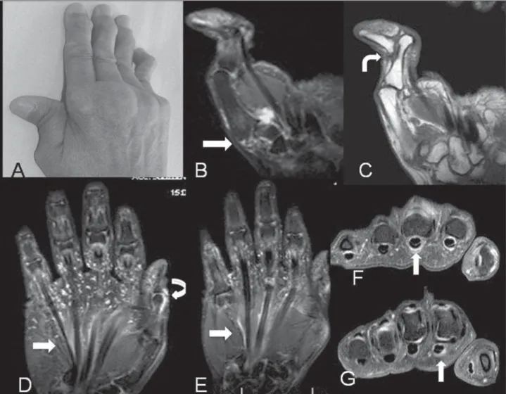
synovial and tendinous involvement that is typical of this condition. In spite of the typi-cal presence of synovial thickening and post-contrast enhancement, the exuberant pannus caused by RA is not characterized, in general with a more subtle involvement being observed, even in the most deform-ing presentations of the disease. In 2003, Ostendorf et al. demonstrated by means of MRI the presence of edematous tenosyno-vitis and synotenosyno-vitis in the hands of most of their 14 lupus patients, three of them de-fined as JA(24). In other recent study
devel-oped in the authors’ institution(25), 20
pa-tients who had met the criteria for JA of the hands were submitted to MRI and the pres-ence of flexor tenosynovitis was demon-strated in more than 90% of the cases, as
well as some degree of subarticular erosion in about 50% of the patients. This demon-strates that the absence of erosion may not be a pathognomonic finding of JA, as pre-viously thought under the radiological point of view, and that new imaging meth-ods can provide relevant information on this pattern of involvement not only in the assessment of hands but also of other joints (Figure 3).
Ultrasonography is a good method for recognizing and following-up synovitis, tenosynovitis and erosion, but it has been underutilized and is poorly recognized in the radiological community as a method to evaluate rheumatological diseases in gen-eral.
Rhupus hand and mild deforming arthropathy
Despite its usual non-erosive nature, some patients develop an erosive form of disease similar to RA. The term rhupus has started being employed to describe this condition because, generally, such patients meet simultaneously the criteria for classi-fication of both SLE and RA(26). There has
been a lot of discussion about to which extent ruphus hand represents a subgroup of lupus arthropathy or an association be-tween both diseases. The possibility of overlapping between these two diseases has been estimated in 1%(27). From the
imaging point of view, the condition pre-sents the typical rheumatoid arthritis
mities with erosive involvement generally already identified at plain radiography, similarly to the findings of RA.
Mild deforming arthropathy would be a deforming, but more subtle, modality of involvement, without erosion and not meeting the criteria for classification as JA. Ulnar deviation and “swan-neck” defor-mity predominate, with no defordefor-mity at the level of the thumb and in the feet. Such sub-group has not been unanimously recog-nized by all the authors.
INFECTION
Infectious involvement is observed in the course of the disease, particularly in the urinary and respiratory tracts, with a preva-lence > 50%, constituting one of the main causes of death and hospital admissions among these patients(28). From the
muscu-loskeletal point of view, septic arthritis and osteomyelitis predominate, although not much frequently. Articular involvement may be observed either in a single or in multiple joints, and is primarily caused by gram-negative bacteria, particularly species
of Salmonellae and Staphylococcus
aureus, generally of hematogenic origin(29).
As regards osteomyelitis, its pathogenesis
in this context is also multifactorial and is related to the infectious organism viru-lence, to the underlying disease, to the patient’s immunological status as well as to the type and location of the involved bone, with S. aureus as the main causative agent in these cases(30). So, the distribution of the
infectious agents resembles the one ob-served in sickle-cell disease(31).
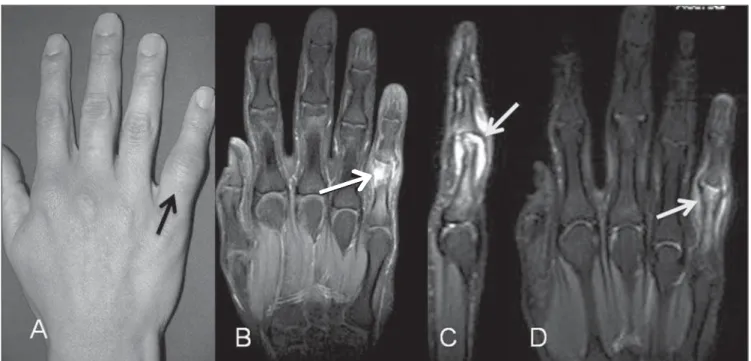
Because of the chronic use of corticoids in such patients, the signs and symptoms of infection are frequently masked and the process generally presents a chronic and indolent course. In case of persistent monoarticular arthritis, the absence of a clinical response to the therapy with corti-coids or other immunosuppressive drugs should raise the suspicion of an underlying infectious process. Depending on the phase of the disease, the different imaging meth-ods can recognize such complications and the radiologist plays a critical role in the identification of these cases (Figure 4).
SPONTANEOUS TENDINOUS RUPTURE
Spontaneous tendinous rupture is a rare clinical condition. Early in the last century, McMaster demonstrated that an artificial
rupture around 75% of the tendinous thick-ness would be unlikely to determine a full thickness rupture in the absence of a basal pathological process(32). Collagen diseases
and use of corticoids, whether in associa-tion or not, would be the necessary condi-tions. In RA, for example, such complica-tion is known and is particularly related to the local mechanical alteration as a result of bone erosion determining a secondary tendinous laceration.
In lupus patients, the etiology of this condition is still to be completely known and would be related to local trauma, basal chronic inflammatory process and use of corticoids(33), and no data on the actual
in-cidence of this alteration is found in the literature. Contrary to RA, where tendinous ruptures occur almost always in the hands, in SLE such ruptures are most frequently seen in the lower limbs, affecting the quad-riceps and, particularly, the patellar and Achilles tendons, also in association with a mechanical component. Corticoid therapy is known to be a predisposing factor and is present in almost all the reports in the lit-erature. Corticoids action is related to an antimycotic effect and fibroblasts inhibi-tion, with collagenase stimulus and conse-quential structural fiber disorganization(34).
Figure 4. Septic arthritis of proximal interphalangeal joint of the fifth finger. Lupus patient with pain and focal increase in volume (A). Coronal and sagittal MRI T2-weighted images with fat suppression (B,C) demonstrating medullary edema in bone borders (black arrow on B) and strain caused by articular capsule fluid in the fifth finger proximal interphalangeal joint (white arrow on C). On the sagittal image, erosion of the corresponding proximal phalanx is also well characterized. Coronal, contrast-enhanced MRI T1-weighted image (D) demonstrating contrast enhancement surrounding the bone, synovia and soft tissues of this area.
→
→
→
Recent evidences demonstrate that this type of SLE complication may be related to a predominance of the primary inflam-matory process in the tendinous sheaths leading to a focal tendinous weakness and later rupture(25). A recent systematic review
developed in the authors’ institution has demonstrated that JA is present in at least 35% of cases of spontaneous tendinous rupture in patients with SLE (35).
OSTEONECROSIS
Osteonecrosis is a cause of morbidity and dysfunction in lupus patients with a variable incidence according to the differ-ent authors, with a prevalence ranging from 2% to 30%(36). Generally, osteonecrosis
affects multiple sites, most frequently
in-volving the femoral head (> 70% of cases) and should be always considered in cases where other areas of infarct or necrosis are identified(37). In the context of the disease,
its etiology is multifactorial and still re-mains controversial in many cases, with several potential risk factors being consid-ered such as presence of Raynaud’s phe-nomenon, vasculitis, hyperlipidemia and, probably, presence of antiphospholip-ides(38). However, high-dose corticoid
therapy (> 20 mg/day) is undoubtedly the mail determining factor(39). In lupus
pa-tients, osteonecrosis determines pain pre-viously to the joint destruction, differently from RA that occurs synchronically with joint destruction by the synovial inflamma-tory disease, affecting both the femoral head and the acetabular margin.
Plain radiography is generally normal in the early phase of the disease, and the pres-ence of subchondral sclerosis already infers irreversible joint damage. Magnetic reso-nance imaging is the method of choice in the early diagnosis of osteonecrosis, allowing the application of therapeutic intervention(40)
aimed at preventing articular collapse and the secondary degenerative disease that is the most common complication (Figure 5). Computed tomography and scintigraphy are less accurate methods and do not iden-tify lesions in the early phase of the disease.
MYOSITIS AND SOFT TISSUE CALCIFICATIONS
In 5% to 10% of lupus patients inflam-matory myopathy is also observed, but
laboratory findings of muscular disease may be present in up to 50% of cases(41).
The pattern of inflammatory myositis asso-ciated with SLE is similar to the one of id-iopathic myositis that, many times, is as severe as the primary form of the disease, making it difficult to define it as myositis secondary to SLE or as an overlapping between SLE and classical myositis(42).
Myopathy associated with use of drugs would be even more common in SLE. Ad-ditionally to corticoids which are the drugs most frequently associated with non-in-flammatory myopathy, some rare cases of myopathy secondary to the use of chloro-quine have been described(43). The
diagno-sis is generally achieved by means of clini-cal examination and laboratory tests, rarely requiring confirmation by muscular biopsy. As in the cases of classical myositis, MRI is the imaging method of choice, in spite of its low specificity. This method aids in the differential diagnosis, follow-up of therapeutic response, and is useful to de-fine the biopsy site. Typically, MRI dem-onstrates increased signal intensity on T2-weighted and STIR sequences as a result of the intracellular increase in the amount of fluid or inflammatory infiltrate associated with the increase in muscle volume(44). In
2000, a Brazilian study with 13 lupus pa-tients assessed by MRI demonstrated prin-cipally predominance of muscle atrophy(45).
As in other collagen diseases, soft tis-sue calcifications are also identified in SLE, but they are poorly frequent in these cases(46,47)
.Precipitating factors like
nephri-tis, use of alphacalcidol and even diuretic drugs have been recently related to this condition(48).
The radiological study clearly demon-strates such calcifications and, in case of dubious diagnosis, additionally to calcifi-cations, MRI can identify an eventual as-sociation with inflammatory involvement of soft tissues.
INSUFFICIENCY FRACTURE
Many factors such as renal failure, amenorrhea, early menopause, chronic in-flammatory cytokines and mainly chronic use of corticoids are involved in the gen-esis of osteoporosis in SLE(49). Particularly
in patients with SLE, the latter is a
deter-Figure 6. Acute insufficiency fracture of vertebral body. A 47-year-old lupus patient under corticoid therapy. Sagittal MRI T1-weighted (A) and FSE T2-weighted (B) images demonstrate partial T12 upper plateau collapse (arrows). The edema becomes better characterized on the STIR sequence (C), associated with a local reactional inflammatory process with contrast uptake on the T1-weighted image (D).
mining factor in the development of insuf-ficiency fractures of the spine and other sites (particularly lower limbs), with preva-lence of upper osteoporotic vertebral frac-tures of > 20% in a recent study(50). This
finding demonstrates the necessity that special attention is paid in the imaging, particularly radiological (that is the method most frequently utilized) evaluation of these patients, appreciating the findings of osteopenia and the vertebral bodies mor-phology. Magnetic resonance imaging is the method of choice in the early phases of the disease, when conventional radiogra-phy is not a diagnostic method yet. Classi-cal findings of medullary bone edema are observed on the sequences with fat sup-pression, with linear areas of low signal intensity inside (Figure 6). Scintigraphy is also sensitive in the detection of such frac-tures, but it is not always very specific.
DISCUSSION
Musculoskeletal involvement is fre-quently observed in lupus disease and may be an early indicator of disease activity. The degree of involvement may range from a subtle arthralgia to marked presentations of deforming arthropathy, besides tendinous ruptures and other forms of involvement such as myopathy, osteonecrosis,
insuffi-ciency fractures and infection, the latter being most frequently associated with the adopted treatment.
Although deforming arthritis (JA) is classically reversible, it is important to highlight its relevance in the musculoskel-etal involvement of SLE, as it may be largely confused with the clinical picture of RA. In this context, the radiologist plays an extremely relevant role, calling the atten-tion of the clinician to this hypothesis rep-resented by plain radiographic findings corresponding to significant deformities and subluxation and the virtual absence of erosions.
Another significant contribution of the radiological evaluation of the musculosk-eletal system in patients with SLE was achieved with a more frequent utilization of MRI so that the development of avascu-lar necrosis secondary to prolonged corti-coid therapy is early detected, allowing the adoption of measures aimed at avoiding bone collapse.
evaluating the relevance of ultrasonogra-phy for patients with JA, besides determin-ing the distribution and severity of alter-ations in these patients as compared with lupus patients with arthritis, but without JA.
Thus, with a deeper knowledge on ra-diological findings in patients with SLE, the radiologist takes over a differentiated role in the follow-up of these patients as he/she already does in cases of rheumato-logical conditions such as RA.
REFERENCES
1. Lebowitz R, Schumacher HR Jr. Articular mani-festations of systemic lupus erythematosus. Ann Intern Med. 1971;74:911–21.
2. Tan EM, Cohen AS, Fries JF, et al. The 1982 re-vised criteria for the classification of systemic lupus erythematosus. Arthritis Rheum. 1982;25: 1271–7.
3. Dubois EL, Tuffanelli DL. Clinical manifestations of systemic lupus erythematosus. JAMA. 1964; 190:104–11.
4. Reilly PA, Evison G, McHugh NJ, et al. Arthropa-thy of hands and feet in systemic lupus erythema-tosus. J Rheumatol. 1990;17:777–84. 5. Aptekar RG, Lawless OJ, Decker JL. Deforming
non-erosive arthritis of the hand in systemic lu-pus erythematosus. Clin Orthop Relat Res. 1974; (100):120–4.
6. Sierra-Jimenez G, Sanchez-Ortiz A, Aceves-Avila FJ, et al. Tendinous and ligamentous derange-ments in systemic lupus erythematosus. J Rheu-matol. 2008;35:2187–91.
7. Bywaters EGL. Jaccoud’s syndrome: a sequel to the joint involvement of systemic lupus erythe-matosus. Clin Rheum Dis. 1975;1:125–48. 8. van Vugt RM, Derksen RH, Kater L, et al.
De-forming arthropathy or lupus and rhupus hands in systemic lupus erythematosus. Ann Rheum Dis. 1998;57:540–4.
9. Manthorpe R. The man behind the syndrome. Sigismond Jaccoud. With his 23d lecture he be-came part of medical history. Lakartidningen. 1992;89:1585–6.
10. Amano H, Furuhata N, Tamura N, et al. Hypo-complementemic urticarial vasculitis with Jaccoud’s arthropathy and valvular heart disease: case report and review of the literature. Lupus. 2008;17:837–41.
11. Ballard M, Meyer O, Adle-Biassette H, et al. Jaccoud’s arthropathy with vasculitis and primary Sjögren’s syndrome. A new entity. Clin Exp Rheumatol. 2006;24(2 Suppl 41):S102–3. 12. Ben Miled-M’Rad K, M’Rad S, Kchir M, et al.
Caroli’s disease and Jaccoud’s arthropathy. Ann Gastroenterol Hepatol (Paris). 1993;29:107–9. 13. Bradley JD, Pinals RS. Jaccoud’s arthropathy in
scleroderma. Clin Exp Rheumatol. 1984;2:337– 40.
14. Conrozier T, Balblanc JC, Chapard R, et al. Jaccoud’s arthritis and angioimmunoblastic
lym-phadenopathy. Rev Rhum Mal Osteoartic. 1990; 57:423–5.
15. Santiago MB, Galvão V. Jaccoud arthropathy in systemic lupus erythematosus: analysis of clini-cal characteristics and review of the literature. Medicine (Baltimore). 2008;87:37–44. 16. Caznoch CJ, Esmanhotto L, Silva MB, et al.
Pa-drão de comprometimento articular em pacientes com lúpus eritematoso sistêmico e sua associa-ção com presença de fator reumatóide e hipere-lasticidade. Rev Bras Reumatol. 2006;46:261–5. 17. De la Sota M, Maldonado Cocco JA. Jaccoud’s arthropathy in knees in systemic lupus erythema-tosus. Clin Rheumatol. 1989;8:416–7. 18. Siam AR, Hammoudeh M. Jaccoud’s arthropathy
of the shoulders in systemic lupus erythematosus. J Rheumatol. 1992;19:980–1.
19. Morley KD, Leung A, Rynes RI. Lupus foot. Br Med J (Clin Res Ed). 1982;284:557–8. 20. Spronk PE, ter Borg EJ, Kallenberg CG. Patients
with systemic lupus erythematosus and Jaccoud’s arthropathy: a clinical subset with an increased C reactive protein response? Ann Rheum Dis. 1992;51:358–61.
21. Paredes JG, Lazaro MA, Citera G, et al. Jaccoud’s arthropathy of the hands in overlap syndrome. Clin Rheumatol. 1997;16:65–9.
22. Galvão V, Atta AM, Sousa Atta ML, et al. Profile of autoantibodies in Jaccoud’s arthropathy. Joint Bone Spine. 2009;76:356–60.
23. Pastershank SP, Resnick D. “Hook” erosions in Jaccoud’s arthropathy. J Can Assoc Radiol. 1980; 31:174–5.
24. Ostendorf B, Scherer A, Specker C, et al. Jaccoud’s arthropathy in systemic lupus erythematosus: dif-ferentiation of deforming and erosive patterns by magnetic resonance imaging. Arthritis Rheum. 2003;48:157–65.
25. Ribeiro DS, Galvão V, Fernandes JL, et al. Mag-netic resonance imaging of Jaccoud’s arthropathy in systemic lupus erythematosus. Joint Bone Spine. 2010;77:241–5.
26. Fernández A, Quintana G, Rondón F, et al. Lupus arthropathy: a case series of patients with rhupus. Clin Rheumatol. 2006;25:164–7.
27. Mawson AR. Are rheumatoid arthritis and sys-temic lupus erythematosus inversely related dis-eases? Med Hypotheses. 1985;18:377–86. 28. Petri M. Infection in systemic lupus
erythemato-sus. Rheum Dis Clin North Am. 1998;24:423–56. 29. Chen JY, Luo SF, Wu YJ, et al. Salmonella septic arthritis in systemic lupus erythematosus and other systemic diseases. Clin Rheumatol. 1998; 17:282–7.
30. Cuchacovich R, Gedalia A. Pathophysiology and clinical spectrum of infections in systemic lupus erythematosus. Rheum Dis Clin North Am. 2009; 35:75–93.
31. Epss CH, Bryant DD, Coles MJ, et al. Osteomy-elitis in patients who have sickle-cell disease. Di-agnosis and management. J Bone Joint Surg Am. 1991;73:1281–94.
32. McMaster PE. Tendon and muscle rupture. Clini-cal and experimental studies on the causes and lo-cation of subcutaneous ruptures. J Bone Joint Surg. 1933;15:705–22.
33. Kissel CG, Sundareson AS, Unroe BJ. Spontane-ous Achilles tendon rupture in a patient with sys-temic lupus erythematosus. J Foot Surg. 1991;30: 390–7.
34. Houck JC, Patel YM. Proposed mode of action of corticosteroids on the connective tissue. Nature. 1965;206:158–60.
35. Alves EM, Macieira JC, Borba E, et al. Sponta-neous tendon rupture in systemic lupus erythema-tosus: association with Jaccoud’s arthropathy. Lupus. 2010;19:247–54.
36. Mont MA, Glueck CJ, Pacheco IH, et al. Risk fac-tors for osteonecrosis in systemic lupus erythe-matosus. J Rheumatol. 1997;24:654–62. 37. Cozen L, Wallace DJ. Avascular necrosis in
sys-temic lupus erythematosus: clinical associations and a 47-year perspective. Am J Orthop (Belle Mead NJ). 1998;27:352–4.
38. Campos LM, Kiss MH, D’Amico EA, et al. Antiphospholipid antibodies and antiphospho-lipid syndrome in 57 children and adolescents with systemic lupus erythematosus. Lupus. 2003; 12:820–6.
39. Zizic TM, Marcoux C, Hungerford DS, et al. Cor-ticosteroid therapy associated with ischemic ne-crosis of bone in systemic lupus erythematosus. Am J Med. 1985;79:596–604.
40. Alves EM, Angrisani AT, Santiago MB. The use of extracorporeal shock waves in the treatment of osteonecrosis of the femoral head: a systematic review. Clin Rheumatol. 2009;28:1247–51. 41. Isenberg D. Myositis in other connective tissue
disorders. Clin Rheum Dis. 1984;10:151–74. 42. Garton MJ, Isenberg DA. Clinical features of
lu-pus myositis versus idiopathic myositis: a review of 30 cases. Br J Rheumatol. 1997;36:1067–74. 43. Richter JG, Becker A, Ostendorf B, et al. Differ-ential diagnosis of high serum creatine kinase levels in systemic lupus erythematosus. Rheu-matol Int. 2003;23:319–23.
44. Adams EM, Chow CK, PremkumarA, et al. The idiopathic inflammatory myopathies: spectrum of MR imaging findings. Radiographics. 1995;15: 563–74.
45. Hilário MO, Yamashita H, Lutti D, et al. Juvenile idiopathic inflammatory myopathies: the value of magnetic resonance imaging in the detection of muscle involvement. Sao Paulo Med J. 2000;118: 35–40.
46. Souza RAS, Rangel LV, Souza HFS, et al. Lúpus eritematoso cutâneo e calcinose universalis. Rev Bras Reumatol. 2000;40:18–20.
47. Souza HFS, Souza RAS, Rangel LV, et al. Calci-ficação intracerebral em lúpus eritematoso sistê-mico. Rev Bras Reumatol. 2001;41:123–6. 48. Okada J, Nomura M, Shirataka M, et al.
Preva-lence of soft tissue calcifications in patients with SLE and effects of alfacarcidol. Lupus. 1999;8: 456–61.
49. Panopalis P, Yazdany J. Bone health in systemic lupus erythematosus. Curr Rheumatol Rep. 2009; 11:177–84.