Prevalence of Hypertrophic Cardiomyopathy in
Fetuses of Mothers with Gestational Diabetes
before Initiating Treatment
Prevalência de miocardiopatia hipertró
fi
ca em
fetos de mães com diabetes gestacional antes do
tratamento
Carolina Rossi Palmieri
1Mona Adalgisa Simões
2Jean Carl Silva
3Anelise Darabas dos Santos
4Mariana Ribeiro e Silva
4Bruna Ferreira
41Health and Environment Master Program, Universidade da Região de Joinville (Univille), Joinville, Santa Catarina, Brazil
2Department of Neonatology, Maternidade Darcy Vargas, Joinville, Santa Catarina, Brazil
3Department of Medicine, Universidade da Região de Joinville (Univille), Joinville, Santa Catarina, Brazil
4Universidade da Região de Joinville (Univille), Joinville, Santa Catarina, Brazil
Rev Bras Ginecol Obstet 2017;39:9–13.
Address for correspondence Carolina Rossi Palmieri, Programa de Pós-graduação em Saúde e Meio Ambiente, Universidade da Região de Joinville (Univille), Rua Blumenau, 26, bairro América, Joinville/SC, CEP 89204-248, Brazil (e-mail: [email protected]).
Keywords
►
gestational diabetes
►
fetal
echocardiography
►
fetus
►
heart diseases
►
hypertrophic
cardiomyopathy
Abstract
Objective
To evaluate the prevalence of hypertrophic cardiomyopathy (HCM) in fetuses of
pregnant women with gestational diabetes mellitus (GDM) in the beginning of the
treatment.
Methods
A cross-sectional study was performed between July 1, 2013, and
Decem-ber 20, 2013, in a public maternity clinic in southern Brazil. The subjects were 63
fetuses of mothers with gestational diabetes, with a single pregnancy and no other
associated pathologies. We diagnosed HCM through a fetal echocardiography before
treatment and evaluated the maternal and fetal characteristics.
Results
The average age of the pregnant women was 32.32 (
6.2) years, and the average
gestational age at the time of the evaluation was 30.59 (
2.27) weeks. The interventricular
septum thickness showed a standard deviation of more than two in 50.8% of the fetuses
(95% con
fi
dence interval [95%CI]: 38.1
–
63.5%). The left ventricular wall thickness showed a
standard deviation of more than 2 in 13 (20.6%) fetuses (95%CI: 11.1
–
30.2%). The HCM was
con
fi
rmed in 54% of the fetuses (95%CI: 41.3
–
65.1%). The fetal abdominal circumference
was normal in 46 (73%) fetuses, and 50% of these fetuses had HCM.
Conclusion
The prevalence of hypertrophic cardiomyopathy in fetuses of pregnant
women with GDM before treatment was of 54% (95%CI: 41.3
–
65.1%).
received
July 5, 2016
accepted
December 15, 2016
DOIhttp://dx.doi.org/ 10.1055/s-0037-1598602.
ISSN 0100-7203.
Copyright © 2017 by Thieme-Revinter Publicações Ltda, Rio de Janeiro, Brazil
Introduction
Gestational diabetes mellitus (GDM) is defined as glucose intolerance with onset or diagnosis during pregnancy. It affects7% of all pregnancies, resulting in over 200 thou-sand cases per year.1
Previous studies have shown that maternal hyperglyce-mia may lead to fetal hypertrophic cardiomyopathy (HCM)2 and cardiac diastolic function impairment,3 among other alterations.
Hypertrophic cardiomyopathy is characterized by an in-creased ventricular wall thickness, which leads to a decrease in the left ventricular cavity size, to a potential reduction in the ventricular systolic performance, and to the impairment of the diastolic function because of the decreased ventricular filling. These alterations can lead to heart failure and death.4 Cardiac complications due to congenital heart malforma-tion and ventricular hypertrophy are the major causes of morbidity and mortality in fetuses and newborns of mothers with GDM.5
The incidence of HCM, especially interventricular septal hypertrophy (IVSH), varies between 10% and 71%.6–8 An increase in the ventricular wall thickness may also be involved in the cardiac changes observed in fetuses of mothers with GDM, but septal hypertrophy is more studied because of the higher number of insulin receptors in the septum of the heart.9
Prenatal HCM is diagnosed through intrauterine two-di-mensional echocardiography, and, based on the results, physi-cians may provide appropriate postnatal care and follow-up for cardiac hypertrophy progression during the gestational
period.10 Some authors claim that fetal echocardiography should be recommended to all pregnant women with GDM.11 The aim of the present study was to assess the prevalence of HCM in fetuses of pregnant women with GDM before their treatment using fetal echocardiography.
Methods
The present cross-sectional study included 63 fetuses of mothers with GDM, and was conducted before the mothers initiated their treatment in a public maternity clinic at the southern region of Brazil between July 1 2013 and Decem-ber 20 2013.
The study was conducted according to the local regula-tions for good clinical practices, specifically those of the resolution of the Brazilian National Health Council (CNS, in the Portuguese acronym, resolution no. 466/12), after ap-proval from the Research Ethics Committee (CEP011/12) of the institution. The principal researcher and collaborators were responsible for providing information to the patients. None of the authors were associated with the companies that provided the equipment or their competitors, and did not receive any kind of support.
The study included singleton fetuses of pregnant women diagnosed with GDM, without malformations and other diseases that could interfere with the fetal development. The diagnosis of GDM was based on the criteria provided by the American Diabetes Association, that is, blood glucose level92 mg/dl (fasting),180 mg/dl (1 hour after plasma glucose) and153 mg/dl (2 hours after plasma glucose), and an ultrasound was performed between the 24th and
Palavras chave
►
diabetes gestacional
►
ecocardiogra
fi
a fetal
►
feto
►
cardiopatias
►
miocardiopatia
hipertró
fi
ca
Resumo
Objetivo
Avaliar a prevalência de miocardiopatia hipertró
fi
ca em fetos de gestantes
com diabetes mellitus gestacional antes do início do tratamento.
Métodos
Foi realizado um estudo de corte transversal, no período de 1
ode julho
de 2013 até 20 de dezembro de 2013, em uma maternidade pública. Foram objetos
do estudo 63 fetos de gestantes portadoras de diabetes mellitus gestacional (DMG),
em gestação única e sem outras patologias associadas. Foi realizada ecocardiogra
fi
a
fetal antes do início do tratamento do diabetes. O diagnóstico de miocardiopatia
hipertró
fi
ca (MH) foi realizado quando a medida do septo interventricular ou da parede
ventricular estava superior a dois desvios-padrão. O desfecho primário avaliado foi
presença de MH.
Resultados
As gestantes apresentavam idade média de 32,32 (
6,2) anos, e a idade
gestacional média no momento da avaliação foi de 30,59 (
2,27) semanas. A medida
do septo interventricular estava acima de 2 desvios-padrão em 50,8% (intervalo de
con
fi
ança de 95% [IC95%]: 38,1
–
63,5%). A parede do ventrículo esquerdo estava acima
de 2 desvios-padrão em 13 fetos, totalizando 20,6% (IC95%: 11,1
–
30,2%). A MH estava
presente em 54% dos fetos (IC95%: 41,3
–
65,1%). A circunferência abdominal fetal
estava normal em 46 (73%) fetos, e destes, 50% apresentavam MH.
28th gestational weeks. Fetuses diagnosed with malforma-tions after their inclusion in the study were then excluded.
The fetal abdominal circumference (FAC) was determined as the average of three measurements obtained at the stomach, umbilical vein, and liver levels. The FAC percentile for the gestational age was calculated according to the method reported by Hadlock et al.12Interventricular septum and left ventricular wall thickness were determined through fetal Doppler echocardiography using two-dimensional se-quential analysis for a longitudinal transverse section at the left atrioventricular (bicuspid) valve tip level using the M-mode.13The test was performed before the patients initiated the treatment for GDM.14Septum and/or left ventricular wall thickness values showing a standard deviation of more than 2 according to the gestational age were considered abnormal, that is, consistent with thefindings of HCM.15–17The fetal echocardiographs were obtained by the same technician using the HD 7XE (Philips®, Amsterdam, Netherlands) equipment.
The maternal data assessed were age, parity, pre-pregnancy body mass index (BMI¼W/H2), and gestational age at the time of inclusion in the study. The fetal data assessed were FAC percentile and interventricular septum and left ventricular wall thickness. The primary and secondary outcomes assessed were the presence of HCM and FAC respectively.
All variables were descriptively analyzed, with quantita-tive variables expressed as means and standard deviations.
Data were statistically analyzed using the Statistical Pack-age for the Social Sciences (SPSS, IBM-SPSS, Inc., Chicago, IL, EUA) software, version 21.0. Continuous (quantitative) varia-bles were expressed as means and standard deviations. Cate-gorical (qualitative) variables were expressed as absolute and relative frequencies. The 95% confidence interval (95%CI) for the frequency was calculated using the one-samplet-test.
Results
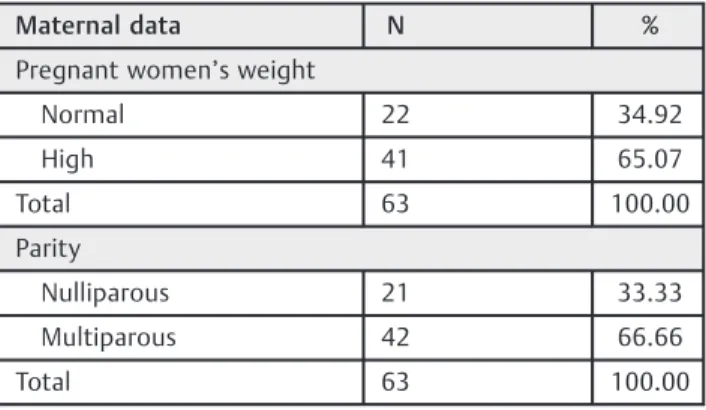
Sixty-three pairs (pregnant woman and her fetus) were selec-ted, and none was excluded. The average age of the pregnant women was 32.32 (6.2) years, and the average gestational age at the time of the evaluation was 30.59 (2.27) weeks. The majority of the pregnant women had multiple gestations (66.66%), and a pre-pregnancy BMI>25 kg/m2 (65.07%); the average BMI was 27.33 (5.64) kg/m2. Maternal data are shown in►Table 1.
The interventricular septum thickness showed a standard deviation of more than 2 in 50.8% of the fetuses, between 1 and 2 in 38.09% of the fetuses, and less than one in 11.11% of the fetuses (95%CI: 38.1–63.5%). The left ventricular wall thickness showed a standard deviation of more than 2 in 13 fetuses (20.6%) (95%CI: 11.1–30.2%), and HCM was confirmed in 54% of the fetuses (95%CI: 41.3–65.1%). Only 2 (5.88%) of the 34 fetuses with HCM exhibited alterations in the left ventricular wall, but none of them exhibited alterations in the septum.
The FAC was normal (between the 10th and the 75th percentiles) in most fetuses (73.01%), and it increased in 26.99% of them. There were no cases with a FAC below the
10th percentile. Half of fetuses with normal abdominal circumferences exhibited HCM. The majority of the fetuses (64.7%) with a FAC greater than the 75th percentile exhibited HCM. Fetal data are shown in►Table 2.
Discussion
Cardiomyopathies represent 8–11% of the cardiovascular abnormalities diagnosed during pregnancy. After birth, car-diomyopathies are diagnosed in only 3% of newborns with cardiovascular diseases, and diastolic dysfunction is associ-ated with a higher risk of postnatal mortality.18
In a previous study that used septal and left ventricular wall thickness measures at birth as reference, it was reported that these values were increased in 69% of the fetuses of mothers with GDM;17 this value is slightly above the CI Table 1 Epidemiological data on pregnant women with gestational diabetes mellitus
Maternal data N %
Pregnant women’s weight
Normal 22 34.92
High 41 65.07
Total 63 100.00
Parity
Nulliparous 21 33.33
Multiparous 42 66.66
Total 63 100.00
Note:Absolute number (N) and percentage (%).
Table 2 Fetal data on interventricular septum, left ventricular wall, and abdominal circumference measurements
Fetal data N %
Interventricular septum
<1 SD 7 11.1
SD between 1 and 2 24 38.0
>2 SD 32 50.8
Total 63 100.0
LV wall
>2 SD 13 20.6
Fetal AC
Normal 46 73.0
Increased 17 27.0
Total 63 100.0
Abbreviations: AC, abdominal circumference; LV, left ventricular; SD, standard deviation.
obtained in our study. In the present study, we found that 54% (95%CI: 41.3–65.1%) of the fetuses evaluated before the treatment for GDM were positive for HCM.
Hypertrophic cardiomyopathy affects the right ventricle and the posterior left ventricular wall, but the septal hyper-trophy is more evident because of the large number of insulin receptors in the septum of the heart.9,19,20 In our study, we observed that only 2 of the 34 fetuses with HCM exhibited alterations in the left ventricular wall, but none of them exhibited alterations in the septum.
The fetuses of mothers with GDM are at a risk of develop-ing HCM even when the mothers exhibit good capillary glycemic control.16,21Other authors have also not found a relationship between the glycemic control detected using maternal glycosylated hemoglobin and the presence of HCM.22,23These results show the limited existing knowledge about this disease.
Hyperinsulinemia is related to fetal macrosomia, and an increased FAC is directly related to higher fetal weight, and fetal weight is related to HCM. Moreover, the rate of HCM incidence in newborns that are large for their gestational age (LGA) can reach 38%.24 Considering that fetuses with FAC above the 75th percentile tend to be LGA, we found a higher percentage of HCM (64.7%) in these fetuses.
In our study, the fetuses were evaluated before initiating the treatment for GDM. Therefore, we do not know the effect of this treatment on HCM; a potential reduction would justify an incidence decline from 54% to 20%2after delivery.
Hypertrophic cardiomyopathy was also observed in 50% of the fetuses with normal FAC, which reflects the occurrence of interventricular septum alterations prior to FAC altera-tions. This result also suggests the diagnosis of poor maternal glycemic control even before FAC alterations.
Information on the postnatal consequences of HCM is insufficient in the current literature.21There are evidences for the effects of diabetes on the cardiac function of adult patients.25The abnormalities and injuries studied in adults are not easy to assess in fetuses because there is no evidence of the effects after the short period of exposure during pregnancy.26Furthermore, a large majority of newborns is asymptomatic at birth, and if hypertrophy is present, it spontaneously regresses in a few months.18
Routine referral to fetal echocardiography for pregnant women with a history of diabetes mellitus has already been established according to some authors;24the severity of the disease has also been established in this group.4 Some authors recommend that all pregnant women with GDM be submitted to fetal echocardiography until a group with a higher risk for HCM is identified.11
Because of the small sample size of the present study, several inferences could not be drawn from the results; however, the high prevalence and early occurrence of HCM in this population indicate that this is one of thefirst effects of maternal diabetes on fetuses. This observation justifies further assessments of this disease, including its onset, manifestations, behavior during maternal treatment, and the perinatal and adult repercussions.
Conflicts of Interest
The research was conducted at the Neonatology Centre of Maternidade Darcy Vargas, in Joinville, Santa Catarina, Brazil. Authors have no conflicts of interest to declare.
References
1 American Diabetes Association. Diagnosis and classification of diabetes mellitus. Diabetes Care 2012;35(Suppl 1):S64–S71
2 Åman J, Hansson U, Östlund I, Wall K, Persson B. Increased fat mass and cardiac septal hypertrophy in newborn infants of mothers with well-controlled diabetes during pregnancy. Neo-natology 2011;100(02):147–154
3 Jaeggi ET, Fouron JC, Proulx F. Fetal cardiac performance in uncomplicated and well-controlled maternal type I diabetes. Ultrasound Obstet Gynecol 2001;17(04):311–315
4 Park MK. Pediatric cardiology. 4th ed. St. Louis: Mosby; 2002. Primary myocardial disease; p. 267–392
5 Demiroren K, Cam L, Oran B, et al. Echocardiographic measure-ments in infants of diabetic mothers and macrosomic infants of nondiabetic mothers. J Perinat Med 2005;33(03):232–235
6 Stuart A, Amer-Wåhlin I, Gudmundsson S, Marsál K, Thuring A, Källen K. Ductus venosus bloodflow velocity waveform in dia-betic pregnancies. Ultrasound Obstet Gynecol 2010;36(03): 344–349
7 Dimitriu AG, Russu G, Stamatin M, Jităreanu C, Streangă V. [Clinical and developmental aspects of cardiac involvement in infant of diabetic mother]. Rev Med Chir Soc Med Nat Iasi 2004; 108(03):566–569
8 Hagemann LL, Zielinsky P. [Prenatal study of hypertrophic cardi-omyopathy and its association with insulin levels in fetuses of diabetic mothers]. Arq Bras Cardiol 1996;66(04):193–198
9 Fouda UM, Abou ElKassem MM, Hefny SM, Fouda RM, Hashem AT. Role of fetal echocardiography in the evaluation of structure and function of fetal heart in diabetic pregnancies. J Matern Fetal Neonatal Med 2013;26(06):571–575
10 García-Díaz L, Coserria F, Antiñolo G. Hypertrophic cardiomyo-pathy due to mitochondrial disease: prenatal diagnosis, manage-ment, and outcome. Case Rep Obstet Gynecol 2013;2013:472356 11 Zielinsky P. Role of prenatal echocardiography in the study of hypertrophic cardiomyopathy in the fetus. Echocardiography 1991;8(06):661–668
12 Hadlock FP, Deter RL, Harrist RB, Park SK. Estimating fetal age: computer-assisted analysis of multiple fetal growth parameters. Radiology 1984;152(02):497–501
13 Allan L. Technique of fetal echocardiography. Pediatr Cardiol 2004;25(03):223–233
14 Hendler I, Blackwell SC, Bujold E, et al. Suboptimal second-trimester ultrasonographic visualization of the fetal heart in obese women: should we repeat the examination? J Ultrasound Med 2005;24(09):1205–1209, quiz 1210–1211
15 Lopes L. Ecocardiografia fetal. São Paulo: Revinter; 2015 16 Zielinsky P, Piccoli AL Jr. Myocardial hypertrophy and dysfunction
in maternal diabetes. Early Hum Dev 2012;88(05):273–278
17 Han SS, Wang G, Jin Y, et al. Investigating the mechanism of hyperglycemia-induced fetal cardiac hypertrophy. PLoS One 2015;10(09):e0139141
18 Pedra SR, Smallhorn JF, Ryan G, et al. Fetal cardiomyopathies: pathogenic mechanisms, hemodynamic findings, and clinical outcome. Circulation 2002;106(05):585–591
19 Garg S, Sharma P, Sharma D, Behera V, Durairaj M, Dhall A. Use of fetal echocardiography for characterization of fetal cardiac struc-ture in women with normal pregnancies and gestational diabetes mellitus. J Ultrasound Med 2014;33(08):1365–1369
21 Garcia-Flores J, Jañez M, Gonzalez MC, Martinez N, Espada M, Gonzalez A. Fetal myocardial morphological and functional changes associated with well-controlled gestational diabetes. Eur J Obstet Gynecol Reprod Biol 2011;154(01):24–26
22 Vela-Huerta MM, Vargas-Origel A, Olvera-López A. Asymmetrical septal hypertrophy in newborn infants of diabetic mothers. Am J Perinatol 2000;17(02):89–94
23 Behle I, Zielinsky P, Zimmer LP, Pontremoli M, Risch JN. [Glycosylated hemoglobin levels and cardiac abnormalities in fetuses of diabetic mothers]. Rev Bras Ginecol Obstet 1998;20(05):237–243 Portuguese.
24 Vural M, Leke L, Mahomedaly H, Maingourd Y, Kremp O, Risbourg B. Should an echocardiographic scan be done routinely for infants of diabetic mothers? Turk J Pediatr 1995;37(04): 351–356
25 Fonarow GC. Managing the patient with diabetes mellitus and heart failure: issues and considerations. Am J Med 2004;116(Suppl A) 76S–88S