Laryngeal chondroradionecrosis following radiotherapy
Condroradionecrose de laringe após radioterapia
Giulianno Molina Melo, TCBC-SP1,2; Paula DeMeTrio Souza1,2; luiz CaSTro BaSToS Filho1; Murilo CaTaFeSTa neveS1; KleBer SiMõeS Do eSPiriTo SanTo3; onivalDo CervanTeS1; MárCio aBrahão1.
INTRODUCTION
T
he incidence of laryngeal cancer in 2012 was approximately 138,102 cases worldwide1,2. In theUS alone, the estimation was for 13,430 new cases in 2016, with a death toll of 3,620. In Brazil, laryngeal cancer represents 2% of all cancers, with 8,000 new cases per year, causing 3,000 annual deaths3,4.
It mainly affects men, aged 50-70, smokers and chronic alcoholics, but other etiologies such as HPV, pharyngo-laryngopharyngeal reflux and occupational risks are also described5-8. As to histopathology, 95%
of carcinomas are of squamous cells (SCC), followed by minor salivary glands carcinomas and sarcomas. Anatomically, 60% to 70% are glottic, 20% to 30%, supraglottic, and 5% are located in the sub-glottis9, 10.
Current forms of treatment for the patient with laryngeal carcinoma include surgery, radiotherapy and chemotherapy, associated or not. Radiotherapy is a treatment modality for several neoplasms, and can be indicated for three purposes: curative, palliative and symptomatic, the latter being less indicated. Compared with surgery, radiotherapy alone or in combination with chemotherapy was effective for the treatment of laryngeal cancer in its early stages, with similar disease-free survival, which in the 1990s gave rise to the
organ-preservation protocol that was widely used, including for advanced stages (III and IV) of the tumor. However, new evidence in the literature has shown that there is no benefit of radiotherapy at these advanced stages, both in overall survival and local control. On the contrary, there was an increase in treatment complications of a dysfunctionalized organ11, 12.
In particular for laryngeal cancer, radiotherapy causes damage and complications in almost 100% of irradiated patients, which are major or minor, transient or permanent, and may occur during the application or even months or years after the end of treatment13-15. Isolated
or associated radiotherapy inflicts alterations on the peri-laryngeal and peri-laryngeal tissues that cause local hypoxia, decrease the vascularization and the cellular population, with consequent decrease in the production of collagen and fibrin, favoring the appearance of fissures, fistulas and chronic wounds that do not heal. These changes, if left untreated and resolved, lead to the loss of the organ, with continuous broncho-aspiration, requiring rescue surgery, even in the absence of neoplasia12,13,16. Thus,
with the increase in the indication of radiotherapy, there was also an increase in the rate of its complications, including laryngeal edema with hoarseness, dyspnea and bronchoaspiration, radiodermatitis (grades I and II), skin necrosis (grade III), simple chondritis, chondritis with
1 - Federal University of São Paulo, Head and Neck Surgery Discipline, Department of Otorhinolaryngology and Head and Neck Surgery, São Paulo, SP, Brazil. 2 - São Paulo Portuguese Beneficence Hospital, Head and Neck Surgery Service, São Paulo, SP, Brazil. 3 - São Paulo Portuguese Beneficence Hospital, Pathology Service, São Paulo, SP, Brazil.
A B S T R A C T
Objective: to study larynx chondroradionecrosis related to radiotherapy and chemotherapy treatment and provide a treatment flow-chart. Methods: retrospective study with clinical data analysis of all larynx cancer patients admitted in a two tertiary hospital in a five years period. Results: from 131 patients treated for larynx cancer, 28 underwent chemoradiotherapy with curative intent and three of them presented chondroradionecrosis. They were treated with hiperbaric oxigen therapy and surgical debridment following our flowchart, preserving the larynx in all. Conclusions: the incidence of chondroradionecrosis as a complication of chemoradiotherapy in our series was 10,7% and the treatment with hiperbaric oxigen therapy, based in our flowchart, was effective to control this complication.
necrosis (chondroradionecrosis) and cartilage exposure to the airway.
Chondroradionecrosis as a complication has a variable incidence, ranging from 1% to 5.3%, resulting in rescue laryngectomies in up to 25% of cases17-19. It clinically progresses to dysphagia,
odynophagia, fistula formation, hoarseness, partial or total dysfunction of the larynx, loss of airway protection with airway obstruction and bronchospasm. Deemed a severe complication, it can lead to death if untreated, a complete recovery being rare19, 20. The
main risk factors for onset of chondroradionecrosis are persistence of smoking, postoperative infection, atherosclerosis, laryngopharyngeal reflux, diabetes and local trauma15, 21, 22.
To guide treatment, some authors proposed a classification of the laryngeal radionecroses based on symptoms, showing a direct relation between the amount of radiation and the severity of tissue reaction23,24. They noted that for grades I and II
the treatment was eminently clinical and that for grades III and IV, where chondritis was severe or chondroradionecrosis had already ensued, treatment was usually surgical (Tables 1 and 2). Some older articles postulate that once chondroradionecrosis occurs, treatment will always be surgical, through total laryngectomy, since there is no way to partially preserve the larynx20,22. However, recently other
authors have described conservative management of chondroradionecrosis with hyperbaric chamber (or Table 1. Chandler’s Classification for laryngeal radionecrosis
Symptoms Signs Treatment
Grade I Slight hoarseness Slight dryness
Slight edema Telengiectasis
Symptomatic care: humidification, anti-reflux therapy, smoking cessation
Grade II Moderate hoarseness, moderate dryness
Mild impairment of vocal cord mobility, moderate edema and hyperemia of vocal cord
Symptomatic care + steroids, antibiotics
Tracheotomy or laryngectomy, if necessary
Grade III
Severe hoarseness with dyspnea
Moderate odynophagia and dysphagia
Severe impairment of vocal cord mobility or fixation of one cord, marked edema, skin changes
Symptomatic care + steroids, antibiotics
Tracheotomy or laryngectomy, if necessary
Grade IV
Respiratory distress, severe odynophagia, weight loss, dehydration
Fistula, fetororis, fixation of skin to larynx, airway obstruction, fever
Tracheostomy and/or laryngectomy
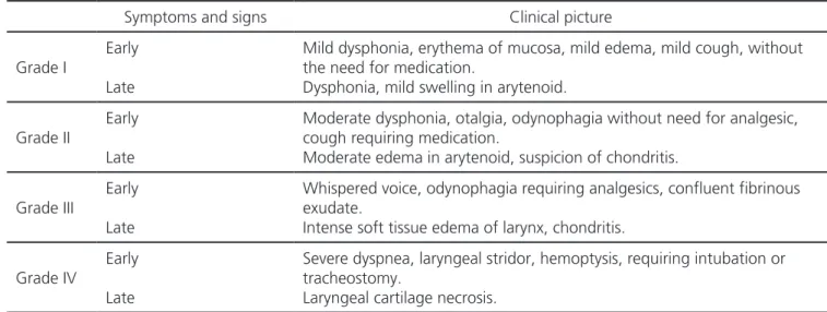
Table 2. Classification of RTOG (Radiation Therapy Oncology Group) for laryngeal radionecrosis21.
Symptoms and signs Clinical picture
Grade I
Early
Late
Mild dysphonia, erythema of mucosa, mild edema, mild cough, without the need for medication.
Dysphonia, mild swelling in arytenoid.
Grade II
Early
Late
Moderate dysphonia, otalgia, odynophagia without need for analgesic, cough requiring medication.
Moderate edema in arytenoid, suspicion of chondritis.
Grade III
Early
Late
Whispered voice, odynophagia requiring analgesics, confluent fibrinous exudate.
Intense soft tissue edema of larynx, chondritis.
Grade IV
Early
Late
Severe dyspnea, laryngeal stridor, hemoptysis, requiring intubation or tracheostomy.
oxygen therapy) air humidifiers, systemic antibiotics and corticosteroids21, 24-26. According to Abe et al.27, the use
of the hyperbaric chamber was effective to maintain larynx function, being an option for the treatment of chondroradionecrosis. Dequanter et al.28 demonstrated
that of 16 patients who underwent rescue surgery and who used oxygen therapy, 14 displayed success and considered hyperbaric oxygen therapy as a powerful and effective ally for the treatment of postoperative fistulas. Allen et al.29, in their review of patients with
laryngeal cancer treated with radiotherapy, propose a follow-up with positron emission tomography (PET-CT) to avoid local trauma and include oxygen therapy as a non-surgical alternative to larynx preservation.
This article aims to study laryngeal condroradionecrosis as a complication of radiotherapy for the treatment of laryngeal cancer and to propose a treatment flow chart with the use of hyperbaric chamber.
METHODS
We reviewed the clinical data of all patients with laryngeal and hypopharyngeal carcinomas consecutively admitted to the Head and Neck Surgery services of two tertiary hospitals, with histological confirmation, in the five-year period from January 2009 to January 2014. There was authorization from the Hospital Ethics in Research Committee to carry out this work, registered at Plataforma Brasil under number 37230414.4.0000.5483.
We gathered data on epidemiology, stage of the neoplasia, date of initial and rescue surgeries, initial treatment proposed and treatment for recurrence when applicable, treatment complications, data regarding treatment of complications and the date of the last visit. Inclusion criteria were patients with laryngeal or hypopharyngeal SCC. Exclusion criteria were patients already suffering from larynx or hypopharynx local recurrence with biopsy confirmation and patients referred for palliative care.
RESULTS
In the analyzed period, 131 patients with larynx or hypopharynx SCC were treated in both
institutions. Of these, 28 (21.4%) were referred for radiotherapy associated or not with chemotherapy as the first treatment, due to refusal of surgery, clinical comorbidities or high surgical risk. Of the 28 patients, three (10.7%) developed laryngeal chondroradionecrosis, clinically proven and with biopsy, none of them with tumor recurrence. Table 3 shows these three patients’ clinical data. All were submitted to neck computed tomography (NCT) to rule out local recurrences. NCT also revealed alterations in all patients, such as persistent edema of the laryngeal mucosa after the end of treatment and cartilage changes such as displacement, collapse or resorption of one or more of them (Figure 1). Computed tomography of the thorax showed no metastases in any patient.
Videolaryngoscopy demonstrated in-tense edema of the laryngeal scaffold, with purulent exudate and fibrin, areas of necrosis to a greater or less degree, with cartilage exposure, and glottis lumen decrease of more than 70%, which determined the indication of tracheostomy in one patient (Figure 2). In all three patients, we performed suspension laryngoscopy in the operating room, with biopsies of the affected site, which were negative for neoplasia recurrence, both in the frozen section and in the final anatomopathological examination.
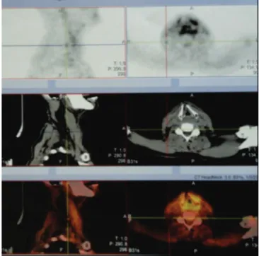
PET-CT was performed in all three patients and showed local edema with laryngeal cartilage alteration and a maximum Standardized Uptake Value (SUV) of less than 3.5, rendering a negative or unlikely recurrence result, and suggestive of inflammatory process, which was confirmed by the negative guided biopsies (Figure 3).
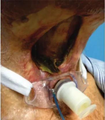
The treatment consisted of antibiotic therapy with Gram positive, Gram negative and anaerobic coverage with a third generation cephalosporin and clindamycin, corticosteroids, analgesics, proton pump inhibitors, air humidification and local care with adequate oxygen supply. We also performed suspension laryngoscopies to remove endolaryngeal exposed cartilage in patient 1 and an external surgical approach for removal of exposed cartilage and skin necrosis with pectoral flap rotation for coverage in patient 2 (Figure 4). All underwent hyperbaric chamber oxygen therapy with 100% O2 at 2 ATM pressure, one hour per session, in 20 to 40 sessions, depending on the case and after re-evaluation in the postoperative period.
There were no deaths in the postoperative period or related to the clinical treatment and we performed a weekly follow-up with videolaryngoscopy. We observed healing with complete epithelization of the necrotic area and remission of edema in all patients, within two months. We found no evidence of local recurrence. The larynx was preserved in all patients and only one patient had the tracheostomy maintained. We performed no total laryngectomy due to complications or aspiration.
DISCUSSION
Initial carcinomas of the larynx and hypopharynx can be treated equally by surgery or
Table 3. Clinical data of the study patients with laryngeal chondronecrosis post radio and chemotherapy.
Number/ Gender/ Age
Primary/ Initial stage
Radio Dose (Gray)
Chemo (months)Time Symptoms necrosisLocal Classification Chandler’s Treatment Result
1/M/62 Glottis (PVD)
T3N0M0 70 CIS 10
Pain, halitosis, moderate dysphonia
Arytenoid
right II
ATB, Cort, Umid, CH,
deb
Full resolution
2/M/78 Glottis (PVE)
T3N0M0 63 CIS 36
Dysphagia, bleeding, Dyspnea
Epiglottis, thyroid cartilage
IV
ATB, Cort, Umid, CH,
debri
Partial resolution
3/M/45
Supraglottis (PAE) T3N0M0
68 CIS 14
Dysphonia, dyspnea, halitosis, PA
PAE,
arytenoid III
ATB, Cort, Umid, CH, traq, cirur
Full resolution
Figure 2. Videolaryngoscopy of laryngeal chondronecrosis.
by radiotherapy in combination with chemotherapy, with similar results in local control and survival. Recent studies demonstrate local control with radiotherapy for T1 glottis neoplasias ranging from 82% to 93%, preserving the larynx in 89% to 96% of cases, and for T2 tumors, 57% to 82% for local control, and preservation of the larynx in 73% to 82%30. The rate
of relapse with surgical treatment is small and depends on factors such as: involvement of the anterior commissure due to its difficult access site for adequate resection; the ideal laryngeal exposure during the surgical procedure for the various open laryngectomy techniques and for endolaryngeal or trans-oral laser resections; and conditions inherent to the patient31.
From 1998 on, there was a change in the treatment of laryngeal cancer, with the institution of radiotherapy associated or not with chemotherapy, with the intention of preserving organs, indicated mainly for stages III (T3)11,32. However, the extension
of the indication to the other stages increased the radiotherapy complication rates33,34. Despite the
current guidelines, there is still no consensus on the best treatment strategy12,35. Radiation as a therapy
induces an environment with local hypoxic alterations,
stimulating the formation of tissue fibrosis, endarteritis with blood vessel obliteration, local lymphatic vessels obstruction and tissue and neoplastic necrosis36. Edema
then develops by lymphatic obstruction and increased reactive lymphovascular permeability, promoting an increase in local tissue pressure and decreasing blood flow. Microfissures of the perichondrium (both internal and external) appear, exposing the cartilage in its deep face to the bacteria of the aero-digestive tract. Infection sets in, leading to chondroradionecrosis, which is then considered a late complication18, 25, 37.
The first report of larynx chondronecrosis due to radiotherapy was by Goodrich and Lenz, in 194838, and since then not many cases have been
reported, up to the present time a total of seventy-seven patients, without including those of the present study. There was also no consensus for the creation of a prospective study involving the treatment of chondroradionecrosis. The incidence of 10.7% of chondroradionecrosis in our cases is somewhat elevated when compared with the literature data, but can be explained by differences in the radiotherapy techniques of the different services17-19.
Since the clinical signs and symptoms of chondroradionecrosis can be many, it was necessary to create severity ratings to guide treatment23,24.
However, these classifications failed because they did not distinguish chondroradionecrosis from local recurrence with greater clarity. Symptoms of chondroradionecrosis are most often indistinguishable from the symptoms of local relapses (or persistence), and control should be done periodically through videolaryngoscopy, biopsies and imaging tests, such as NCT, magnetic resonance imaging, and more recently PET-CT21, 25.
The tomographic findings of laryngeal changes during or shortly after radiotherapy are not able to rule out local recurrence. The findings are often nonspecific and the data in the literature are confusing, except for citing that the likelihood of relapse is greater within one year and that after this period the chance of chondroradionecrosis is higher37,39-41. According
to Hermans et al.40, the local recurrence rate after
radiotherapy is about 10% to 20% for T1 / T2 lesions and 40% to 50% for T3 / T4 lesions.
Since recurrences are much more frequent than chondroradionecrosis, deep biopsies should be performed for restaging, although they may worsen the infection and local necrosis (14, 37). Currently, this diagnostic dilemma can be resolved through PET-CT, in which tumor activity is evident in the larynx if relapse occurs, but examination should be done only three months after the end of radiotherapy to eliminate the interference of actinic inflammation42-44. All our
three chondroradionecrosis patients underwent PET-CT, with a negative result for tumor recurrence, a fact evidenced by the performed biopsies.
Although this is a small sample, given the rarity of chondroradionecrosis, we can verify that PET-CT has been highly accurate, in accordance with findings of a recent systematic review of 1,476 articles on imaging exams, of which eight focused on PET-CT. The authors demonstrated a sensitivity of 89% and specificity of 74% of the PET-CT to differentiate between tumor recurrence and local actinic late alterations. It should therefore be the exam of choice to avoid unnecessary CT scans and laryngoscopies with biopsies42,43,45.
The various treatments proposed for chondroradionecrosis included the use of antibiotic therapy, air humidifiers, corticosteroids, antireflux drugs (proton pump blockers, prokinetics) and hyperbaric chamber (HBC), the latter being in varied regimens. The use of HBC is approved for the treatment of complications of radiotherapy by the American Society of Diving and Hyperbaric Medicine and by the Brazilian Society of Hyperbaric Medicine. The first report of its use for the treatment of necrosis by radiotherapy was in 1976 and, in 1987, its use was first described for laryngeal necrosis46-48. The principle
of HBC is to increase the partial pressure of O2 from 21% present in ambient air to 100% in a pressurized chamber, transferring this oxygenation to the tissues and promoting healing49. A common fear concerning
the use of HBC refers to the possibility of tumor growth during treatment, which is why we have to rule out disease relapse, although some studies fail to demonstrate this characteristic50,51. The review of
the literature demonstrates the effectiveness of the treatment of laryngeal chondroradionecrosis with
HBC, with fistula closure, high index of laryngeal preservation (without laryngectomies) and eventually decannulation of patients, despite reports of HBC failure in up to 22% of cases classified as Chandler IV24,25,41,46. In our patients, there was no HBC failure,
achieving 100% success for clinical treatment, with epithelization of the cartilage necrotic or exposed area and avoiding rescue laryngectomy. The HBC regimen applied 100% O2, at a 2-ATM pressure, of one hour per session, in 20 to 40 sessions, with results comparable to those of other authors19,24,52.
To date, there is no randomized trial comparing the treatment of laryngeal chondroradionecrosis by HBC with other clinical forms of treatment, unlike what exists for mandible bone necrosis53. We did not
find any randomized study with the key words “larynx chondronecrosis”, “laryngeal chondroradionecrosis”, “laryngeal necrosis”, “laryngeal radionecrosis”. Most of the current literature is composed of case reports or series of cases, describing, to date, only 77 patients.
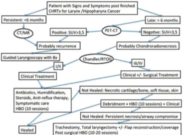
With this small series of three cases, we attempt to contribute with the scarce literature to understand this disease, which seems to be more common than we imagine. We propose an algorithm (Figure 5) to guide the treatment of laryngeal chondroradionecrosis with the use of the hyperbaric chamber, based on the data of the current literature. We emphasize, however, that one should apply it with caution, evaluating the different clinical characteristics of each case.
REFERENCES
1. GLOBOCAN [Internet]. Lyon (FR): IARC; 2012. Larynx - Estimated incidence, all ages: male 2012; [cited 2016 mar 22]. [Available from: http://glo- bocan.iarc.fr/old/summary_table_sitehtml.asp?se- lection=11100&title=Larynx&sex=1&type=0&win- dow=1&africa=1&america=2&asia=3&europe=4&o- ceania=5&build=6&sort=0&submit=%C2%A0Execu-te%C2%A0
2. Moura MA, Bergmann A, Aguiar SS, Thuler LC. The magnitude of the association between smoking and the risk of developing cancer in Brazil: a multicenter study. BMJ Open. 2014;4(2):e003736.
3. NIH. National Cancer Institute [Internet]. Bethesda (MD): National Cancer Institute; 2016. [cited 2016 mar 23] Cancer Stat Fact Sheets: larynx cancer. 2016 Available from: https://seer.cancer.gov/statfacts/html/ laryn.html
4. Instituto Nacional de Câncer José Alencar Gomes da Silva. Coordenação de Prevenção e Vigilância. Estima-tiva 2014: Incidência de câncer no Brasil. Rio de Janei-ro: INCA; 2014.
5. Hashibe M, Brennan P, Benhamou S, Castellsague X, Chen C, Curado MP, et al. Alcohol drinking in never users of tobacco, cigarette smoking in never drinkers, and the risk of head and neck cancer: pooled analysis in the International Head and Neck Cancer Epidemio-logy Consortium. J Natl Cancer Inst. 2007;99(10):777-89. Erratum in: J Natl Cancer Inst. 2008;100(3):225. Fernandez, Leticia [added].
6. Gillison ML, Alemany L, Snijders PJ, Chaturvedi A, Steinberg BM, Schwartz S, et al. Human papilloma-virus and diseases of the upper airway: head and neck cancer and respiratory papillomatosis. Vaccine. 2012;30 Suppl 5:F34-54.
7. Galli J, Cammarota G, Calò L, Agostino S, D´Ugo D, Cianci R, et al. The role of acid and alkaline reflux in laryngeal squamous cell carcinoma. Laryngoscope. 2002;112(10):1861-5.
8. Brown T, Darnton A, Fortunato L, Rushton L; British Occupational Cancer Burden Study Group. Occupa-tional cancer in Britain. Respiratory cancer sites: larynx, lung and mesothelioma. Br J Cancer. 2012;107 Suppl 1:S56-70.
9. Reidenbach MM. Borders and topographic rela-tions of the cricoid area. Eur Arch Otorhinolaryngol. 1997;254(7):323-5.
10. Reidenbach MM. Borders and topographic rela-tionships of the paraglottic space. Eur Arch Otorhi-nolaryngol. 1997;254(4):193-5.
11. Hillman RE, Walsh MJ, Wolf GT, Fisher SG, Hong WK. Functional outcomes following treatment for advanced laryngeal cancer. Part I--Voice preserva-tion in advanced laryngeal cancer. Part II--Laryngec-tomy rehabilitation: the state of the art in the VA System. Research Speech-Language Pathologists. Department of Veterans Affairs Laryngeal Can-cer Study Group. Ann Otol Rhinol Laryngol Suppl. 1998;172:1-27.
12. Lefebvre J. Surgery for Laryngopharyngeal SCC in the Era of Organ Preservation. Clin Exp Otorhino-laryngol. 2009;2(4):159-63.
13. Lederman M. Radiation therapy in cancer of the larynx. JAMA. 1972;221(11):1253-4.
14. Fitzgerald PJ, Koch RJ. Delayed radionecrosis of the larynx. Am J Otolaryngol. 1999;20(4):245-9.
15. Varghese BT, Paul S, Elizabeth MI, Somanathan T, Elizabeth KA. Late post radiation laryngeal chondro-necrosis with pharyngooesophageal fibrosis. Indian J Cancer. 2004;41(2):81-4.
16. Weber RS, Berkey BA, Forastiere A, Cooper J, Maor
Objetivo: estudar a condroradionecrose de laringe por complicação de radio-quimioterapia para tratamento do câncer de laringe e propor um fluxograma de tratamento com a utilização de câmara hiperbárica. Métodos: estudo retrospectivo de pacientes portadores de carcinoma de laringe admitidos em dois hospitais terciários num período de cinco anos. Resultados: de 131 pacientes portadores de câncer de laringe, 28 foram submetidos à radio e quimioterapia exclusiva e destes, três evoluíram com condroradionecrose. O tratamen-to destes pacientes foi realizado com câmara hiperbárica e com desbridamentratamen-to cirúrgico, conforme proposição do fluxograma. Todos os pacientes tiveram a laringe preservada. Conclusão: a incidência de condroradionecrose de laringe por complicação de radioterapia e quimioterapia em nossa casuística foi de 10,7% e o tratamento com oxigenoterapia hiperbárica, com base no nosso fluxograma, foi efetivo no controle desta complicação.
Descritores: Oxigenoterapia. Neoplasias Laríngeas. Necrose. Radioterapia Adjuvante. Complicações Intraoperatórias.
M, Goepfert H, et al. Outcome of salvage total laryn-gectomy following organ preservation therapy: the Radiation therapy Oncology Group trial 91-11. Arch Otolaryngol Head Neck Surg. 2003;129(1):44-9. 17. Sebelik ME. Chondroradionecrosis of the larynx in
patients treated with organ preservation therapy. Otolaryngol Head Neck Surg. 2004;131(2):63-4. 18. Rowley H, Walsh M, McShane D, Fraser I,
O’Dwyer TP. Chondroradionecrosis of the larynx: still a diagnostic dilemma. J Laryngol Otol. 1995;109(3):218-20.
19. Cukurova I, Cetinkaya EA. Radionecrosis of the larynx: case report and review of the literature. Acta Otorhinolaryngol Ital. 2010;30(4):205.
20. Oppenheimer RW, Krespi YP, Einhorn RK. Manage-ment of laryngeal radionecrosis: animal and clinical experience. Head Neck. 1989; 11(3):252-6.
21. Hernando M, Hernando A, Calzas J. [Laryngeal chondronecrosis following radiotherapy and con-current chemotherapy]. Acta Otorrinolaringol Esp. 2008;59(10):509. Spanish.
22. Sancho E, Escorial O, Cortés JM, Rivas P, Millán J, Vallés H. [Laryngeal chondro-radionecrosis]. Acta Otorhinolaryngol Esp. 2003;54:123-6. Spanish. 23. Chandler JR. Radiation fibrosis and necrosis of the
larynx. Ann Otol Rhinol Laryngol. 1979;88(4 Pt 1):509-14.
24. Filntisis GA, Moon RE, Kraft KL, Farmer JC, Scher RL, Piantadosi CA. Laryngeal radionecrosis and hyperbaric oxygen therapy: report of 18 cases and review of the literature. Ann Otol Rhinol Laryngol. 2000;109(6):554-62.
25. Roh JL. Chondroradionecrosis of the larynx: diag-nostic and therapeutic measures for saving the or-gan from radiotherapy sequelae. Clin Exp Otorhino-laryngol. 2009;2(3):115-9.
26. Ferguson BJ, Hudson WR, Farmer JC Jr. Hyperba-ric oxygen therapy for laryngeal radionecrosis. Ann Otol Rhinol Laryngol. 1987;96(1 Pt 1):1-6.
27. Abe M, Shioyama Y, Terashima K, Matsuo M, Hara I, Uehara S. Successful hyperbaric oxygen therapy for laryngeal radionecrosis after chemoradiotherapy for mesopharyngeal cancer: case report and literature review. Jpn J Radiol. 2012;30(4):340-4.
28. Dequanter D, Jacobs D, Shahla M, Paulus P, Aubert
C, Lothaire P. The effect of hyperbaric oxygen thera-py on treatment of wound complications after oral, pharyngeal and laryngeal salvage surgery. Undersea Hyperb Med 2013;40(5):381-5.
29. Allen CT, Lee CJ, Merati AL. Clinical assessment and treatment of the dysfunctional larynx after radiation. Otolaryngol Head Neck Surg 2013;149(6):830-9. 30. Ambrosch P, Fazel A. Functional organ
preser-vation in laryngeal and hypopharyngeal cancer. GMS Curr Top Otorhinolaryngol Head Neck Surg. 2011;10:Doc02.
31. Peretti G, Piazza C, Cocco D, De Benedetto L, Del Bon F, Redaelli De Zinis LO, et al. Transoral CO(2) laser treatment for T(is)-T(3) glottic cancer: the Uni-versity of Brescia experience on 595 patients. Head Neck 2010;32(8):977-83.
32. Calais G. TPF: a rational choice for larynx preserva-tion? Oncologist. 2010;15 Suppl 3:19-24.
33. Quer M, León X. [Organ preservation in laryngeal and hypopharyngeal cancer]. Acta Otorrinolaringol Esp. 2007;58(10):476-82. Spanish.
34. Beauvillain C, Mahé M, Bourdin S, Peuvrel P, Ber-gerot P, Rivière A, et al. Final results of a randomi-zed trial comparing chemotherapy plus radiotherapy with hemotherapy plus surgery plus radiotherapy in locally advanced respectable hypopharyngeal carci-nomas. Laryngoscope. 1997;107(5):648-53.
35. Lefebvre JL, Andry G, Chevalier D, Luboinski B, Col-lette L, Traissac L, et al. Laryngeal preservation with induction chemotherapy for hypopharyngeal squa-mous cell carcinoma: 10-year results of EORTC trial 24891. Ann Oncol. 2012;23(10):2708-14.
36. DiCarlo AL, Maher C, Hick JL, Hanfling D, Dainiak N, Chao N, et al. Radiation injury after a nuclear de-tonation: medical consequences and the need for scarce resources allocation. Disaster Med Public he-alth Prep. 2011;5 Suppl 1:S32-44.
37. Baba Y, Kato Y, Ogawa K. Unusual computed to-mography findings of radionecrosis after chemora-diation of stage IV hypopharyngeal cancer: a case report. J Med Case Rep. 2011;5:25.
38. Goodrich WA, Lenz M. Laryngeal chondronecrosis following roentgen therapy. Am J Roentgenol Ra-dium Ther. 1948;60(1):22-8.
Curschmann J, Stauffer E. Radionecrosis or tumor recurrence after radiation of laryngeal and hypo-pharyngeal carcinomas. Otolaryngol Head Neck Surg. 2006;135(6):838-43.
40. Hermans R, Pameijer FA, Mancuso AA, Parsons JT, Mendenhall WM. CT findings in chondrora-dionecrosis of the larynx. AJNR Am J Neuroradiol. 1998;19(4):711-8.
41. Allen CT, Lee CJ, Merati AL. Clinical assessment and treatment of the dysfunctional larynx after radiation. Otolaryngol Head Neck Surg. 2013;149(6):830-9. 42. McGuirt WF, Greven KM, Keyes JW Jr, Williams DW
3rd, Watson N. Laryngeal radionecrosis versus re-current cancer: a clinical approach. Ann Otol Rhinol Laryngol. 1998;107(4):293-6.
43. Gupta T, Master Z, Kannan S, Agarwal JP, Ghsoh--Laskar S, Rangarajan V, et al. Diagnostic perfor-mance of post-treatment FDG PET or FDG PET/CT imaging in head and neck cancer: a systematic re-view and meta-analysis. Eur J Nucl Med Mol Ima-ging. 2011;38(11):2083-95.
44. Greven KM, Williams DW 3rd, Keyes JW Jr, Mc-Guirt WF, Harkness BA, Watson NE Jr, et al. Distin-guishing tumor recurrence from irradiation sequelae with positron emission tomography in patients tre-ated for larynx cancer. Int J Radiat Oncol Biol Phys. 1994;29(4):841-5.
45. Brouwer J, Hooft L, Hoekstra OS, Riphagen II, Cas-telijns JA, de Bree R, et al. Systematic review: accu-racy of imaging tests in the diagnosis of recurrent laryngeal carcinoma after radiotherapy. Head Neck. 2008;30(7):889-97.
46. Ferguson BJ HW, Farmer JC. Hyperbaric oxygen the-rapy for laryngeal radionecrosis. Ann Otol Rhinol Laryngol. 1987;96(1 Pt 1):1-6.
47. Narozny W, Kuczkowski J, Mikaszewski B. Radione-crosis or tumor recurrence after radiation: importan-ce of choiimportan-ce for HBO. Otolaryngol Head Neck Surg. 2007;137(1):176-7.
48. Hart GB, Mainous EG. The treatment of radiation necrosis with hyperbaric oxygen (OHP). Cancer. 1976;37(6):2580-5.
49. Jallali N, Withey S, Butler PE. Hyperbaric oxygen as adjuvant therapy in the management of necrotizing fasciitis. Am J Surg. 2005;189(4):462-6.
50. Sun TB, Chen RL, Hsu YH. The effect of hyperba-ric oxygen on human oral cancer cells. Undersea Hyperb Med. 2004;31(2):251-60.
51. Feldmeier J, Carl U, Hartmann K, Sminia P. Hyperba-ric oxygen: does it promote growth or recurrence of malignancy? Undersea Hyperb Med. 2003;30(1):1-18.
52. Hsu YC, Lee KW, Ho KY, Tsai KB, Kuo WR, Wang LF, et al. Treatment of laryngeal radionecrosis with hyperbaric oxygen therapy: a case report. Kaohsiung J Med Sci. 2005;21(2):88-92.
53. Shaw RJ, Dhanda J. Hyperbaric oxygen in the ma-nagement of late radiation injury to the head and neck. Part I: treatment. Br J Oral Maxillofac Surg. 2011;49(1):2-8.
Received in: 03/01/2017
Accepted for publication: 30/03/2017 Conflict of interest: none.
Source of funding: none.
Mailing address:
Giulianno Molina Melo