ABSTRACT
Sao Paulo Med J. 2008;126(6):329-32.
ORIGINAL AR
TICLE
Joice Matos Biselli
Daniela Brumati
Vivian Fernanda Frigeri
Bruna Lancia Zampieri
Eny Maria Goloni-Bertollo
Érika Cristina Pavarino-Bertelli
A80G polymorphism of reduced
folate carrier 1 (
RFC1
) and C776G
polymorphism of transcobalamin
2 (
TC2
) genes in Down’s syndrome
etiology
Genetics and Molecular Biology Research Unit, Department of Molecular
Biology, Faculdade de Medicina de São José do Rio Preto (Famerp), São José
do Rio Preto, São Paulo, Brazil
CONTEXT AND OBJECTIVE: There is evidence that polymorphisms of genes involved in folate metabolism may be associated with higher risk that mothers may bear a Down’s syndrome (DS) child. This study therefore had the objective of investigating the A80G polymorphism of the reduced folate carrier 1 (RFC1) gene and the C776G polymorphism of the transcobalamin 2 (TC2) gene as maternal risk factors for DS among Brazilian women.
DESIGN AND SETTING: Analytical cross-sectional study with control group, at Faculdade de Me-dicina de São José do Rio Preto (Famerp). METHODS: Sixty-seven mothers of DS individuals with free trisomy 21, and 113 control moth-ers, were studied. Molecular analysis of the polymorphisms was performed by means of the polymerase chain reaction with restriction frag-ment length polymorphism (PCR-RFLP), followed by electrophoresis on 2% agarose gel. RESULTS: The frequencies of the polymorphic alleles were 0.51 and 0.52 for RFC1 80G, and 0.34 and 0.34 for TC2 776G, in the case and control groups, respectively. Thus, there were no differences between the groups in relation to either the allele or the genotype frequency, for both polymorphisms (P = 0.696 for RFC1
A80G; P = 0.166 for TC2 C776G; P = 0.268 for combined genotypes).
CONCLUSION: There was no evidence of any association between the RFC1 A80G and TC2
C776G polymorphisms and the maternal risk of DS in the sample evaluated.
KEY WORDS: Down syndrome. Polymorphism, genetic. Folic acid. Nondisjunction, genetic. Transcobalamins.
INTRODUCTION
Down’s syndrome (DS) or trisomy 21 is a genetic disease associated with abnormal chromosomal segregation.1 Free trisomy 21 is
found in 95% of DS cases and is due to chro-mosome 21 nondisjunction, which in most cases occurred during maternal meiosis.2
The risk factors for meiotic nondisjunc-tion are not very clear, except for advanced maternal age.3,4 Folic acid has an important
role in the process of genetic material distri-bution during cell division, because of its im-portance to cellular methylation reactions.5
The folate metabolism is responsible for synthesizing S-adenosylmethionine (SAM), the main donor of methyl groups for cellu-lar methylation reactions.6 It has been shown
that DNA methylation is important for main-taining centromeric chromatin stability and plays an important role in chromosomal segregation.7,8
Polymorphisms of genes that encode en-zymes involved in folate metabolism have been associated with the etiology of DS.9-12 A
polymorphism of the reduced folate carrier 1 (RFC1) gene consisting of an adenine-to-gua-nine substitution at position 80 (A80G) has been associated with altered concentrations of products derived from the folate metabol-ic pathway.13,14 This has been indicated as a
maternal risk factor for DS, in combination with other polymorphisms involved in this metabolism.9,11 The RFC1 gene encodes the
reduced folate carrier 1 protein, which plays a role in folic acid absorption, thereby trans-porting 5-methyltetrahydrofolate, the meta-bolically active form of folate, into a variety of cells.15
Polymorphisms of genes encoding coba-lamin-transporting proteins such as transco-balamin 2 (TC2) may interfere with the avail-ability of this vitamin in the organism.16,17
Co-balamin (vitamin B12) plays an important role in folate metabolism because of its action as cofactor for methionine synthase (MTR) en-zyme.18 Thus, genetic variants in the TC2
gene possibly have an infl uence on cellular methylation reactions and on the risk that the mother may bear a DS child. The continuing studies on the etiology of DS are motivated by the importance of this subject for fami-lies, given the repercussions from the birth of a child with DS.
OBJECTIVE
This study had the objective of investigat-ing the RFC1 A80G and TC2 C776G poly-morphisms as maternal risk factors for DS.
METHODS
This was an analytical cross-sectional study with a control group carried out at Faculdade de Medicina de São José do Rio Preto (Famerp). After informed consent had been obtained, peripheral blood samples were taken from 67 mothers of DS individuals with free trisomy 21 (case group) and 113 mothers of individuals without the syndrome (control group). Mothers of DS individuals with trans-location or mosaicism were not included in the study. The average age among the mothers at the time of blood collection was 36.7 ± 10.4 years in the case group and 40.5 ± 8.2 years in the control group.
For molecular analysis, DNA was ex-tracted from peripheral blood leukocytes, as described by Miller et al.19 The RFC1 A80G
polymorphism was investigated by means of the polymerase chain reaction with restric-tion fragment length polymorphism (PCR-RFLP), using the primer sequences described by Födinger et al.20 and the enzyme CfoI to
330
Sao Paulo Med J. 2008;126(6):329-32.
performed by means of PCR-RFLP using the forward primer 5’- CAT CAG AAC AGT GCG AGA GG –3’ and the reverse primer 5’- GTG CCA GAC AGT CTG GGA AG –3’, and the enzyme ScrF1 to recognize the polymorphic site.21 The resulting fragments
from enzyme digestion were then subjected to electrophoresis on 2% agarose gel.
The chi-squared test was used for sta-tistical analysis on genotype frequencies. Comparisons of maternal age between the groups was carried out by using Student’s t test; P values ≤ 0.05 were taken to be statisti-cally significant.
RESULTS
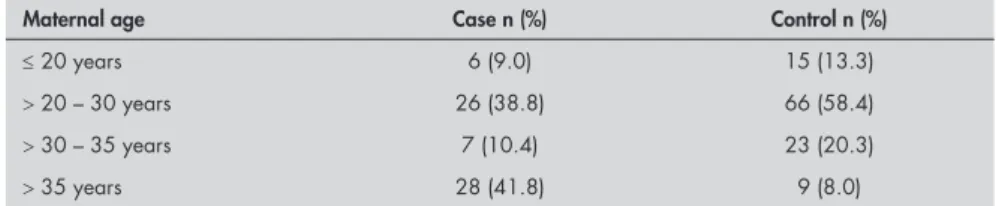
Table 1 presents the distribution of the groups according to maternal age. The mean maternal age in the case group was 32 ± 8.6 years, and in the control group, it was 27.4 ± 5.5 years (P < 0.0001).
The frequencies of the polymorphic alleles
RFC1 80G and TC2 776G were 0.51 and 0.34 in the case group and 0.52 and 0.34 in the control group, respectively. The genotype dis-tribution of the polymorphisms is presented in Table 2. There were no significant differences in genotype distribution between the groups (P = 0.696 for RFC1 A80G; P = 0.166 for
TC2 C776G).
Analysis of the combined genotypes of the two polymorphisms did not show any statistically significant differences between the groups (P = 0.268). Nor did the genotypes differentiate between the groups when only
women with maternal age under 35 years were considered (P = 0.714 for RFC1 A80G; P = 0.166 for TC2 C776G; P = 0.759 for combined genotypes).
DISCUSSION
Several technological advances in molecu-lar cytogenetics, making it possible to identify most chromosomal aberrations (both struc-tural and numerical) and the genes respon-sible for some diseases, have been achieved.22
Nevertheless, although the chromosomal basis of DS has been well characterized, the etiology of chromosome 21 nondisjunction remains unclear.
Among the risk factors associated with DS occurrence, advanced maternal age is the best established factor.3,4 In fact, it has been
shown that the frequency of chromosomal aberrations becomes greater with advancing maternal age,3,24,25 especially with regard to
trisomy of chromosomes 13, 18 and 21 in women who give birth when over 35 years old.26 According to data from the Latin
American Collaborative Study of Congenital Malformations (ECLAMC), 40% of live births with DS were born from mothers who were between 40 and 44 years old, although women of this age account for only 2% of all births.27 In our study, the mean maternal
age was significantly higher in the case group than in the control group. This corroborates with data in the literature regarding the association between maternal age and the risk of DS.
Occurrences of births of children with DS among young mothers suggests that advanced maternal age is not the only risk factor involved. Investigation of genetic poly-morphisms that lead to abnormalities in folate metabolism products is the approach furthest investigated at the present time.28
James et al.29 reported that mothers with
DS children have abnormal folate metabolism. They suggested that a variant of the methyl-enetetrahydrofolate reductase gene (MTHFR
C677T), which regulates cellular methylation reactions, could lead to DNA hypomethyla-tion and consequently to chromosomal seg-regation errors. Other genes involved in the folate metabolic pathway have been investi-gated as maternal risk factors for DS, such as
RFC1, MTR, methionine synthase reductase (MTRR), cystathionine B-synthase (CBS). Evidence indicating the contribution of vari-ants of these genes towards the maternal risk of bearing a DS child has been put forward.9-12
However, the few studies that have evaluated the influence of RFC1 A80G poly-morphism, including a previous study by our group,9 did not observe any association
between this variant per se and the maternal risk of bearing a DS child,9,10,30 thereby
cor-roborating the present study. Higher maternal risk of DS has been observed in the presence of this polymorphism, in combination with other genetic polymorphisms relating to folate metabolism, such as MTHFR C677T,
MTHFR A1298C and MTR A2756G.9,10 This
may be due to the small impact of RFC1 A80G polymorphism on the affinity and transport efficiency of the variant enzyme in relation to the wild-type enzyme.31
With regard to TC2 C776G polymor-phism, the present study is, to our knowledge, the first to investigate the contribution of polymorphisms of the cobalamin-transport-ing gene (an important cofactor for folate metabolism), in relation to the maternal risk of bearing a DS child. There is evidence for an association between this genetic variant and the maternal risk of bearing a child with neural tube defects,32 which are influenced
by genetic determinants involved in folate metabolism.
Evidence showing a higher frequency of DS cases in families with a risk of neural tube defects, and vice versa,33 strengthens the
notion that the same genetic determinants of folate metabolism influence both disorders.34
However, in the present study, no associa-tion was observed between the TC2 C776G polymorphism and the maternal risk of bearing a DS child. It is widely accepted that
Table 2. Genotype distribution of the RFC1 A80G and TC2 C776G polymorphisms between the case (n = 67) and control (n = 113) groups
Genotypes Case n (%) Control n (%)
RFC1 A80G
AA 14 (20.9) 30 (26.5)
AG 33 (49.3) 49 (43.4)
GG 20 (29.9) 34 (30.1)
TC2 C776G
CC 32 (47.8) 48 (42.5)
CG 24 (35.8) 54 (47.8)
GG 11 (16.4) 11 (9.7)
Table 1. Distribution of the case (n = 67) and control (n = 113) groups according to maternal age
Maternal age Case n (%) Control n (%)
≤ 20 years 6 (9.0) 15 (13.3)
> 20 – 30 years 26 (38.8) 66 (58.4)
> 30 – 35 years 7 (10.4) 23 (20.3)
331
Sao Paulo Med J. 2008;126(6):329-32.
supplementation or fortification with folic acid reduces the risk of neural tube defects,35,36
and it is believed that supplementation with cobalamin in combination with folate could contribute towards reducing this risk.18,32
CONCLUSION
No evidence for an association between
RFC1 A80G and TC2 C776G polymorphisms and the maternal risk of bearing a DS child was observed in this study. Thus, further
stud-ies including these and other polymorphisms involved in folate metabolism could provide a better understanding of the role of genetic variants in the etiology of the chromosomal nondisjunction that results in DS.
1. Oliver TR, Feingold E, Yu K, et al. New insights into human nondisjunction of chromosome 21 in oocytes. PLoS Genet. 2008;14;4(3):e1000033.
2. Jyothy A, Kumar KS, Mallikarjuna GN, et al. Parental age and the origin of extra chromosome 21 in Down syndrome. J Hum Genet. 2001;46(6):347-50.
3. Gusmão FA, Tavares EJ, Moreira LM. Idade materna e sín-drome de Down no Nordeste do Brasil. [Maternal age and Down syndrome in Northeast Brazil]. Cad Saude Publica. 2003;19(4):973-8.
4. Beiguelman B, Krieger H, Silva LM. Maternal age and Down syndrome in Southeastern Brazil. Rev Bras Genet = Braz J Genet. 1996;19(4):637-40.
5. Fenech M. Micronutrients and genomic stability: a new para-digm for recommended dietary allowances (RDAs). Food Chem Toxicol. 2002;40(8):1113-7.
6. Finkelstein JD, Martin JJ. Homocysteine. Int J Biochem Cell Biol. 2000;32(4):385-9.
7. D’Alessio AC, Szyf M. Epigenetic tête-à-tête: the bilateral rela-tionship between chromatin modifications and DNA methyla-tion. Biochem Cell Biol. 2006;84(4):463-76.
8. Sciandrello G, Caradonna F, Mauro M, Barbata G. Arsenic-induced DNA hypomethylation affects chromosomal instability in mammalian cells. Carcinogenesis. 2004;25(3):413-7. 9. Biselli JM, Goloni-Bertollo EM, Zampieri BL, Haddad R,
Eberlin MN, Pavarino-Bertelli EC. Genetic polymorphisms involved in folate metabolism and elevated plasma concentra-tions of homocysteine: maternal risk factors for Down syndrome in Brazil. Genet Mol Res. 2008;7(1):33-42.
10. Coppedè F, Marini G, Bargagna S, et al. Folate gene polymor-phisms and the risk of Down syndrome pregnancies in young Italian women. Am J Med Genet A. 2006;140(10):1083-91. 11. da Silva LR, Vergani N, Galdieri L de C, et al. Relationship
between polymorphisms in genes involved in homocysteine metabolism and maternal risk for Down syndrome in Brazil. Am J Med Genet A. 2005;135(3):263-7.
12. Acácio GL, Barini R, Bertuzzo CS, Couto EC, Annichino-Bizzacchi JM, Júnior WP. Methylenetetrahydrofolate reductase gene polymorphisms and their association with trisomy 21. Prenat Diagn. 2005;25(13):1196-9.
13. Fillon-Emery N, Chango A, Mircher C, et al. Homocysteine concentrations in adults with trisomy 21: effect of B vitamins and genetic polymorphisms. Am J Clin Nutr. 2004;80(6):1551-7. 14. Chango A, Emery-Fillon N, de Courey GP, et al. A polymor-A polymor-phism (80G->A) in the reduced folate carrier gene and its
as-sociations with folate status and homocysteinemia. Mol Genet Metab. 2000;70(4):310-5.
15. Nguyen TT, Dyer DL, Dunning DD, Rubin SA, Grant KE, Said HM. Human intestinal folate transport: cloning, expression, and distribution of complementary RNA. Gastroenterology. 1997;112(3):783-91.
16. Miller JW, Ramos MI, Garrod MG, Flynn MA, Green R. Transcobalamin II 775G>C polymorphism and indices of vita-min B12 status in healthy older adults. Blood. 2002;100(2):718-20.
17. Afman LA, Lievers KJ, van der Put NM, Trijbels FJ, Blom HJ. Single nucleotide polymorphisms in the transcobalamin gene: relationship with transcobalamin concentrations and risk for neural tube defects. Eur J Hum Genet. 2002;10(7):433-8. 18. Yamada K, Gravel RA, Toraya T, Matthews RG. Human
methionine synthase reductase is a molecular chaperone for human methionine synthase. Proc Natl Acad Sci U S A. 2006;103(25):9476-81.
19. Miller SA, Dykes DD, Polesky HF. A simple salting out proce-dure for extracting DNA from human nucleated cells. Nucleic Acids Res. 1988;16(3):1215.
20. Födinger M, Dierkes J, Skoupy S, et al. Effect of glutamate carboxypeptidase II and reduced folate carrier polymorphisms on folate and total homocysteine concentrations in dialysis patients. J Am Soc Nephrol. 2003;14(5):1314-9. 21. Pietrzyk JJ, Bik-Multanowski M. 776C>G polymorphism of
the transcobalamin II gene as a risk factor for spina bifida. Mol Genet Metab. 2003;80(3):364.
22. Coelho IS. Doenças cromossômicas. Available from: http://www. ufv.br/dbg/BIO240/DC14.htm. Accessed in 2008 (Sep 10). 23. Barros ACSD, Zugaib M, Pereira PP, Guglielmi GF, Durante
AA. Gestantes de pelo menos 45 anos de idade: considerações sobre 40 casos. [Pregnant women of 45 or more. Considerations on 40 cases]. J Bras Ginecol. 1984;94(1/2):33-6. 24. Hansen JP. Older maternal age and pregnancy outcome: a review
of the literature. Obstet Gynecol Surv. 1986;41(11):726-42. 25. Kiely JL, Paneth N, Susser M. An assessment of the effects of
maternal age and parity in different components of perinatal mortality. Am J Epidemiol. 1986;123(3):444-54. 26. Schupp T. Espaço médico: idade materna avançada.
Avail-able from: http://drauziovarella.ig.com.br/espaco/espaco. asp?doe_id=25. Accessed in 2008 (Sep 10).
27. Castilla EE, Lopez-Camelo JS, Paz JE. Atlas geográfico de las malformaciones congénitas en sudamérica. Rio de Janeiro: Fiocruz; 1995.
28. Eskes TK. Abnormal folate metabolism in mothers with Down syndrome offspring: review of the literature. Eur J Obstet Gynecol Reprod Biol. 2006;124(2):130-3.
29. James SJ, Pogribna M, Pogribny IP, et al. Abnormal folate metabolism and mutation in the methylenetetrahydrofolate reductase gene may be maternal risk factors for Down syndrome. Am J Clin Nutr. 1999;70(4):495-501.
30. Chango A, Fillon-Emery N, Mircher C, et al. No association between common polymorphisms in genes of folate and homo-cysteine metabolism and the risk of Down’s syndrome among French mothers. Br J Nutr. 2005;94(2):166-9.
31. Whetstine JR, Gifford AJ, Witt T, et al. Single nucleotide poly-morphisms in the human reduced folate carrier: characterization of a high-frequency G/A variant at position 80 and transport properties of the His(27) and Arg(27) carriers. Clin Cancer Res. 2001;7(11):3416-22.
32. Guéant-Rodriguez RM, Rendeli C, Namour B, et al. Transco- Transco-balamin and methionine synthase reductase mutated polymor-phisms aggravate the risk of neural tube defects in humans. Neurosci Lett. 2003;344(3):189-92.
33. Barkai G, Arbuzova S, Berkenstadt M, Heifetz S, Cuckle H. Frequency of Down’s syndrome and neural-tube defects in the same family. Lancet. 2003;361(9366):1331-5.
34. Guéant JL, Guéant-Rodriguez RM, Anello G, et al. Genetic determinants of folate and vitamin B12 metabolism: a common pathway in neural tube defect and Down syndrome? Clin Chem Lab Med. 2003;41(11):1473-7.
35. De Wals P, Tairou F, Van Allen MI, et al. Reduction in neural-tube defects after folic acid fortification in Canada. N Engl J Med. 2007;357(2):135-42.
36. Pitkin RM. Folate and neural tube defects. Am J Clin Nutr. 2007;85(1):285S-288S.
Acknowledgements: The authors thank Prof. Dr. José Antônio Cordeiro for his assistance in the statistical analysis and Prof.a
Adília Maria Pires Sciarra for her assistance in translating this article
Sources of funding: Fundação de Amparo à Pesquisa do Estado de São Paulo (Fapesp) — 04/15944-5; Coordenação de Aperfeiçoamento de Pessoal de Nível Superior (Capes) — CGPP 046/2006); Conselho Nacional de Desenvol-vimento Científico e Tecnológico (CNPq) — 3210/2006 and 3211/2006
Conflict of interest: Not declared Date of first submission: December 9, 2007 Last received: June 23, 2008
Accepted: November 4, 2008
332
Sao Paulo Med J. 2008;126(6):329-32.
AUTHOR INFORMATION
Joice Matos Biselli, MSc. Genetics and Molecular Biology Research Unit, Department of MolecularBiology, Faculdade de Medicina de São José do Rio Preto (Famerp), São José do Rio Preto, São Paulo, Brazil.
Daniela Brumati, BSc. Nurse, Faculdade de Medicina de São José do Rio Preto (Famerp), São José do Rio Preto, São Paulo, Brazil.
Vivian Fernanda Frigeri, BSc. Nurse, Faculdade de Medicina de São José do Rio Preto (Famerp), São José do Rio Preto, São Paulo, Brazil.
Bruna Lancia Zampieri, MSc. Student. Genetics and Mo-lecular Biology Research Unity, Faculdade de Medicina de São José do Rio Preto (Famerp), São José do Rio Preto, São Paulo, Brazil.
Eny Maria Goloni-Bertollo, PhD. Genetics and Molecular Biology Research Unit, Department of MolecularBiology, Faculdade de Medicina de São José do Rio Preto (Famerp), São José do Rio Preto, São Paulo, Brazil.
Érika Cristina Pavarino-Bertelli, PhD. Genetics and Molecular Biology Research Unit, Department of MolecularBiology, Faculdade de Medicina de São José do Rio Preto (Famerp), São José do Rio Preto, São Paulo, Brazil.
Address for correspondence Érika Cristina Pavarino Bertelli
Faculdade de Medicina de São José do Rio Preto (Famerp)
Unidade de Pesquisa em Genética e Biologia Molecular (UPGEM)
Av. Brigadeiro Faria Lima, 5.416 — Bloco U-6 São José do Rio Preto (SP) — Brasil CEP 15090-000
Tel. (+55 17) 3201-5720 Fax. (+55 17) 3201-5708 E-mail: [email protected]
Copyright © 2008, Associação Paulista de Medicina
RESUMO Polimorfismos do gene carregador de folato reduzido(RFC1) A80G e transcobalamina 2(TC2) C776G na etiologia da síndrome de Down
CONTEXTO E OBJETIVO: Considerando as evidências de que polimorfismos em genes envolvidos no metabolismo do folato podem estar associados ao risco materno elevado para a síndrome de Down (SD), o objetivo deste estudo foi investigar os polimorfismos A80G do genecarregador de folato reduzido 1(RFC1) e C776G do gene transcobalamina 2(TC2) como fatores de risco maternos para a SD em mulheres brasileiras.
TIPO E ESTUDO LOCAL: Estudo transversal analítico com grupo controle, realizado na Faculdade de Me-dicina de São José do Rio Preto (Famerp).
MÉTODOS: Foram avaliadas 67 mães de indivíduos com trissomia livre do 21 e 113 mães de indivíduos sem a síndrome. A análise molecular dos polimorfismos foi realizada pela técnica de reação em cadeia da polimerase/polimorfismo de comprimento fragmentos de restrição (PCR-RFLP), seguida por eletroforese em gel de agarose 2%.
RESULTADOS: As freqüências dos alelos polimórficos foram de 0,51 e 0,52 para RFC1 80G e 0,34 e 0,34 para TC2 776G nos grupos caso e controle, respectivamente. Assim, não houve diferença nas freqüências alélicas e genotípicas para ambos os polimorfismos entre os grupos (P = 0,696 para RFC1 A80G; P = 0,166 para TC2 C776G; p = 0,268 para genótipos combinados).
CONCLUSÃO: Não há evidência de associação entre os polimorfismos RFC1 A80G e TC2 C776G e o risco materno para a SD na casuística avaliada.