Rogério Castilho JACINTO(a)
Giane LINHARES-FARINA(b)
Otávio da Silva SPOSITO(b)
César Henrique ZANCHI(c)
Maximiliano Sérgio CENCI(c)
(a) Universidade Federal de Pelotas – UFPel, Dental School, Endodontic Division, Pelotas, RS, Brazil.
(b) Universidade Federal de Pelotas – UFPel, Dental School, Department of Semiology and Clinics, Pelotas, RS, Brazil.
(c) Universidade Federal de Pelotas – UFPel, Dental School, Department of Restorative Dentistry, Pelotas, RS, Brazil.
Influence of 2% chlorhexidine on pH,
calcium release and setting time of a
resinous MTA-based root-end filling
material
Abstract: The addition of chlorhexidine (CHX) to a resinous experimental Mineral Trioxide Aggregate (E-MTA) based root-end
illing material is an alternative to boost its antimicrobial activity. However, the inluence of chlorhexidine on the properties of this material is unclear. The aim of this study was to evaluate the inluence of 2% chlorhexidine on the pH, calcium ion release and setting time of a Bisphenol A Ethoxylate Dimethacrylate/Mineral Trioxide Aggregate (Bis-EMA/MTA) based dual-cure experimental root-end illing material (E-MTA), in comparison with E-MTA without the addition of CHX and with conventional white MTA (W-MTA). The materials were placed in polyethylene tubes, and immersed in deionized water to determine
pH (digital pH meter) and calcium ion release (atomic absorption
spectrometry technique). The setting time of each material was analyzed using Gilmore needles. The data were statistically analyzed at a signiicance level of 5%. E-MTA + CHX showed an alkaline pH in the 3 h period of evaluation, the alkalinity of which decreased but remained as such for 15 days. The pH of E-MTA + CHX was higher than the other two materials after 7 days, and lower after 30 days (p < 0.05).
All of the materials were found to release calcium ions throughout
the 30 days of the study. The addition of CHX increased the calcium ion release of E-MTA to levels statistically similar to W-MTA. E-MTA showed shorter initial and inal setting time, compared with W-MTA
(p < 0.05). The addition of 2% CHX to MTA prevented setting of the
material. The addition of CHX to E-MTA increased its pH and calcium ion release. However, it also prevented setting of the material.
Keywords: Chlorhexidine; Hydrogen-Ion Concentration; Dental Materials.
Introduction
Periapical surgery usually consists of root-end resection and root-end illing to seal the communication between the apical tissues and the root canal system.1 Root-end illing materials should prevent leakage, promote antimicrobial activity, and provide an effective environment for healing of the apical tissues.2,3 Mineral Trioxide Aggregate (MTA) as a root-end
illing material has proved to be the most effective material in preventing leakage3 and stimulating tissue repair.4
Declaration of Interests: The authors certify that they have no commercial or associative interest that represents a conflict of interest in connection with the manuscript.
Corresponding Author: Rogério Castilho Jacinto
E-mail: [email protected]
DOI: 10.1590/1807-3107BOR-2015.vol29.0036
Submitted: Apr 30, 2014
Chlorhexidine (CHX), a cationic antimicrobial agent frequently used as a root canal irrigant5 or
as an inter-appointment root canal dressing,6 has
been added to conventional MTA as a sterile water substitute.7 Studies have shown that MTA mixed with
CHX produces a biocompatible material – insofar as it
created weak inlammatory responses characterized by the presence of a ibrous connective tissue capsule
in the subcutaneous tissues of rats8 – with enhanced antimicrobial activity.9
Although MTA has many favorable properties that support its clinical use, there are also several drawbacks, e.g., it is a material of dificult manipulation.
Moreover, the extended setting time of MTA may cause the material to be washed out of the cavity during root-end surgery. The addition of light-curable
resinous monomers to MTA has been proposed to
improve its properties and reduce its setting time.10
However, this addition to MTA cements inhibits MTA calcium ion release, impairing its ability to stimulate tissue repair.11,12
Chlorhexidine not only acts as an antimicrobial
agent but also has been shown to increase the calcium
release of calcium hydroxide medication.13 The
addition of chlorhexidine to a resinous MTA-based root-end material could enhance its antimicrobial
activity and improve its ability to release calcium ions, thus increasing the success rate of root-end endodontic surgery. However, there is no data in the
literature in respect to the effect of CHX addition on the properties of resinous MTA-based root-end
material. Therefore, the objective of this study was to investigate the inluence of 2% CHX on pH, calcium ion release and setting time of Bisphenol A Ethoxylate Dimethacrylate/Mineral Trioxide (Bis-EMA/MTA) based root-end illing material.
Methodology
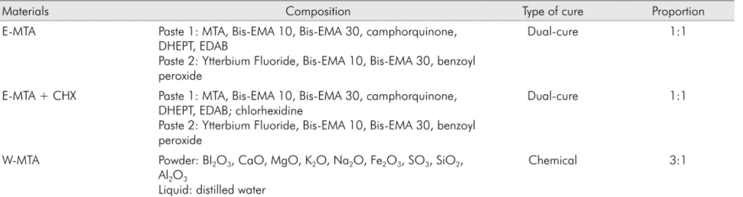
The compositions of the experimental root-end
illing material (E-MTA) (containing Bis-EMA/MTA), of the E-MTA + 2% CHX powder, and of the white-MTA (W-MTA) are shown in Table 1.
W-MTA (Angelus, Londrina, Brazil) was prepared following the manufacturer’s instructions. E-MTA was prepared using equal parts of Paste 1 and Paste 2.
Calcium ion release and pH evaluation The materials were manipulated and inserted in single, open-ended polyethylene tubes with 1.0 mm internal diameter and 10.0 mm length, using a lentulo spiral (Dentsply Maillefer, Toronto, Canada). E-MTA and E-MTA + CHX were cured for 40 seconds using a light-cure unit (Ultralux-Dabi Atlante, Ribeirão Preto, Brazil). After the tubes were illed, they were weighed to ensure standardization of the amount of cement in each tube. Five specimens of each material were prepared. Each specimen was immediately immersed in test tubes containing 10 mL of deionized water (Permution, Curitiba, Brazil). The tubes were then sealed with Parailm (American National Can, Menasha, USA) and incubated at 37°C (Farmen, São Paulo, Brazil), and kept as such throughout the study. Previous to the immersion of the specimens, the pH and calcium ion concentration of the deionized water was veriied (attesting pH 7.0). All laboratory equipment was previously treated with nitric acid to avoid interference in the results. Evaluations were performed at periods of 3 hours, 24 hours, 7 days, 15 days and 30 days. The specimens were carefully transferred to new tubes with fresh deionized water after each measurement.
The measurement of pH was performed with a pH
meter (Quimis Q400A, Diadema, Brazil). The release of
calcium ions was measured using an atomic absorption
spectrophotometer (AA6300, Shimadzu, Tokyo, Japan).
The conditions for using the appliance were determined
following the manufacturer’s instructions: wavelength of 422.70 nm, gap of 0.5 nm, lamp current of 10 mA, and slightly reduced stoichiometry, and were maintained by an air-supported acetylene low of 2.0 L per minute. A lanthanum chloride solution at 1 g/L was used to eliminate the interference of phosphates and sulfates, as well as the possible formation of refractory oxides. A standard stock solution of 100 mg/L was diluted in water to produce the following concentrations: 0.5 mg/L, 1 mg/L, 1.5 mg/L, and 2.0 mg/L. The results were calculated according to a standard curve, established on the basis of solutions with predeined calcium concentrations.
Setting Time
The setting time was determined according to
Table 2. pH values (mean ± standard deviation, n = 5 for each material) of soaking water after immersion of the samples in different evaluation periods (3 h to 30 days).
Periods W-MTA E-MTA E-MTA+CHX
3 h 9.92(±0.26)a 9.32(±0.27)a 9.63(±0.32)a
24 h 10.06(±0.55)a 7.86(±0.78)b 7.83(±0.84)b
7 d 7.02(±0.14)b 6.25(±0.20)c 7.35(±0.12)a
15 d 8.05(±0.67)a 6.63(±0.13)b 7.35(±0.16)a
30 d 7.78(±0.13)a 7.04(±0.20)b 6.75(±0.06)c
Different lowercase letters indicate significant differences between groups. (p ≤ 05)
The materials were mixed and placed in stainless
steel rings with a 10 mm internal diameter and 2 mm height. Three stainless steel rings were illed with each material and stored in an incubator at 37°C and 95% relative humidity. Next, a 113.4 g Gillmore needle was used to determine the initial setting times, and a 453.6 g Gillmore needle, to determine the inal setting times. This procedure was repeated every 60 s until the material set. In both analyses, the setting times
were recorded at the moment in which the needle
failed to leave a complete circular indentation on the surface of the specimen.
Statistical analysis
The assumptions of equality variances and normal distribution of errors were checked for all the response variables tested, and those that were not suitable were transformed. pH and calcium data were ranked, transformed and analyzed using two-way repeated measures of variance (ANOVA), followed by the Holm-Sidak method. Setting time was analyzed using one-way ANOVA followed by the Holm-Sidak method. Statistical analysis was carried out using
the SigmaStat® software package (Version 3.5 for Windows®, Systat Software Corporation, San Jose, USA). Values of p < .05 were considered signiicant.
Results
W-MTA showed an alkaline pH during the 30 days of the experiment. E-MTA showed an alkaline pH in the irst 24 h and then acid pH at the 7- and 15-day evaluations, ending with a neutral pH at the 30-day evaluation. E-MTA + CHX showed an alkaline pH in the 3 h evaluation period; although the level decreased, it remained alkaline for 15 days. Table 2 shows the mean and standard deviation pH values for each group. No statistical differences were observed between the 3 materials in the 3 h period. The pH of E-MTA + CHX
was higher than that of the other two materials after
7 days, and lower after 30 days (p < 0.05).
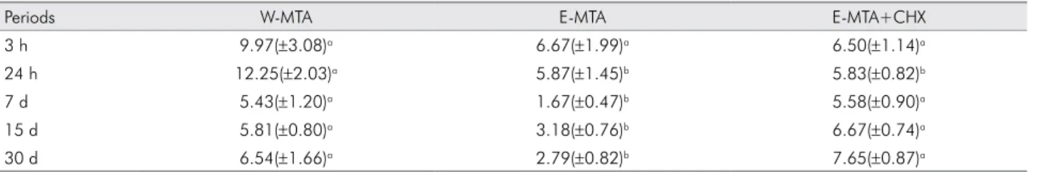
All of the materials released calcium ions
throughout the 30 days of the study. Table 3 presents the mean and standard deviation values of calcium ion release in different periods up to 30 days. Statistical differences were observed between E-MTA and W-MTA (p < 0.05), and between E-MTA and
Table 1. Composition of materials used in the study.
Materials Composition Type of cure Proportion
E-MTA Paste 1: MTA, Bis-EMA 10, Bis-EMA 30, camphorquinone, DHEPT, EDAB
Paste 2: Ytterbium Fluoride, Bis-EMA 10, Bis-EMA 30, benzoyl peroxide
Dual-cure 1:1
E-MTA + CHX Paste 1: MTA, Bis-EMA 10, Bis-EMA 30, camphorquinone, DHEPT, EDAB; chlorhexidine
Paste 2: Ytterbium Fluoride, Bis-EMA 10, Bis-EMA 30, benzoyl peroxide
Dual-cure 1:1
W-MTA Powder: BI2O3, CaO, MgO, K2O, Na2O, Fe2O3, SO3, SiO2,
Al2O3
Liquid: distilled water
Chemical 3:1
E-MTA + CHX (p < 0.05). In general, the addition of
CHX increased the calcium ion release of E-MTA to
levels statistically similar to W-MTA.
Table 4 shows the mean setting time for all
the materials. E-MTA showed immediate setting, whereas W-MTA showed a mean final setting of 81.67 minutes. E-MTA + CHX remained unset even after a 7-day incubation period.
Discussion
The present in vitro study demonstrated the inluence of 2% CHX on pH, calcium ion release and setting time of a resinous experimental MTA (E-MTA).
The formulation of the experimental material includes
Bis-EMA, a high-molecular-weight monomer that does not require diluents, which could result in a material with less cytotoxic effects. Linhares et al.14 evaluated
the same experimental material and found that the addition of CaCl2 to E-MTA improved the release of calcium ions. In the present study, the addition of CHX increased the pH and calcium ion release of E-MTA, although it prevented the setting of this material.
The study model using cylindrical polyethylene tubes was chosen to standardize the amount of material used in each sample. Atomic absorption spectrophotometry has been used successfully in
earlier studies for analyzing calcium ions,15,16 whereas
the setting time evaluation using Gillmore needles is a standard test method that has been used previously to evaluate the setting time of MTA-based materials.15,17
In the present study, W-MTA showed the highest pH values. It is well-established that MTA promotes an alkaline pH in a physiological solution.11,15,18 The
alkalization of the medium, promoted by MTA in a surgical environment, activates alkaline phosphatase – an important enzyme for the formation of mineralized tissue and consequent apical healing.19 Overall, E-MTA
presented the lowest pH values, whereas the addition
of CHX to this material allowed the pH to remain
alkaline for 15 days. A possible explanation is that E-MTA had already set as of the irst immersion in water, whereas E-MTA + CHX had not set during the experiment, thus allowing hydroxyl ions to be released into the solution.
Structural characteristics of MTA allow a continuous
hydration reaction, forming calcium hydroxide,
which dissociates and releases calcium ions into the
medium. Calcium ion release has been reported for both conventional MTA,20 and light-curable MTA.21 Although all materials tested in the present study released calcium ions, E-MTA showed a statistically lower release of these ions than W-MTA.
The basis for the biological properties of MTA has
been attributed to the production of hydroxyapatite
when the released calcium ions come into contact
with tissue luids.22 The low calcium release shown by E-MTA could reduce the ability of the material to stimulate tissue mineralization. However, the addition
of CHX to E-MTA increased the calcium ion release to
levels statistically similar to those of W-MTA. When CHX is associated with Ca(OH)2, it has a potential to
induce the formation of para-chloroaniline, but may
Table 3. Calcium released (mean ± standard deviation, expressed as ppm, n = 5 for each material) in soaking water after immer-sion of the samples in different evaluation periods (3 h to 30 days).
Periods W-MTA E-MTA E-MTA+CHX
3 h 9.97(±3.08)a 6.67(±1.99)a 6.50(±1.14)a
24 h 12.25(±2.03)a 5.87(±1.45)b 5.83(±0.82)b
7 d 5.43(±1.20)a 1.67(±0.47)b 5.58(±0.90)a
15 d 5.81(±0.80)a 3.18(±0.76)b 6.67(±0.74)a
30 d 6.54(±1.66)a 2.79(±0.82)b 7.65(±0.87)a
Different lowercase letters indicate significant differences between groups. (p ≤ 05)
Table 4. Initial and final setting time (mean ± standard deviation, n = 3 for each material) determined by Gillmore needles at
37oC and 98% relative humidity. E-MTA was light-cured for 40 s.
Materials Initial setting time (min) Final setting time (min)
W-MTA 9.33±1.15b 81.67±7.63b
E-MTA 0.66±0a 0.66±0a
E+2 CHX - c -c
also produce reactive oxygen species (ROS), which play a critical role in the cellular wall and membrane structure of microorganisms.23 Moreover, it has been
shown that the association between a mixture of
MTA and 0.12% CHX has biocompatible effects when implanted subcutaneously in rats.8 It is important to point out that, in the present study, CHX powder
was added to the material instead of CHX solution
or gel, in order to avoid the interference of vehicles normally associated with CHX, such as NATROSOL.
Nevertheless, the addition of CHX to E-MTA affected the setting of the material. An extended setting time poses a disadvantage for root-end fillings, insofar as it facilitates leakage and cement dislodgement during apical surgery.24
The addition of antibacterial agents to dental
materials has frequently resulted in changes in physical properties or in bond strength,25 which
may affect the clinical performance of these materials. It has been observed that the cationic
properties of CHX interfere with the setting mechan isms of glass ionomer cements26 and
resins.27 Moreover, Kogan et al.28 showed that MTA
mixed with CHX gel did not set until the end of
a 4-hour observation period.
The CHX molecules added to E-MTA could have remained enclosed within linear polymer networks upon light curing. The addition of drugs to copolymers has been seen to interfere with the polymerizing process.29 Consequences of clinical use of an unset
resinous MTA could include dissolution of the material
into the surgical cavity, and release of monomers that would exert a cytotoxic effect. As such, the results of this study showed that CHX should not be added to light-curable MTA.
Conclusion
In conclusion, the addition of CHX to E-MTA increased its pH and calcium ion release. However, it prevented the setting of the material. Therefore,
the addition of CHX to light-curable MTA derails its
clinical use as a root-end illing material.
Acknowledgements
We would like to thank Prof. Mariana Antunes Vieira and Prof. Anderson Schiwingel Ribeiro of the Chemical Institute, and the staff of the Centro de Desenvolvimento e Controle em Biomateriais – CDC-Bio of the Universidade Federal de Pelotas – UFPel, RS.
1. Von Arx T, Hänni S, Jensen SS. Clinical results with two different methods of root-end preparation and filling in apical surgery: mineral trioxide aggregate and adhesive resin composite. J Endod. 2010 Jul;36(7):1122-9.
2. Torabinejad M, Hong CU, Pitt Ford TR, Kettering JD. Antibacterial effect of some root end filling materials. J Endod. 1995 Aug;21(8):403-6.
3. Post LK, Lima FG, Xavier CB, Demarco FF, Gerhardt-Oliveira M. Sealing ability of MTA and amalgam in different root-end preparations and resection bevel angles: an in vitro evaluation using marginal dye leakage. Braz Dent J. 2010;21(5):416-9.
4. Otani K, Sugaya T, Tomita M, Hasegawa Y, Miyaji H, Tenkumo T, et al. Healing of experimental apical periodontitis after apicoectomy using different sealing materials on the resected root end. Dent Mater J. 2011;30(4):485-92.
5. Mohammadi Z, Abbott PV. The properties and applications of chlorhexidine in endodontics. J Endod.2009 Apr;42(4):288-302. 6. Tavares WL, Brito LC, Henriques LC, Oliveira RR, Maciel KF, Vieira LQ, et al. The impact of chlorhexidine-based
endodontic treatment on periapical cytokine expression in teeth. J Endod. 2013 Jul;39(7):889-92.
7. Stowe TJ, Sedgley CM, Stowe B, Fenno JC. The effects of chlorhexidine gluconate (0.12%) on the antimicrobial properties of tooth-colored ProRoot mineral trioxide aggregate. J Endod. 2004 Jun;30(6):429-31.
8. Sumer M, Muglali M, Bodrumlu E, Guvenc T. Reactions of connective tissue to amalgam, intermediate restorative material, mineral trioxide aggregate, and mineral trioxide aggregate mixed with chlorhexidine. J Endod. 2006 Nov;32(11):1094-6. 9. Bidar M, Naderinasab M, Talati A, Ghazvini K, Asgari S,
Hadizadeh B, et al. The effects of different concentrations of chlorhexidine gluconate on the antimicrobial properties of mineral trioxide aggregate and calcium enrich mixture. Dent Res J (Isfahan). 2012 Jul;9(4):466-71.
10. Gandolfi MG, Taddei P, Siboni F, Modena E, Ciapetti G, Prati C. Development of the foremost light-curable calcium-silicate MTA cement as root-end in oral surgery. Chemical-physical properties, bioactivity and biological behavior. Dent Mater. 2011 Jul;27(7):e134-57.
11. Duarte MA, Demarchi AC, Yamashita JC, Kuga MC, Fraga SC. pH and calcium ion release of 2 root-end filling materials. Oral Surg Oral Med Oral Pathol Oral Radiol Endod. 2003 Mar;95(3):345-7.
12. Okabe T, Sakamoto M, Takeuchi H, Matsushima K. Effects of pH on mineralization ability of human dental pulp cells. J Endod. 2006 Mar;32(3):198-201.
13. Signoretti FG, Gomes BP, Montagner F, Barrichello Tosello F, Jacinto RC. Influence of 2% chlorhexidine gel on calcium hydroxide ionic dissociation and its ability of reducing endotoxin. Oral Surg Oral Med Oral Pathol Oral Radiol Endod. 2011 May;111(5):653-8.
14. Linhares GS, Cenci MS, Kanabach CB, Oliz CM, Vieira MA, Ribeiro AS, et al. Evaluation of pH and calcium ion release of a dual-cure bisphenol A ethoxylate dimethacrylate/mineral trioxide aggregate-based root-end filling material. J Endod. 2013 Dec;39(12):1603-6.
15. Duarte MA, Aguiar KA, Zeferino MA, Vivan RR, Ordinola-Zapata R, Tanomaru-Filho M, et al. Evaluation of the propylene glycol association on some physical and chemical properties of mineral trioxide aggregate. Int Endod J. 2012 Jun;45(6):565-70. 16. Duarte MA, Midena RZ, Zeferino MA, Vivan RR, Weckwerth
PH, Santos F, et al. Evaluation of pH and calcium ion release of calcium hydroxide pastes containing different substances. J Endod. 2009 Sep;35(9):1274-7.
17. Cavenago BC, Pereira TC, Duarte MA, Ordinola-Zapata R, Marciano MA, Bramante CM, et al. Influence of powder-to-water ratio on radiopacity, setting time, pH, calcium ion release and a micro-CT volumetric solubility of white mineral trioxide aggregate. Int Endod J. 2014 Feb;47(2):120-6. 18. Massi S, Tanomaru-Filho M, Silva GF, Duarte MA, Grizzo
LT, Buzalaf MA, et al. pH, calcium ion release, and setting
time of an experimental mineral trioxide aggregate-based root canal sealer. J Endod. 2011 Jun;37(6):844-6.
19. Modareszadeh MR, Di Fiore PM, Tipton DA, Salamat N. Cytotoxicity and alkaline phosphatase activity evaluation of endosequence root repair material. J Endod. 2012 Aug;38(8):1101-5. 20. Tanomaru-Filho M, Chaves Faleiros FB, Sacaki JN, Hungaro
Duarte MA, Guerreiro-Tanomaru JM. Evaluation of pH and
calcium ion release of root-end filling materials containing calcium hydroxide or mineral trioxide aggregate. J Endod. 2009 Oct;35(10):1418-21.
21. Vivan RR, Zapata RO, Zeferino MA, Bramante CM, Bernardineli N, Garcia RB, et al. Evaluation of the physical and chemical properties of two commercial and three experimental root-end filling materials. Oral Surg Oral Med Oral Pathol Oral Radiol Endod. 2010 Aug;110(2):250-6. 22. Camilleri J, Sorrentino F, Damidot D. Investigation of the
hydration and bioactivity of radiopacified tricalcium silicate cement, Biodentine and MTA Angelus. Dent Mater. 2013 May;29(5):580-93.
23. Yeung SY, Huang CS, Chan CP, Lin CP, Lin HN, Lee PH, et al. Antioxidant and pro-oxidant properties of chlorhexidine and its interaction with calcium hydroxide solutions. Int Endod J. 2007 Nov;40(11):837-44.
24. Parirokb M, Torabinejad M. Mineral trioxide aggregate: a comprehensive literature review – Part III: Clinical applications, drawbacks, and mechanism of action. J Endod. 2010 Mar;36(3):400-13.
25. Egilmez F, Ergun G, Cekic-Nagas I, Vallittu PK, Lassila LV. Bond strength of self-adhesive resin cements to dentin after antibacterial and chelating solution treatment. Acta Odontol Scand. 2013 Jan;71(1):22-31.
26. Castilho AR, Duque C, Negrini TC, Sacono NT, Paula AB, Costa CAS, et al. In vitro and in vivo investigation of the biological and mechanical behaviour of resin-modified glass-ionomer cement containing chlorhexidine. J Dent. 2013 Feb;41(2):155-63.
27. Anusavice KJ, Zhang NZ, Shen C. Controlled release of chlorhexidine from UDMA-TEGDMA resin. J Dent Res. 2006 Oct;85(10):950-4.
28. Kogan P, He J, Glickman GN, Watanabe I. The effects of various additives on setting properties of MTA. J Endod. 2006 Jun;32(6):569-72.