1Department of Microbiology and Immunology; 2Department of Neurology, Medical School, University of Campinas, Campinas SP,
Brazil (UNICAMP). Financial support: FAPESP (grant # 00/07703-7, 01/12827-0); CNPq (grant # 300375/84-87) and FAEP-UNICAMP. Received 6 December 2004, received in final form 13 June 2005. Accepted 4 August 2005.
Dra. Leonilda M.B. Santos - Departamento de Microbiologia e Imunologia - Instituto de Biologia / UNICAMP - 13083-970 Campinas SP - Brazil. E-mail: [email protected]
Multiple sclerosis (MS) is a disorder of the cen-tral nervous system (CNS) characterized by perivas-cular inflammation and demyelination in the white m a t t e r. The etiology and pathogenesis of MS is u n-known, although it is a complex phenomenon in-volving both genetic and environmental aspects. These forces interact to produce individual suscep-tibility to the disease and influence its course1. Al-though MS is a disease of the CNS, the peripheral blood and cere b rospinal fluid (CSF) of patients con-tain activated autoreactive T cells recognizing my-elin components such as mymy-elin basic protein ( M B P ) , p roteolipid protein (PLP), myelin associated glyco-p rotein and myelin oligodendrocyte glycoglyco-pro t e i n
( M O G )2. Activated T lymphocytes also produce cy-tokines that modulate the immune response either positively or negatively. Some of these cytokines have pro i n f l a m m a t o ry effects enhancing inflam-m a t o ry reactions; whereas others have anti-inflainflam-m- anti-inflam-m a t o ry pro p e rties. The cytokines produced by Th1 cells, such as tumor necrosis factor alpha (TNFα) and interf e ron gamma (IFNγ), may promote the p ro g ress of the disease3 , 4, while those pro d u c e d by the Th2/Th3 subsets, such as interleukin 10 (IL10) and transforming growth factor beta (TGFβ), may limit it5. Some authors have also re p o rted the im-p o rtant contribution of antibodies in the initia-tion of myelin sheath damage in MS6.
CYTOKINES AND INTRATHECAL IgG SYNTHESIS
IN MULTIPLE SCLEROSIS PATIENTS
DURING CLINICAL REMISSION
Carlos Otávio Brandão
1, Heloísa Helena Ruocco
2,
Alessandro dos Santos Farias
1, Celina Oliveira
1,
Dannie Eiko Maeda Hallal-Longo
1, Sandra Regina Mirandola
1,
Elaine Conceição Oliveira
1, Fernando Cendes
2,
Benito Pereira Damasceno
2, Leonilda Maria Barbosa Santos
1ABSTRACT - Cytokines and intrathecal IgG synthesis were determined in the cerebrospinal fluid (CSF) and sera to evaluate inflammatory activity in multiple sclerosis (MS) patients during clinical remission. Although the disease was stable, there had been a significant increase of pro i n f l a m m a t o ry cytokines such as TNFα
and IFNγin the CSF and serum, with no significant changes of IL12 and IL10 production. The changes in t h e cytokine production patterns were associated with an increase of leukocytes in the CSF, as well as the pre s-ence of oligoclonal bands suggesting intrathecal IgG synthesis. These results suggest that even when the disease is clinically silent, one can observe inflammatory activity in these MS patients.
KEY WORDS: multiple sclerosis; cerebrospinal fluid; cytokines; intrathecal IgG synthesis.
Citocinas e síntese intratecal de IgG em pacientes com esclerose múltipla durante remissão clínica
RESUMO - Os níveis de citocinas e síntese intratecal de IgG foram dosados no líquido cefalorr a q u i d i a n o (LCR) e soro, com o objetivo de avaliar a atividade inflamatória em pacientes com esclerose múltipla durante remissão clínica. Foram detectados níveis elevados de citocinas pró-inflamatórias (TNFαe IFNγ) no LCR e s o ro, sem alterações significativas na produção de IL12 e IL10. O perfil de produção das citocinas pró-infla-matórias estava associado ao aumento de leucócitos no LCR, assim como a presença de bandas oligoclona-is IgG sugerindo síntese intratecal de IgG. Estes resultados sugerem que mesmo quando a doença está clini-camente silenciosa, a atividade inflamatória está presente nestes pacientes.
For diagnosis, magnetic resonance imaging ( M R I ) has shown great potential as an indicator of disea-se activity in patients with MS7, with the use of CSF analysis receiving less attention. However, the CSF analysis is still a valuable tool and in association w i t h imaging data provide reliable information about the inflammatory status of a patient.
In the present study, we assessed serum and CSF levels of cytokines, leukocytes count and intrathe-cal IgG synthesis to evaluate inflammatory activit y in a subgroup of untreated MS Brazilian patients during clinical remission.
METHOD
Patients – Patients were re c ruited at the hospital of the University of Campinas, located in Campinas, SP, Bra-zil, during the period of 2002-2004. A total of 58 indivi-duals (42 women and 16 men) were included in the stu-d y. At that time, the instu-divistu-duals were stu-divistu-destu-d into thre e g roups: I. 23 clinically definite MS patients (6 men, 17 wo-men, the mean age was 30) with a re l a p s i n g - re m i t t i n g f o rm of disease (RRMS) according to Poser criteria8; II. 16 patients with other neurologic diseases (3 men, 13 wo-men, the mean age was 33): 4 with motor neuron disea-se, 2 with here d i t a ry sensory-motor neuro p a t h y, 2 with Parkinson´s disease, 4 with epilepsy, 2 Alzheimer´s disease, 2 with cere b rovascular disease; III. 19 individuals (7 men, 12 women, the mean age was 31 years) with backache or headache and who had no evidence of organic neuro l o g-ical disease, studied as a healthy control group. The pati-ents with clinically definite MS who were seen in our MS clinic were studied. MRI and laboratory investigations w e re perf o rmed during the initial contact for diagnosis purposes and the determination that the patients were in remission, without treatment. The delay between first attack and first consultation was 4.2 ± 3.8 years. The Ex-panded Disability Status Scale (EDSS) score was determ i n e d at lumbar puncture. None of the patients had re c e i v e d c o rt i c o s t e roids or other immunosuppressive drugs during a period of at least 6 months prior to donating CSF and blood for the study. The study was approved by the Medical Ethics Committee of the hospital, and inform e d consent was obtained.
Samples – All specimens were obtained for the ro u-tine determination of CSF leukocyte count, IgG synthe-sis and oligoclonal IgG bands. Paired samples of seru m and CSF were stored at - 80oC until these exams were p e rf o rmed. The CSF (10 mL) was taken and cell counting done quickly before sampling.
L a b o r a t o ry pro c e d u res – IgG and albumin in the CSF and serum were measured by nephelometry (BNII; Dade Behring, Marburg, Germany), and Link Indexes (LI) were calculated according to the literature9. Oligoclonal IgG bands were identified by isoelectrofocusing10.
Antigen, antibody and recombinant cytokines – T h e IL12, IFNγ, TNFα, and IL10 were quantified using com-m e rcial kits frocom-m Biosource International, Nivelles, Bel-gium. All measure was made at the single occasion in o rder to minimize the intra assay variability. Briefly, 96-well microtiter plates were coated with 1-2 ρg/mL of c a p t u re monoclonal antibody for each cytokine in 0.1M N a H C O3(pH= 8.5) and allowed to incubate overn i g h t at 4oC. Following blocking with 3% dry milk in PBS at room temperature for 2 h, samples and standard re c o m-binant IFNγ, TNFα, IL10 and IL12 were added again and incubated overnight at 4º C. Then, 0.5-2.0 ρg/mL of bio-tinylated detection monoclonal antibody for human IL12, IFNγ, TNFαand IL10 were added, followed by the addition of avidin-peroxidase 1/400 (Sigma Chem.- USA) and the peroxidase substrate. A stop solution was used to obtain OD determined at 492 nm.
Imaging – MRIs were perf o rmed, using a 2.0 Te s l a s y stem (Elscint, Pre s t i g eR). All exams covered the whole brain using 6.0 mm slice thick and 1.2 mm inter- s l i c e gap. MRI acquisition parameters for images used for analysis were: T1 sagittal images and T2 axial.
Statistics – The statistical significance of the results was d e t e rmined using a non-parametric analysis of variance, and a Kru s k a l - Wallis test and Spearman Rank test. A pv a l-ue smaller than 0.05 was considered to be significant.
RESULTS
Twenty three patients with MS were evaluat-ed (Table 1). Brain MRI showevaluat-ed abnormalities with demyelinating lesions in 23 (100 %) and pre s e n c e of gadolinium-enhancing lesions in 5 (21 %) of the 23 MS patients. These patients showed CSF hyper-cytosis and intrathecal IgG synthesis. Twenty-one (91.3 %) MS patients were oligoclonal bands (OCB) positive.
CSF data and patients characteristics of the g roups are shown in Table 2. The percentage of patients with hypercytosis was increased in the MS g roup when compared with the group II (other n e u-rologic diseases; p= 0.004) and group III (healthy controls; p= 0. 0001).
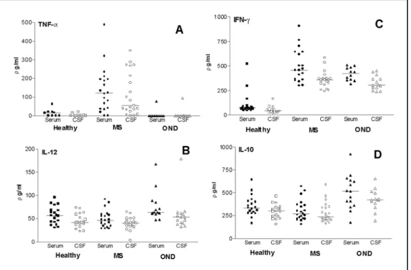
T N Fαlevels in the CSF exceeded those in the seru m for 10/23 patients, suggesting an intrathecal syn-thesis (Fig 1A). There was a positive corre l a t i o n between the number of leukocytes in the CSF and the level of TNFα(R2= 0.6874, p= 0.001).
Production of IL12p40 in CSF and serum – The means for MS patients were 49.1 ± 18.3 and 39.4 ± 13.2 pg/mL for serum and CSF, respectively; whe-reas for the OND group they were 76.2 ± 32.2 and 58.1 ± 35.1 pg/mL; for the healthy controls these means were 57.4 ± 18.8 and 45.4 ± 15.3 pg/mL. No significant diff e rence (p>0.05) were observed bet-ween the three groups (Fig 1 B).
P roduction of IFNγin CSF and serum patients with MS and, other neurological disorders, as well as healthy contro l s– The means for MS patients w e re 500.9 ± 169.5 and 359.0 ± 85.1 pg/mL in se-rum and CSF respectively; whereas for the OND g roup, these were 418.6 ± 67.0 and 325.2 ± 70.5 pg/mL; for the healthy controls these mean were 102.6 ± 102.7 and 54.1 ± 39.6 pg/mL. A significant i n c rease (p< 0.001) in the IFNγwas observed in both CSF and serum of patients with both MS and OND (Fig 1C).
P roduction of IL-10 in CSF and serum – The m e a n s for MS patients were 304.6 ± 112.0 and 287.6 ± Table 1. Clinical and laboratory data of relapsing-remitting multiple sclerosis group.
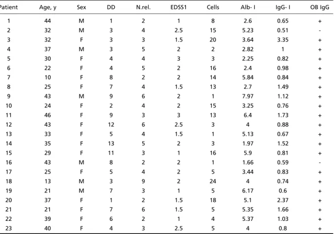
Patient Age, y Sex DD N.rel. EDSS1 Cells Alb- I IgG- I OB IgG
1 44 M 1 2 1 8 2.6 0.65 +
2 32 M 3 4 2.5 15 5.23 0.51
-3 32 F 3 3 1.5 20 3.64 3.35 +
4 37 M 3 5 2 2 2.82 1 +
5 30 F 4 4 3 3 2.25 0.82 +
6 22 F 4 5 2 16 2.4 0.98 +
7 10 F 8 2 2 14 5.84 0.84 +
8 25 F 7 4 1.5 13 2.7 1.49 +
9 43 M 9 6 2 1 7.97 1.12 +
10 24 F 2 4 2 15 3.25 0.76 +
11 46 F 9 3 3 13 6.4 1.73 +
12 43 F 12 6 2.5 3 4 0.88 +
13 33 F 5 4 1.5 1 5.13 0.67 +
14 35 F 13 5 2 3 1.97 1.52 +
15 29 F 11 3 1 16 5.9 0.81 +
16 43 M 8 2 2 1 1.66 0.59
-17 25 F 5 4 2 5 3.44 0.83 +
18 13 M 3 9 2 24 4 0.74 +
19 21 M 7 3 1 5 6.17 0.6 +
20 37 F 1 2 1.5 18 5.1 2.37 +
21 21 F 7 6 1.5 5 5.35 1.66 +
22 39 F 6 2 1 4 5.37 1.03 +
23 40 F 4 3 2.5 5 4 0.8 +
DD, duration of disease; EDSS1, Expanded Disability Status Scale at the time of the study; Alb-I, albumin index; IgG-I, IgG index; OB IgG, oligo-clonal IgG bands; N.rel., number of relapses; y, years.
Table 2. Patients characteristics, number of CSF leukocytes, intrathecal IgG synthesis index and oligoclonal bands.
Patients Age, y (mean± SD) Te, y Sex, M/F Cells IgG-index OB IgG+
MS (23) 30± 9 5 ± 3 6/ 17 9.13 ± 7.05 1.12 ± 0.66 21/ 23
ODN (16) 31± 10 6 ± 3 3/ 13 4.19 ± 5.55 0.53 ± 0.13 1/ 16
HC (19) 37 ± 16 - 6/ 13 1.47 ± 0.77 0.45 ± 0.12 0/ 19
113.7 pg/mL for serum and CSF, respectively; whe-reas for the OND group they were 507.6 ± 187.1 and 424.3 ± 113.7 pg/mL for serum and CSF, re s p e c-tively; for the healthy controls, these means were 354.6 ± 111.4 and 296.2 ± 74.8 pg/mL. No signifi-cant diff e rences were found between the patients with MS and healthy controls (p>0.05), where a s the OND group of patients revealed significantly higher levels of IL 10 (p=0.006) (Fig 1D).
DISCUSSION
The purpose of this investigation was to evalu-ate inflammatory activity in MS patients during clinical remission. Our results showed that patients with MS which is identified stable phase of the dis-ease reveal incrdis-ease in the secretion of pro - i n f l a m-m a t o ry cytokines in both serum-m and CSF associat-ed with the increase in the intrathecal synthesis of IgG and the number of leukocytes in the CSF. T h i s study demonstrated that patients with stable RRMS have an elevated number of leukocytes in the CSF when compared with patients with other non-in-f l a m m a t o ry diseases onon-in-f CNS and healthy contro l s . Other studies suggested that the presence of white blood cells in the CSF is a good predictor of the activity of MS, since after two years, patients with a high number of white blood cells in the CSF had more relapses than those in control group11.
Parallel to the increase of the number of cells there is also the presence of oligoclonal bands in 91% of studied MS patients. These results are in a g reement with previous Brazilian re p o rt s1 2 , 1 3. Al-though the antibody specificity in the oligoclonal bands is still enigmatic, as suggested by re p o rts in the literature, a lack of intrathecal synthesis of ol-igoclonal IgG bands is related to short lasting and benign course of MS14.
The B-cell proliferation, diff e rentiation and an-tibody production is coordinated by the helper T cells and the cytokines they produce. Although Th1 and Th2 cells are the major sources of their respective cytokines, many others cells within and outside the immune system also produce these cytokines, contributing to an overall Th1 and Th2 cytokine pattern. It was thus decided to quantify both pro and anti-inflammatory cytokines in the CSF and the serum, independent of their source.
The cytokines produced in the early phase of i n f l a m m a t o ry response, such as IL12, contribute to the development of Th1 immune response. Con-flicting results re g a rding IL12 production have, h o w e v e r, been observed in MS. An increase in the production of this cytokine has been observed in progressive MS15and increased frequencies of IL-12 secreting monocytes appear to correlate with Fig 1. Production of cytokines (TNFα, IL 12, IFNγand IL 10 ) in the CSF and sera from MS patients, OND (oth
the presence of active brain lesions detected by M R I1 6. Serum levels of IL12, however, have been found to be similar in MS patients and controls17. In the present study, as well, no change in the pro-duction of this cytokine was observed in the seru m or CSF of MS patients. It seems that, blood monocy-tes must be stimulated in order to produce detec-table amounts of IL12, but since we did not use activated cells, the production of IL12 may have been too low to be detected in the ELISA assay.
The IFNγof MS patients, on the other hand, showed significant increase in the CSF and serum over that of healthy controls, although no diff e re n-ce was observed in relation to the OND group. Ini-t i a l l y, inIni-terf e rons were Ini-tesIni-ted as Ini-therapeuIni-tic agenIni-ts for MS because of their antiviral pro p e rties and it was felt that MS might be due to persistent viral infection. However, a pathogenic role for MS pa-tients who received recombinant IFNγt re a t m e n t in MS has been re p o rt e d1 8. Our data support pre v i-ous ones which demonstrated that IFNγhas potent p ro i n f l a m m a t o ry response, including the ability to induce the production of other pro i n f l a m m a t o-ry cytokines1 9. In the animal model, the pro d u ct i o n of isotypes of IgG, such as IgG2a is induced by the I F Nγ. In our findings, no information is given about the subclass of IgG, which is increased in the CSF of MS patients, but it is possible that the incre a s e in intrathecal IgG production is due to the incre a s e in intrathecal synthesis of IFNγ.
In addition to the production of IFNγ, there was a parallel increase of TNFαin the serum and CSF. A p p roximately 40 % of the MS patients pre s e n t e d levels of TNFαin the CSF equal to or greater than that in the serum, suggesting the intrathecal syn-thesis of this cytokine. TNFαhas been described as a major cytokine in this demyelinating disease, since it has been demonstrated to be myelinotoxic2 0 - 2 3. T N Fαis also a major inducer of endothelial adhe-sion molecules and chemokines, hence, the upre g u-lation TNFαmay have a major effect on the re c ru i t-ment of leukocytes to the CNS2 4. In this study it was possible to show a positive correlation between the number of leukocytes in the CSF and the level of T N Fα. This pro p e rty may explain, at least in part, the i n c reased number of inflammation-perpetuating leukocytes observed in the CSF in MS patients. Elevation in CSF concentrations of soluble ICAM-1 and soluble TNFαreceptor were demonstrated pre-viously in Brazilian MS patients with acute re l a p s-ing form of MS durs-ing exacerbation2 5.
As the inflammatory response develops, the cy-tokine products of Th1 or Th2 lymphocytes pro v i-de mutually inhibitory functions for the diff e re n t i a-tion and effector effect of the re c i p rocal phenoty-pe. IFNγp revent Th2 cell proliferation, whereas I L 1 0 p rofoundly inhibits the synthesis of Th1 cytokines2 6. Reports on the IL-10 production showed that pa-tients with high IL-10 production had significant-ly lower disability scores and lower T2 lesion load2 7. In the present study, we did not observe signifi-cant changes in IL10 levels in MS patients. These data agree with that of studies demonstrating a reduction in IL10 levels in the serum, as well as in the number of IL10-secreting cells in MS pati-e n t s1 9 , 2 8 - 3 0, re i n f o rcing the fact that the pro i n f l a m-m a t o ry response prevails in this group of patients, despite the absence of clinical manifestation.
This study provides evidence of a significant in-c rease in inflammatory ain-ctivity in patients with stable MS over that in the control groups. These observations suggest that an investigation of the inflammatory parameters in the CSF may provide a valuable tool, which would be useful in the in-dication of activity of the disease, thus helping un-derstand the damages caused by the inflammato-ry response.
Acknowledgements -The authors acknowledge
the collaboration of Linda Gentry El-Dash in the linguis-tic revision of the manuscript and of Gislaine C.L. Brito for the technical assistance.
REFERENCES
1. Steinman L. Multiple sclerosis: a coordinated immunological attack against myelin in the central nervous system. Cell 1996;85:299-302. 2. Ota K, Matsui M, Milford EL, et al. T cell recognition of an
immunodo-minant myelin basic protein epitope in multiple sclerosis. Nature 1999; 346:183-187.
3. Sharief MK, Hentges R Association between tumor necrosis factor α
and disease pro g ression in patients with multiple sclerosis. N Engl J Med 1991;325:467-472.
4. Costa PM, Yasuda CL, Scagliusi SM, et al. Pattern of cytokines secre-tion by peripheral blood cells of patients with multiple sclerosis in Brazil. Mult Scler 2000;6:293-299.
5. Nicoletti F, Di Marco R, Patti F, et al. Blood levels of transforming g rowth factor beta 1 are elevated in both re l a p s i n g - remitting and chro n-ic pro g ressive multiple sclerosis patients and are further augmented by treatment with interferon beta. Clin Exp Immunol 1998;113:96-99. 6. Genain CP, Cannella B, Hauser SL, Raine CS. Identification of
autoan-tibodies associated with myelin damage in multiple sclerosis. Nature Med 1999;5:170-175.
7. Miller DH, Grossman RI, Reingold SC, Mcfarland HF. The role of mag-netic resonance techniques in understanding and managing multiple sclerosis. Brain 1998;121:3-24.
8. Poser CM, Paty DW, Scheinberg L, et al. New diagnostic criteria for multiple sclerosis: guidelines for re s e a rch protocols. Ann Neurol 1983; 13:227-231.
10. Keir G, Luxton RW, Thompson EJ. Isoelectric focusing of cere b ro s p i n a l fluid immunoglobulin G: an annotated update. Ann Clin Biochen 1990; 27:436-443.
11. Rudick R, Cookfair D, Simonian N, et al. Cere b rospinal fluid abnor-malities in a phase III trial of Avonex (IFNβ-1a) for relapsing multiple sclerosis. J Neuroimmunol 1999;93:8-14.
12. Puccioni-Sohler M, Passeri F, Oliveira C, Brandão CO, Papaiz-Alvare n g a R. Multiple sclerosis in Brazil. Analysis of cere b rospinal fluid by stan-dard methods. Arq Neuropsiquiatr 1999;57:927-931.
13. Puccioni-Sohler M, Lavrado FP, Bastos RRG, Brandão CO, Papaiz-A l v a renga R. Esclerose múltipla: correlação clínico laboratorial. Papaiz-A rq Neuropsiquiatr 2001;59:89-91.
14. Verjans E, Theys P, Delmotte P, Carton H. Clinical parameters and intrathecal IgG synthesis as prognostic features for multiple sclerosis. J Neurol 1983;229:155-165.
15. van Boxel-Dezaire AH, Hoff SC, van Oosten BW, et al. Decreased inter-leukin-10 and increased interleukin-12p40 mRNA a re associated with disease activity and characterize diff e rent disease stages in multiple sclerosis. Ann Neurol 1999;45:695-703.
16. Makhloufk K, Weiner HL, Khoury SJ. Increased percentage of IL-12+ monocytes in the blood correlates with the presence of active MRI le-sions in MS. J Neuroimmunol 2001;119:145-149.
17. Heesen C, Sieverding F,. Schoser BG, Hadji B, Kunze K. Interleukin 12 is detectable in sera of patients with multiple sclerosis- association with chronic progressive disease course? Eur J Neurol 1999;6:591-596. 18. Panitch HS, Hirsch RL, Schindler J, et al. Treatment of multiple
sclero-sis with gamma interferon: exacerbations associated with activation of the immune system. Neurology 1987;37:1097-1102.
19. Hohnoki K, Inoue A, Koh CS. Elevated serum levels of IFN gamma, IL4 and TNFαunelevated serum levels of IL10 in patients with de-myelinating diseases during the acute stage. J Neuroimmunol 1998; 87:27-32.
20. Buntinx M, Moreels M, Vandenabeele F, et al. Cytokine- induced cell
death in human oligodendroglial cell lines: I. Synergistic effects of
IFN-γand TNF-αon apoptosis. J Neuros Res 2004;76:834-835.
21. Rieckmann P, A l b recht M, Kitze B, et al. Cytokine mRNA levels in mo-nonuclear blood cells from patients with multiple sclerosis. Neuro l o g y 1994;44:1523-1526.
22. Choffon M, Jillard C, Gauthier G, et al. Tumor necrosis factor produc-tion as possible predictor of relapse in patients with multiple sclero s i s . Eur Cytokine Netw 1992;433:523-531.
2 3 . Baraczka K, Nékám K, Pozsonyi T, Szuts I, Ormos G. Investigation of cytokine (tumor necrosis factor-alpha, interleukin-6, interleukin-10) con-centrations in the cere b rospinal fluid of female patients with multiple s c l e rosis and systemic lupus erythematosus. Eur J Neurol 2004;11:37-42. 24. Hartung HP, Reiners K, A rchelos JJ, et al. Circulating adhesion molecu-les and tumor necrosis factor in multiple sclerosis: correlation with magnetic resonance imaging. Ann Neurol 1995;38:186-193. 25. Alves-Leon SV, Batista E, Papaiz-Alvarenga R, Quírico-Santos T.
Deter-mination of soluble ICAM-1 and TNFαR in the cere b rospinal fluid and s e rum levels in a population of Brazilian patients with re l a p s i n g - re m i t-ting multiple sclerosis. Arq Neuropsiquiatr 2001;59:18-22.
26. Mosmann TR, Sad S. The expanding universe of T-cell subsets: Th1, Th2 and more. Immunol Today 1996;138:138-146.
27. P e t e reit HF, Pukrop R, Fazecas F, et al. Low interleukin- 10 pro d u c t i o n is associated with higher disability and MRI lesion load in secondary progressive multiple sclerosis. J Neurol Sci 2003;206:209-214. 28. Filion LG, Graziani-Bowering G, Matusevicius D, Freedman MS.
Mo-nocyte- derived cytokines in multiple sclerosis. Clin Exp Immunol 2003;131:324-334.
29. Huang W-X, Haung P, Link H, Hillert J. Cytokine analysis in multiple sclerosis by competitive RT PCR: a decreased expression of IL 10 and an increased expression of TNFαin chronic pro g ression. Mult Scler 1999;5:342-348.