Expression of neuronal nitric oxide
synthase in the developing superficial
layers of the rat superior colliculus
Laboratório de Neurobiologia Celular e Molecular, Instituto de Biofísica Carlos Chagas Filho,
Universidade Federal do Rio de Janeiro, Rio de Janeiro, RJ, Brasil
A. Giraldi-Guimarães, R.E. Bittencourt-Navarrete and R. Mendez-Otero
Abstract
We investigated the level of expression of neuronal nitric oxide synthase (nNOS) in the retinorecipient layers of the rat superior colliculus during early postnatal development. Male and female Lister rats ranging in age between the day of birth (P0) and the fourth postnatal week were used in the present study. Two biochemical methods were used, i.e., in vitro measurement of NOS specific activity by the conversion of [3H]-arginine to [3H]-citrulline, and analysis of Western blotting immunoreactive bands from superior colliculus ho-mogenates. As revealed by Western blotting, very weak immunoreac-tive bands were observed as early as P0-2, and their intensity increased progressively at least until P21. The analysis of specific activity of NOS showed similar results. There was a progressive increase in enzymatic activity until near the end of the second postnatal week, and a nonsignificant tendency to an increase until the end of the third week was also observed. Thus, these results indicated an increase in the amount of nNOS during the first weeks after birth. Our results confirm and extend previous reports using histochemistry for NADPH-dia-phorase and immunocytochemistry for nNOS, which showed a pro-gressive increase in the number of stained cells in the superficial layers during the first two postnatal weeks, reaching an adult pattern at the end of the third week. Furthermore, our results suggested that nNOS is present in an active form in the rat superior colliculus during the period of refinement of the retinocollicular pathway.
Correspondence
R.M. Otero Instituto de Biofísica
Carlos Chagas Filho, CCS, Bloco G 21941-590 Rio de Janeiro, RJ Brasil
Fax: +55-21-2280-8193 E-mail: [email protected] Research supported by CNPq, PRONEX, and FAPERJ to R. Mendez-Otero and a grant from the MCT to the Millennium Institute for Tissue Bioengineering, Brazil.
Received June 24, 2003 Accepted February 18, 2004
Key words
•Superior colliculus •Nitric oxide synthase •Development •Retinotectal projection
Introduction
Nitric oxide (NO) is a membrane-perme-able gas that was first identified as endothe-lium-derived relaxing factor (1). However, it is also synthesized in the nervous system and functions as an important intercellular mes-senger in neurotransmission, excitotoxicity, plasticity, and control of cerebral blood flow (2-4). The enzyme responsible for NO
regions (7-12). nNOS was first described as a constitutively expressed protein; however, recent studies have shown that nNOS ex-pression levels change in response to a vari-ety of physiological and pathological stimuli (13).
The study of NOS expression during the development of the retinotectal pathway is relevant in view of some evidence indicating a role for NO in the formation of the topo-graphic map in this system. For example, in chickens, the elimination of the ipsilateral ret-inotectal projections is partially dependent on the NO synthesized in the tectum opticum (14). Furthermore, several studies on rodents have suggested that NO might be involved in the topographic establishment of the ipsilat-eral retinotectal projections (15-18).
The presence of NOS-positive cells in the retinorecipient superficial layers of the superior colliculus has been reported in sev-eral mammalian species (19-25). In the rat, a subpopulation of all the different cell types described in the superficial layers of the superior colliculus by the Golgi technique (26) expresses nNOS (21,27,28). Previous studies have also shown that the first NOS-positive cells in these layers appear by the end of the first postnatal week (between the fifth and the seventh postnatal day). Their numbers increase over the following days, reaching an adult pattern of NOS expression at the end of the third week (19,20).
All of these previous studies dealt with qualitative aspects of the expression of nNOS in the superior colliculus. In the present study, we have investigated nNOS expression and activity during the postnatal development of the superficial layers of the rat superior col-liculus by means of quantitative biochemical analysis and Western blotting.
Material and Methods
Animals
We used male and female Lister rats
ranging in age between the day of birth (P0) and the fourth postnatal week. The animals were housed in our colony in a room with controlled temperature and maintained on a 12:12-h light/dark cycle with food and water
available ad libitum. All experimental
proto-cols were approved by the Committee for the Use of Experimental Animals of our institu-tion and followed NIH guidelines.
Chemicals
L-2, 3, 4, 5-[3H]-arginine
monohydro-chloride was obtained from Amersham (São Paulo, SP, Brazil). The ion-exchange resin
DOWEX AG50W-X8 (Na+ form) was
ob-tained from Bio-Rad (Hercules, CA, USA). The enhanced chemiluminescence Western blotting system was obtained from
Amer-sham PharmaciaBiotech (São Paulo, SP,
Brazil), including a secondary antibody conjugated with horseradish peroxidase (HRP). All other reagents and chemicals were purchased from Sigma (St. Louis, MO, USA).
Antibodies
Two anti-nNOS antibodies were used: a monoclonal (mouse) anti-nNOS (Sigma), which was prepared against a fragment (amino acids 1-181) of the N-terminal re-gion of nNOS from the rat brain (29); a polyclonal (rabbit) anti-nNOS (Transduction Laboratories, Lexington, KY, USA), which was prepared against the C-terminal region of nNOS (amino acids 1095-1289). The monoclonal antibody recognizes only the
nNOSα isoform and the polyclonal antibody
recognizes the α, ß and γ isoforms.
Tissue preparation for biochemical analysis
Animals ranging from P0 to P23 were decapitated under deep anesthesia with
so-dium pentobarbital (50 mg/kg, ip) and the
upper layers of the superior colliculus were carefully dissected and washed with cold 0.9% NaCl. They were then weighed and
homogenized at 4oC in five volumes (w/v) of
50 mM Tris buffer, pH 7.0, containing 320 mM sucrose, 1 mM ethylenediaminetetra-acetic acid (EDTA), 1 mM dithiothreitol (DTT), 10 µg/ml leupeptin, 10 µg/ml soy-bean trypsin inhibitor, and 2 µg/ml aproti-nin. For both the enzymatic activity meas-urements and the electrophoretic procedure, each homogenate consisted of the tissue dis-sected from a single animal, corresponding to an experimental sample. The
homoge-nates were then centrifuged at 10,000 g for
20 min at 4oC to mainly remove the blood
vessels and nuclear material. Only the super-natants were used in the assays.
NOS activity
NOS activity was determined by measur-ing the formation of citrulline from L-arginine (30). Briefly, 10-20 µl of each sample was added to Eppendorf microtubes with 50 µl of 50 mM potassium phosphate buffer,
pH 7.2, containing 1.2 mM MCl2, 0.25 mM
CaCl2, 60 mM L-valine, 1.2 mM
L-citrul-line, 25 µM L-arginine, 1 mM DTT, 4 µM flavin adenine dinucleotide, 4 µM flavin
mononucleotide, 120 µM NADPH, and [3
H]-L-arginine (1 mCi/ml, 59 Ci/mmol). After
incubation for 30-45 min at 25oC, the
reac-tion was stopped by the addireac-tion of 500 µl
Dowex resin AG-50WX8 (200-800, Na+
form) (1:1 in water). The microtubes were
then centrifuged at 2,000 g for 5 min to pellet
the resin and an aliquot of the supernatants was removed to measure the emission of ß
radiation from [3H]-L-citrulline by
liquid-scintillation counting. A 10-20-µl aliquot of the same sample was added to microtubes
containing 1 mM EGTA (to detect the Ca2+
-independent NOS activity) or 1 mM EGTA
plus 1 mM Nω-nitro-L-arginine (to provide a
negative control for counting). These prepa-rations were used to obtain the blank values
and no difference between the two blank microtubes was found. The final counting value of each sample was the number of counts less the counts of the blank microtube. Each sample and its respective blank were prepared in duplicate, and the mean was used for the calculations. The protein con-centration of the samples was determined by the technique of Bradford (31) and the enzy-matic activity was reported as picomol of citrulline per mg of total protein per min for each sample.
Data were analyzed statistically by ANOVA and by the Tukey multiple compar-ison post-test.
Electrophoresis (SDS-PAGE) and Western blotting
An aliquot containing 30 µg protein (for immunoblotting using the monoclonal anti-body) or 65 µg protein (for immunoblotting using the polyclonal antibody) from each sample was mixed with electrophoresis sample buffer containing 50 mM Tris-HCl, 2% (w/v) sodium dodecyl sulphate (SDS), 5% (v/v) ß-mercaptoethanol, pH 6.8, and 10% glycerol (v/v). Samples were boiled for 5 min prior to loading onto 10% SDS-polyacrylamide gel for electrophoresis using the Mini-Protean 3 electrophoresis module (BioRad). After separation on the gel, the proteins were transferred to a nitrocellulose membrane (0.45-µm pore) and submitted to the following sequential incubations: 5% milk in 10 mM phosphate-buffered sa-line (PBS) for 1 h, monoclonal (1:2000) or polyclonal (1:1000) anti-nNOS antibodies diluted in the previous solution overnight followed by the secondary antibody (goat anti-mouse or goat anti-rabbit) conjugated to HRP and diluted 1:5000 in 10 mM PBS
and 0.1% nonidet P-40 for 2 h. Afterseveral
additional washes in PBS, immunoreactive
bands were visualized ona film
Histochemistry and immunohistochemistry
Animals were anesthetized with an
over-dose of sodium pentobarbital (50 mg/kg, ip)
and perfused intracardially with cold 0.9% NaCl followed by 4% paraformaldehyde (PF) or 2% glutaraldehyde (GLUT) + 0.5% PF in 100 mM phosphate buffer, pH 7.4 (29). Brains were removed and immersed in 100 mM phosphate buffer containing 20%
su-crose for 24 h at 4oC. After this period, the
midbrain was cut into 30-60-µm sections at -20°C with a CM 1850 cryostat (Leica In-struments GmbH, Heidelberg, Baden-Wurttemberg, Germany). For NADPH-dia-phorase (NADPH-d) histochemistry, the brain sections from animals perfused with one or the other fixative solution were washed twice in 50 mM Tris-HCl buffer, pH 7.4, and reacted as
free-floating sections at 37oC for at least 1 h.
The reaction solution contained 1 mM ß-NADPH (reduced form), 0.5 mM nitroblue tetrazolium dissolved in dimethylsulfoxide (DMSO, at a final concentration of 1:1000 DMSO:buffer, v/v) and 1% Triton X-100 in 50 mM Tris-HCl buffer, pH 7.4. As a control, some sections were incubated without ß-NADPH, with no staining being observed.
For immunohistochemistry, brain sections from animals perfused with 4% PF were washed twice in PBS (100 mM phosphate buffer, pH 7.4, and 0.9% sodium chloride) and then preincubated as free-floating sec-tions in PBS + 0.3% Triton X-100 with 10% normal goat serum for 60 min at room tem-perature. They were then incubated in the same solution supplemented with the mono-clonal anti-nNOS antibody (1:400) for 24 h. Bound primary antibody was visualized by means of a Cy3-conjugated goat anti-mouse antibody (Jackson; 1:1000) incubated for 2 h. Control sections prepared without incuba-tion with the primary antibody did not show any staining.
Sections reacted for NADPH-d were ex-amined under light field illumination and those reacted for immunohistochemistry were
analyzed under fluorescent illumination, us-ing a Zeiss microscope in both cases. Images were obtained with an AxioCam digital cam-era connected to the microscope and the Zeiss Axiovision 3.0 software.
Results
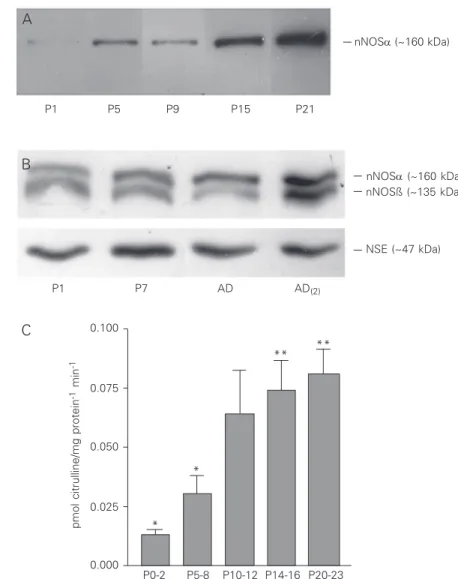
We used the immunoblot and enzyme activity to demonstrate the presence of nNOS in the tissue homogenate obtained from the developing rat superior colliculus to com-plete the histochemical data (19,20). There was a progressive increase in the amount of the enzyme during the first three weeks after birth. Figure 1A shows a representative im-munoblotting using a specific monoclonal
antibody to the nNOSα isoform (estimated
molecular mass: 160 kDa). A very weak immunoreactive band was observed as early as P0-2, with its intensity increasing progres-sively at least until P21, the last age studied in the present investigation. Interestingly, when using a polyclonal antibody that recog-nizes all the isoforms of nNOS, a second
band was observed in addition to the nNOSα
isoform band. This second band was slightly lighter than the first and corresponded to the nNOSß isoform with a molecular mass near 135 kDa (Figure 1B) (32). Although the intensity of this second band varied, it seemed to increases progressively, as observed for
the nNOSα isoform band (Figure 1B). These
results indicate that both isoforms follow the same pattern of expression during develop-ment of the superior colliculus.
enzy-matic activity of nNOS from the first postna-tal days (P0-2) to the third week after birth (P20-23). There was a progressive increase in enzymatic activity until near the end of the second postnatal week, followed by a non-significant tendency to an increase until the end of the third week (Figure 1C). Thus, the results of both the immunoblotting and the catalytic activity measurements indicated an increase in the amount of the nNOS during the first weeks after birth. However, the level of activity seemed to reach a peak first, despite a subsequent increase in the protein level (Figure 1A). Part of the enzymatic
ac-Figure 1. Demonstration of neuronal nitric oxide syn-thase (nNOS) in rat superior colliculus homogenates by immunoblot and enzymatic activity. A, Western blot analysis of nNOS in extracts from the upper layers of the rat superior colliculus obtained at postnatal (P) ages (in days) P1, P5, P9, P15 and P21. Equal amounts of protein were loaded onto each well. The bands which were immunoreactive with monoclonal anti-nNOSα were visualized with a secondary antibody conjugated with horseradish peroxidase. Note a gradual increase in the expression of nNOSα (~160 kDa) over the first postnatal weeks. The illustrated film is representative of two independent experiments, each using different animals. B, Western blot analysis of nNOS in extracts from the upper layers of the rat superior colliculus ob-tained at P1, P7 and during adulthood using a second anti-nNOS antibody that recognizes all nNOS isoforms. In addition to the nNOSα band, note a second band which corresponds to nNOSß (~135 kDa). Despite the variability in the intensity of this second band, as illus-trated in the lines obtained from the two different adults (AD and AD(2)) used in this representative experiment,
nNOSß expression also seemed to increase during post-natal development. The film presented is representa-tive of two independent experiments, each using differ-ent animals. Neuron-specific enolase (NSE, ~47 kDa) was used as a “housekeeping” protein to control load-ing and, as expected, no difference in protein levels was observed. C, NOS activity in extracts from the upper layers of rat superior colliculus, reported as pmol citrul-line/mg protein-1 min-1. The enzymatic activity increased
from the beginning of the first postnatal week (P0-2) to the end of the third postnatal week (P20-23), in agree-ment with the immunoblot results. Maximum NOS ac-tivity was reached in the course of the third postnatal week. Each bar represents the mean ± SEM of enzyme activity for each group: P0-2 (N = 6), P5-8 (N = 10), P10-12 (N = 5), P14-16 (N = 7), and P20-23 (N = 6). ANOVA revealed a significant difference
be-tween all groups (P < 0.001). Tukey’s multiple comparisons post-test revealed a significant difference bebe-tween P0-2 and P10-12 (*P < 0.05), P0-2 and P14-16 (**P < 0.01), P0-2 and P20-23 (**P < 0.01), P5-8 and P14-16 (*P < 0.05), and P5-8 and P20-23 (*P < 0.05).
tivity could be due to the endothelial eNOS although most of the blood vessels were removed during the centrifugation proce-dure. Thus, we checked the possibility that some neurons could express eNOs since the expression of this isoform by neurons has been already described in other regions of the central nervous system (29). eNOS im-munohistochemistry revealed no significant staining at P5, indicating that this isoform does not contribute to the catalytic activity of the NOS observed in the superficial layers of the superior colliculus at this age (data not shown).
nNOSα (~160 kDa) nNOSα (~160 kDa)
NSE (~47 kDa) nNOSß (~135 kDa)
P1 P5 P9 P15 P21
P1 P7 AD AD(2)
0.100
0.075
0.050
0.025
0.000
pmol citrulline/mg protein
-1 min
-1
* *
** **
P0-2 P5-8 P10-12 P14-16 P20-23
A
B
Previous studies using NADPH-d his-tochemistry in the rat superior colliculus (19,20) have reported the appearance of the first NOS-positive cells only at P7. How-ever, with both the immunoblots and radio-enzymatic assays we were able to detect the enzyme and its activity as early as P0 al-though at very low levels. It is possible that the histochemical and
immunohistochemi-cal studies were not sensitive enough to de-tect these low levels of the enzyme. Alterna-tively, it is possible that in this region nNOS staining might be highly sensitive to the fixation protocols used, as reported for other regions of the central nervous system (29,33). We then performed NADPH-d staining us-ing different fixative solutions and nNOS immunohistochemistry. NADPH-d
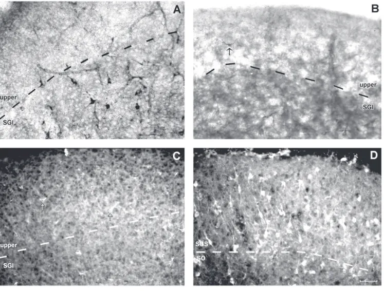
histo-Figure 2. Neuronal nitric oxide synthase (nNOS) histochemistry and immunohistochemistry in the rat superior colliculus. A, NADPH-d histochemistry at P5 after fixation with 4% paraformaldehyde (PF). No stained cells were observed in the upper layers, but a few labelled cells were found in the stratum griseum intermediale (SGI). B, NADPH-d histochemistry at P5 after fixation with 2% glutaraldehyde (GLUT) + 0.5% PF. Note that this fixation procedure increased the sensitivity of the NADPH-d reaction, with some stained cells (arrow) now being observed at this age, earlier than with 4% PF.
chemistry in animals fixed with 4% PF re-vealed no stained cells in the upper layers of the superior colliculus at P5 (Figure 2A), confirming previous results (19,20). How-ever, NADPH-d histochemistry in tissue fixed with 2% GLUT + 0.5% PF showed stained cells in these layers at this age, although in very small numbers (Figure 2). These results were also confirmed by immunohistochem-istry (Figure 2C) which revealed scattered cells in the superior colliculus at P5. Thus, NADPH-d histochemistry after fixation with GLUT or immunohistochemistry was appar-ently more sensitive than NADPH-d his-tochemistry after fixation with 4% PF in detecting the lower levels of nNOS at P5. However, in adulthood the number of stained cells dramatically increased (Figure 2D), as also reported previously (19,21,27,28).
Discussion
We have quantitatively analyzed the ex-pression of nNOS in the superficial layers of the rat superior colliculus during the period of postnatal development. In general, our results agree with previous histochemical studies of the expression of this enzyme (19,20) which showed a progressive increase in the number of stained cells in the collicular retinorecipient layers of the superior collicu-lus during the first two postnatal weeks, reaching an adult pattern of expression at the end of the third week. The total amount of nNOS protein assayed by Western analysis, as well as its catalytic activity measured by the conversion of L-arginine to L-citrulline, also followed a similar pattern during the first postnatal weeks, reaching a maximal amount during the third postnatal week, the last period of postnatal development studied here. We cannot exclude a possible decrease in NOS expression after the third week. Cork et al. (23) observed a decrease after the peak of NOS expression during the postnatal de-velopment of the mouse superior colliculus by histochemical labeling. Nevertheless,
since our biochemical data correlated well with the previous histochemical descriptions for the rat superior colliculus, we would argue that a decrease after the third week is not likely because the previous studies did not show a decrease in NOS staining at this time (19,20).
Our results differ from those observed by Campello-Costa et al. (16). In their study, using a radioenzymatic assay and a his-tochemical assessment of NADPH-d activ-ity from homogenates of the superficial lay-ers of the superior colliculus, they showed that the maximal level of nNOS expression was at postnatal day 5 and that there was a decrease in the enzymatic activity during the following weeks reaching a significantly lower level in adulthood. At present, we are not able to explain the discrepancy with our results obtained with immunochemistry and enzyme activity which did not show any peak at P5. In addition, from their results (16), we would expect to find a larger num-ber of NADPH-positive cells at P5 in tissue sections and also in immunohistochemical staining. However, using the most sensitive variations of these techniques we did not find a significant number of labeled cells at P5. Actually, at this age only very few cells are labeled and the number and intensity of staining increases progressively, reaching a peak only during the third postnatal week.
We also were able to conclude that there is no significant expression of eNOS by the collicular cells at the end of the first postna-tal week since the use of NAPDH-d with GLUT did not show any significant staining at this age. This last possibility was also assessed by immunohistochemistry for eNOS. Similarly, we did not observe any significant staining at P5 (data not shown), confirming that this isoform did not contri-bute to the low catalytic activity of NOS observed in the superficial layers of the su-perior colliculus.
acti-vation of the N-methyl-d-aspartate (NMDA) receptor, and it has been suggested that NO could act as a retrograde messenger in syn-aptic plasticity, influencing both the refine-ment and stabilization of co-active afferent terminals and the induction of long-term changes in synaptic potency (2,34-38). Our results showed that during the first two post-natal weeks, the period when the final reti-notopic map is established in the rat superior colliculus (39), nNOS is expressed at lower levels than in adulthood. However, the po-tential amount of NO synthesis by the nNOS present in this period, as assayed indirectly by the levels of enzymatic activity, seems to be sufficient to participate in the formation of this map, since previous studies with NOS inhibitors have shown that the blockage of NO synthesis impairs the refinement of retinocollicular projections (16,17).
Long-term changes in retinocollicular transmission might be involved in the refine-ment of the retinocollicular projection dur-ing the postnatal development of the rat su-perior colliculus (37). In fact, both long-term depression and long-term potentiation can be induced in the superior colliculus after
stimulation of optic tract fibers in an in vitro
isolated brainstem preparation during the
period of refinement of this pathway (40). In addition, the cited investigators have shown that in the superior colliculus, as in other structures, long-term potentiation is NMDA receptor-dependent (40). Since NO synthe-sis has also been linked to the NMDA recep-tor activity, it is possible that the role of NO in the retinocollicular refinement might be through a mechanism of long-term synaptic strengthening during postnatal development. A role for NO in the induction of long-term potentiation has been suggested by several investigators (2,35), although this hypothesis is still the subject of some controversy (38). Additional studies are needed to identify the involvement of NO in the induction of long-term potentiation in the superior colliculus, which would shed light on the functional roles of this molecule in collicular physiolo-gy.
Acknowledgments
We thank Dr. Leny A. Cavalcante (Insti-tuto de Biofísica Carlos Chagas Filho, Uni-versidade Federal do Rio de Janeiro) for helpful comments about the manuscript and Felipe Marins for technical support.
References
1. Palmer RM, Ferrige AG & Moncada S (1987). Nitric oxide release accounts for the biological activity of endothelium-derived relaxing factor. Nature, 327: 524-526.
2. Dawson TM & Snyder SH (1994). Gases as biological messengers: nitric oxide and carbon monoxide in the brain. Journal of Neurosci-ence, 14: 5147-5159.
3. Schuman EM & Madison DV (1994). Nitric oxide and synaptic func-tion. Annual Review of Neuroscience, 17: 153-183.
4. Cudeiro J & Rivadulla C (1999). Sight and insight - on the physiologi-cal role of nitric oxide in the visual system. Trends in Neuroscien-ces, 22: 109-116.
5. Bredt DS & Snyder SH (1992). Nitric oxide, a novel neuronal mes-senger. Neuron, 8: 3-11.
6. Förstermann U, Schmidt HHHW, Pollock JS, Sheng H, Mitchell JA, Warner TD, Nakane M & Murad F (1991). Isoforms of nitric oxide synthase: characterization and purification from different cell types.
Biochemical Pharmacology, 42: 1849-1857.
7. Mikuzawa K, Vincent SR, McGeer PL & McGeer EG (1989). Distribu-tion of reduced nicotinamide adenine dinucleotide phosphate
dia-phorase-positive cells and fibers in the cat central nervous system.
Journal of Comparative Neurology, 279: 281-311.
8. Bredt DS, Glatt CE, Hwang PM, Fotuhi M, Dawson TM & Snyder SH (1991). Nitric oxide synthase protein and mRNA are discretely local-ized in neuronal populations of the mammalian CNS together with NADPH diaphorase. Neuron, 7: 615-624.
9. Brüning G, Funk U & Mayer B (1994). Immunocytochemical localiza-tion of nitric oxide synthase in the brain of the chicken. NeuroRe-port, 5: 2425-2428.
10. Brüning G, Katzbach R & Mayer B (1995). Histochemical and immu-nocytochemical localization of nitric oxide synthase in the central nervous system of the goldfish, Carassius auratus. I. Journal of Comparative Neurology, 358: 353-382.
13. Förstermann U, Boissel J & Kleinert H (1998). Expressional control of the ‘constitutive’ isoforms of nitric oxide synthase (NOS I and NOS III). FASEB Journal, 12: 773-790.
14. Wu HH, Williams CV & McLoo SC (1994). Involvement of nitric oxide in the elimination of transient retinotectal projection in devel-opment. Science, 265: 1593-1596.
15. Mize RR, Wu HH, Cork RJ & Scheiner CA (1998). The role of nitric oxide in development of patch-cluster system and retinocollicular pathways in the rodent superior colliculus. Progress in Brain Re-search, 118: 134-152.
16. Campello-Costa P, Fosse Jr AM, Ribeiro JC, Paes-de-Carvalho R & Serfaty CA (2000). Acute blockade of nitric oxide synthesis induces disorganization and amplifies lesion-induced plasticity in the rat retinotectal projection. Journal of Neurobiology, 44: 371-381. 17. Vercelli A, Garbossa D, Biasiol S, Repici M & Jhaveri S (2000). NOS
inhibition during postnatal development leads to increased ipsilat-eral retinocollicular and retinogeniculate projections in rats. Euro-pean Journal of Neuroscience, 12: 473-490.
18. Wu HH, Cork RJ, Huang PL, Shuman DL & Mize RR (2000). Refine-ment of the ipsilateral retinocollicular projections is disrupted in double endothelial and neuronal nitric oxide synthase gene knock-out mice. Developmental Brain Research, 120: 105-111.
19. González-Hernández T, Conde-Sendín M, González-González B, Mantolan-Sarmiento B, Perez-González H & Meyer G (1993). Post-natal development of NADPH-diaphorase activity in the superior colliculus and the ventral lateral geniculate nucleus of the rat. Devel-opmental Brain Research, 76: 141-145.
20. Tenório F, Giraldi-Guimarães A & Mendez-Otero R (1995). Develop-mental changes of nitric oxide synthase in the rat superior collicu-lus. Journal of Neuroscience Research, 42: 633-637.
21. Tenório F, Giraldi-Guimarães A & Mendez-Otero R (1996). Morphol-ogy of NADPH-diaphorase-positive cells in the retinoceptive layers of the developing rat superior colliculus. International Journal of Developmental Neuroscience, 14: 1-10.
22. Giraldi-Guimarães A, Tenório F, Brüning G, Mayer B, Mendez-Otero R & Cavalcante LA (1999). Nitric oxide synthase expression in the opossum superior colliculus: a histochemical, immunohistochemi-cal and biochemiimmunohistochemi-cal study. Brain, Behavior and Evolution, 199: 303-313.
23. Cork RJ, Calhouin T, Perrone M & Mize RR (2000). Postnatal devel-opment of nitric oxide synthase expression in the mouse superior colliculus. Journal of Comparative Neurology, 427: 581-592. 24. Scheiner CA, Kratz KE, Guido W & Mize RR (2001). Prenatal and
postnatal expression of nitric oxide in the developing kitten superior colliculus revealed with NADPH diaphorase histochemistry. Visual Neuroscience, 18: 43-54.
25. González-Soriano J, Contreras-Rodríguez J, Martínez-Sainz P, Martín-Palacios S, Marín-García P & Rodríguez-Veiga E (2002). NADPH-diaphorase distribution in the rabbit superior colliculus and co-localization with calcium-binding proteins. Journal of Anatomy, 200: 297-308.
26. Langer TP & Lund RD (1974). The upper layers of the superior colliculus: A Golgi study. Journal of Comparative Neurology, 158: 405-436.
27. González-Hernández T, Conde-Sendín TM & Meyer G (1992). Lami-nar distribution and morphology of NADPH-diaphorase containing neurons in the superior colliculus and underlying periaqueductal grey of the rat. Anatomy and Embryology, 186: 245-250.
28. Soares-Mota M, Henze I & Mendez-Otero R (2001). Nitric oxide synthase-positive neurons in the rat superior colliculus: colocaliza-tion of NOS with NMDAR1 glutamate receptor, GABA and parvalbu-min. Journal of Neuroscience Research, 64: 501-507.
29. Dinerman JL, Dawson TM, Schell MJ, Snowman A & Snyder SH (1994). Endothelial nitric oxide synthase localized to hippocampal pyramidal cells: implications for synaptic plasticity. Proceedings of the National Academy of Sciences, USA, 91: 4214-4218.
30. Assreuy J, Cunha FQ, Liew FY & Moncada S (1992). Feedback inhibition of nitric oxide synthase by nitric oxide. British Journal of Pharmacology, 108: 833-837.
31. Bradford MM (1976). A rapid and sensitive method for the quantita-tion of microgram quantities of protein utilizing the principle of protein-dye-binding. Analytical Biochemistry, 72: 248-254. 32. Batista C, Carneiro K, De Bittencourt-Navarrete RE, Soares-Mota M,
Cavalcante LA & Mendez-Otero R (2003). Nitrergic dendrites in the superficial layers of the rat superior colliculus: Retinal afferents and alternatively spliced isoforms in normal and deafferented animals.
Journal of Neuroscience Research, 71: 455-461.
33. González-Hernandez T, Perez de la Cruz MA & Mantolan-Sarmiento B (1996). Histochemical and immunohistochemical detection of neurons that produce nitric oxide: effect of different fixative param-eters and immunoreactivity against non-neuronal NOS antisera.
Journal of Histochemistry and Cytochemistry, 44: 1399-1413. 34. Daniel H, Hemart N, Jaillard T & Crepel F (1993). Long-term
depres-sion requires nitric oxide and guanosine 3':5' cyclic monophos-phate production in rat cerebellar Purkinje cells. European Journal of Neuroscience, 5: 1079-1082.
35. Son H, Hawkins RD, Martin K, Kiebler M, Huang PL, Fishman MC & Kandel ER (1996). Long-term potentiation is reduced in mice that are doubly mutant in endothelial and neuronal nitric oxide synthase.
Cell, 87: 1015-1023.
36. Holscher C (1997). Nitric oxide, the enigmatic neuronal messenger: its role in synaptic plasticity. Trends in Neurosciences, 20: 298-303. 37. Mize RR & Lo FS (2000). Nitric oxide, impulse activity and neurotro-phins in visual system development. Brain Research, 886: 15-32. 38. Prast H & Philippu A (2001). Nitric oxide as modulator of neuronal
function. Progress in Neurobiology, 64: 51-68.
39. Simon DK & O’Leary DD (1992). Development of topographic order in the mammalian retinocollicular projection. Journal of Neurosci-ence, 12: 1212-1232.