(1) Instituto da Criança “Prof. Pedro de Alcântara”, Hospital das Clínicas, University of São Paulo Medical School, São Paulo, Brazil. (2) Microbiology Section of Central Laboratory, Hospital das Clínicas, University of São Paulo Medical School, São Paulo, Brazil. (3) Laboratory of Virology, Instituto de Medicina Tropical de São Paulo, University of São Paulo Medical School, São Paulo, Brazil.
Correspondence to: Dr. Luiz Vicente Ferreira da Silva Filho, Instituto da Criança/HCFMUSP, Rua Artur de Azevedo, 1690, apt. 802, 05404-004 São Paulo, SP, Brazil. Fax: +55 11 3069-8503. E-mail: [email protected]
USE OF SELECTIVE MEDIUM FOR Burkholderia cepacia ISOLATION IN RESPIRATORY
SAMPLES FROM CYSTIC FIBROSIS PATIENTS
Luiz V.F. da SILVA FILHO(1,3), Luciana de F. VELLOSO(1), Christina N.O. BENTO(2), Edelyn GYTIN(2), Adriana F. TATENO(3), José E. LEVI(3), Joaquim C. RODRIGUES(1) & Sonia R.T.S. RAMOS(1)
SUMMARY
Burkholderia cepacia colonizes cystic fibrosis (CF) patients. We evaluated the impact of the use of aselective medium in the rate of B. cepacia recovery from respiratory samples of CF patients. During a 6-month period, respiratory samples were collected from 106 CF patients and cultivated on selective media including a B. cepacia selective medium. Confirmation of the identity of B. cepacia isolates was carried out by species specific PCR and determination of genomovar status performed by a sequential PCR approach. Results of B. cepacia isolation during this period were compared to the preceding two years, when the sample processing was identical except for the lack of the B. cepacia selective medium. B. cepacia was isolated in 11/257 (4.2%) of thesamples using the selective medium, in contrast with the preceding two years, when it was isolated in 6/1029 samples (0.58%), p < 0.0001. Identity of all 11 isolates was confirmed by PCR and genomovar determination was accomplished in all but one isolate. These results suggest that the use of a selective medium increases recovery rate of B. cepacia from respiratory samples.
KEYWORDS:Burkholderia cepacia, Selective medium, Cystic fibrosis, Sputum, Genomovars.
INTRODUCTION
Cystic fibrosis (CF) is an inherited disease characterized by chronic obstructive lung disease, pancreatic insufficiency and elevated chloride levels in the sweat22,28. Cystic fibrosis patients are particularly susceptible
to infections caused by specific bacterial pathogens such as Pseudomonas aeruginosa, Staphylococcus aureus and Haemophilus influenzae. Despite substantial advances in the prognosis due to antimicrobial therapy and early diagnosis, respiratory disease remain the first cause of death in these patients8.
Burkholderia cepacia, a plant pathogen, was identified in sputum samples from cystic fibrosis patients in the early 80s13,18,23. In some
patients, isolation of this bacteria was associated with the occurrence of a fatal necrotizing pneumonia called B. cepacia syndrome but in the majority of patients it caused an increase of lung function deterioration. The inherent resistance to the majority of antimicrobial drugs and further observations that B. cepacia strains can spread by social contact among patients, resulted in several recommendations for new approaches on microbiological surveillance4,25 and patient cohorting9,15. As a result,
isolation of B. cepacia in sputum from cystic fibrosis patients carries a significant medical and psychosocial burden. Strategies for B. cepacia infection control, however, depend on the accurate identification of this pathogen on the microbiology laboratory.
B. cepacia is a fastidious gram-negative bacillus that can be difficult to isolate, since it usually grows slowly when compared to other organisms frequently found in sputum samples from CF patients, such as P. aeruginosa. B. cepacia is also difficult to identify after isolation, and misidentifications occur very frequently20. Although the use of
selective media is actually recommended for B. cepacia isolation, this is not an homogenous practice among microbiology laboratories involved in the care of cystic fibrosis patients25. Furthermore, recent taxonomic
analyses have demonstrated that B. cepacia is not an unique species, but a complex that comprises six different genomovars or genomic species26,
and this characterization depends mainly on genomic analysis2,16,17,21,29.
The aim of this study was to compare the rate of isolation of B. cepacia during this period with the rate of isolation in the previous two years, when sample processing was identical, except for the lack of the B. cepacia selective medium. Confirmation of the identity of B. cepacia isolates was also examined by species specific PCR2, and genomovar
determination was attempted by a PCR approach described by WHITBY et al.29.
PATIENTS AND METHODS
A total of 106 cystic fibrosis patients (sex: 53M:53F; age: 9m-19y, mean age = 9.77 ± 4.83 years) being treated at the Pediatric Pulmonology Unit of Instituto da Criança of University of São Paulo were studied from September 2000 to April 2001. Diagnosis of CF was based on clinical symptoms and two positive sweat tests or identification of two mutations by genetic analysis, according to international guidelines22.
SAMPLES: During the period of study, samples of sputum or oropharyngeal swabs from CF patients were collected on each patient visit. Sputum samples were collected directly by expectoration in a sterilized plastic receptacle, and throat swabs were collected by direct friction of a sterile cotton swab in the posterior pharynx, if possible after coughing and with concomitant use of a tongue depressor. Sputum samples were initially processed as described by WONG et al.30. Briefly,
an equal volume of a sterile solution of dithithreitol (DTT) 50 µg/mL in phosphate buffered saline with 0.1% of gelatin was added to the sputum samples and after 30 minutes homogenized by vortexing. Oropharyngeal swabs were kept on transport medium (modified Stuart’s bacterial transport medium, Beckton Dickinson, USA) until delivery to the microbiology laboratory within a short period of time (up to four hours). Written informed consent was obtained from the parents. The Ethical Committee of the Institution approved the study.
CULTURE TECHNIQUES: The samples were cultivated on blood agar (Columbia Agar – Oxoid), chocolate agar (GC Agar – Biobrás, São Paulo, Brazil), MacConkey agar (MacConkey Agar – Merck) and B. cepacia selective medium (Burkholderia cepacia medium – Oxoid), incubated at 36 ± 1 °C for a period of 18 to 48 hours. Bacterial identification was performed with VitekÒ system (bioMérieux Vitek Inc., St. Louis, Mo), using Gram-negative – GNI and Gram-positive – GPI cards, and additional biochemical tests for bacterial identification whenever necessary. Results were recorded after 24 h of incubation at 37 oC. All isolates of B. cepacia were transferred to tryptic soy broth
(Tryptic Soy Broth - MerckÒ) with 50% glycerol and stored in a –80 °C freezer.
The results of culture of respiratory samples obtained from the same group of patients in the preceding two years were obtained by retrospective analysis of medical records. During this period, the sample processing was identical, except for the initial processing of sputum samples (addition of dithiothreitol) and the lack of B. cepacia selective medium.
DNA EXTRACTION: DNA of B. cepacia isolates was extracted by the Proteinase K - Phenol Chloroform method24. Bacterial colonies
were suspended in 500 µL of a solution consisting of 200 µg/mL Proteinase K (GIBCO-BRL Gaithesburg, FL, USA); Tris 50 mM pH = 8.0; SDS 0.5% and incubated at 56 oC for 1 h, then boiled for 10 minutes.
This was followed by two steps of organic extraction with Phenol-Chloroform (vol:vol) and DNA precipitation with 2.5 volumes of cold ethanol and 0.1 volume of sodium acetate (3M, pH = 5.2). After centrifugation, the pellet was dried and solubilized in sterile water, and DNA was quantified in a UV spectrophotometer (UltroSpec 3000 UV/ Visible Spectrophotometer - Pharmacia, Uppsala, Sweden).
SPECIES SPECIFIC PCR: Species specific PCR was performed as described by BAUERNFEIND et al.2, using primers Eub-16-1 (AGR
GTT YGA TYM TGG CTC AG) and CeMuVi-16-2457 (CCG RCT GTA TTA GAG CCA) targeting a 463 base pair segment of the 16S ribosomal DNA of B. cepacia. Reactions were done in a final volume of 25 µL containing 100 ng of template DNA, 0.4 µM of each primer, 2 mM of MgCl2, 10 mM Tris HCl (pH 8.0), 50 mM KCl, 250 µM of dNTPs and 1U of Taq DNA polymerase (GIBCO-BRL Gaithesburg, FL, USA). Amplification was carried out using Mastercycler gradient thermocycler (Eppendorf) with an initial denaturation step of 94 oC for 4 minutes and
30 cycles of 94 oC for 1 minute, 56 oC for 1 minute and 72 oC for 1
minute followed by additional 7 minutes at 72 oC. Following
amplification, PCR products were visualized by electrophoresis in 1.5% agarose gel stained with ethidium bromide 0.5 mg/mL, with an UV transilluminator. A negative control (sample with no DNA added) was included in all PCR reactions.
PCR FOR GENOMOVAR DETERMINATION: Determination
of genomovar status of B. cepacia isolates was performed according to the protocol described by WHITBY et al.29, using primers G1-G2, SPR3
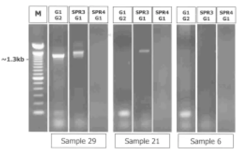
and SPR4. Isolates were submitted to three separate PCR reactions with primers G1-G2, SPR3-G1 and SPR4-G1, and genomovar status was defined according to the algorithm shown in Fig. 1. PCR reactions were performed in 25 µL mixtures containing 200 ng of template DNA, 2 mM of MgCl2, 0.8 µM of each primer, 10 mM Tris HCl (pH 8.0), 50 mM KCl, 200 µM of dNTPs and 1.25U of Taq DNA polymerase (GIBCO-BRL Gaithesburg, FL, USA). Amplification was carried out using Mastercycler gradient thermocycler (Eppendorf) with an initial denaturation step of 95 oC for 5 minutes and 30 cycles of 94 oC for 45
seconds, 66 oC for 45 s and 72 oC for 2 minutes followed by a final
extension step at 72 oC for 10 minutes. Following amplification, PCR
products were visualized by electrophoresis in 0.8% agarose gel stained with ethidium bromide 0.5 mg/mL, with an UV transilluminator. A negative control (sample with no DNA added) was included in all PCR reactions.
STATISTICAL ANALYSIS: The results of B. cepacia isolation during the six month period were compared to the results of preceding two years, when the sample processing was identical except for the lack of a B. cepacia selective media. Comparisons between the results of the two periods used c2 test or two-tailed Fisher exact test, and a p value <
0.05 was considered significant.
RESULTS
During the six month study period, a total of 257 samples were cultured. B. cepacia was isolated in 11/257 (4.2%) of the samples using the B. cepacia selective medium. Cultures were obtained from 9 sputum samples and two oropharyngeal swabs, from 8 patients aged 11 months to 17 years old. In the two years preceding the start of the study, B. cepacia was isolated in 6/1029 samples (0.58%), from 5 patients aged 7 to 16 years old. The difference in the rate of isolation among the two
periods, 11/257 (4.2%) versus 6/1029 (0.6%) was statistically significant (two-tailed Fisher exact test, p < 0.0001), Fig. 2. However, the number of patients characterized as colonized by B. cepacia (i.e., with at least one sample positive for the bacterium)was not significantly different when the two periods were compared (8/106 (7.5%) versus 5/106 (4.7%), c2 test: p = 0.39).
The identity of the 11 isolates of B. cepacia was confirmed by species specific PCR, with all isolates tested resulting in a 463 bp amplicon resultant of amplification of the 16S ribosomal DNA of B. cepacia,as partially shown in Fig. 3.
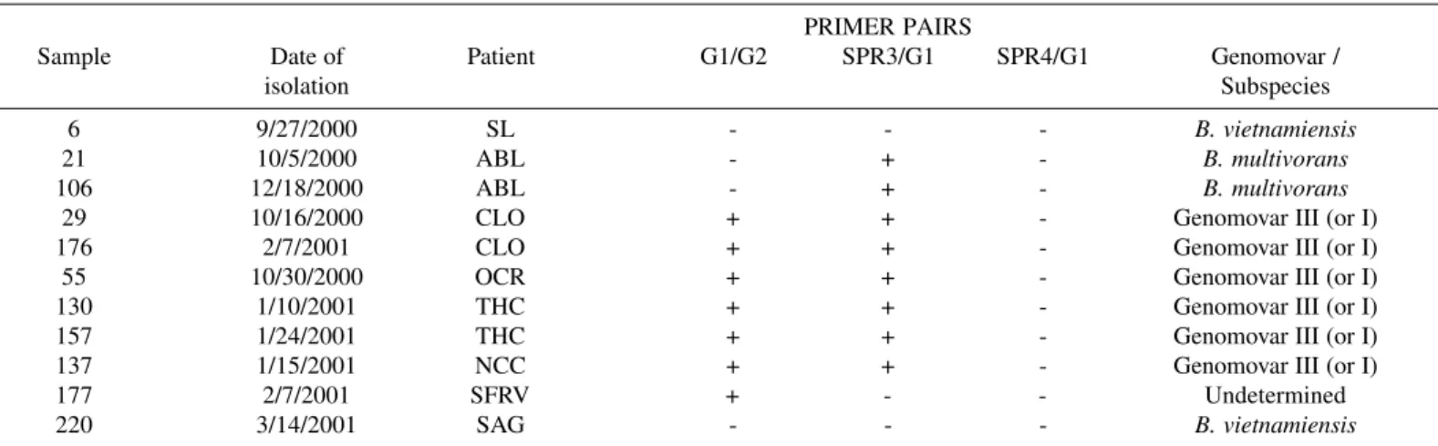
The application of the PCR protocol for B. cepacia genomovar determination classified two strains as Burkholderia vietnamiensis, two strains as Burkholderia multivorans and 4 strains as belonging to genomovars I or III, as shown in Table 1 and illustrated in Fig. 4. PCR reactions resulted in the amplification of DNA fragments of approximately 1.3 kb in at least one of the combination of the three primers in all but two samples, which were assigned as B. vietnamiensis.
Table 1
PCR results of B. cepacia complex genomovar determination, using primers G1, G2, SPR3 and SPR4, as described by WHITBY et al.29. Patient names are
indicated by assorted initials. PCR results are expressed: +, positive PCR; -, negative PCR
PRIMER PAIRS
Sample Date of Patient G1/G2 SPR3/G1 SPR4/G1 Genomovar /
isolation Subspecies
6 9/27/2000 SL - - - B. vietnamiensis
21 10/5/2000 ABL - + - B. multivorans
106 12/18/2000 ABL - + - B. multivorans
29 10/16/2000 CLO + + - Genomovar III (or I)
176 2/7/2001 CLO + + - Genomovar III (or I)
55 10/30/2000 OCR + + - Genomovar III (or I)
130 1/10/2001 THC + + - Genomovar III (or I)
157 1/24/2001 THC + + - Genomovar III (or I)
137 1/15/2001 NCC + + - Genomovar III (or I)
177 2/7/2001 SFRV + - - Undetermined
220 3/14/2001 SAG - - - B. vietnamiensis
Fig. 3 - Species specific PCR of representative B. cepacia strains with primers Eub-16-1/ CeMuVi-16-2457.
Lane 1: B. cepacia strain obtained from the microbiology laboratory repository. Lanes 2 to 10: nine representative strains of B. cepacia isolated during the six month study period. Lane 11: negative control (no DNA added). M: molecular weight marker (100 bp ladder, Pharmacia, Upsalla, Sweden).
8
We didn’t observe PCR products using the primers G1-SPR4 in any of the strains tested (Table 1), and therefore we did not identify strains of B. stabilis or B. cepacia genomovar I. The genomovar status of one isolate could not be determined, as it didn’t result in PCR products of the expected size with primer pair G1-SPR3 while showed positive result with primer pair G1-G2, an unexpected event.
DISCUSSION
B. cepacia is a microorganism that has great significance in cystic fibrosis. Since its recognition as an infecting agent in CF patients in the mid 80s, much have been learned about its importance, transmission and clinical impact, but several questions remain to be answered. The isolation of this organism in respiratory secretions of CF patients still carry a severe psychosocial burden, arising from the precautions to minimize person-to-person transmission and to the increased rates of morbidity and mortality in this patient population15.
Pseudomonas cepacia was originally described by Burkholder in 1950 as the causative agent of bacterial rot of onion bulbs. In 1992, P. cepacia was transferred to the new genus Burkholderia, which currently include 22 bacterial species7. Several additional modifications in the
taxonomy of B. cepacia have occurred in the last decade, with the recognition of marked heterogeneity among B. cepacia strains isolated from different echological niches. B. cepacia was classified as a complex, comprising at least five different genomic species or genomovars26. B.
cepacia genomovar V was identified as the previously described species B. vietnamiensis7 and the name B. multivorans was proposed for strains
belonging to the genomovar II. B. cepacia genomovar IV was subsequently classified as B. stabilis27. Recent taxonomic studies
identified more three members of B. cepacia complex, B. cepacia genomovar VI, B. ambifaria and B. pyrrocinia1,5,6.
Clinical management of respiratory infections in CF patients rely on the identification of organisms cultured from respiratory specimens such
as sputum, oropharyngeal swabs or bronchoalveolar lavage samples, but this apparently simple procedure may not be straightforward. Identification of B. cepacia can be problematic and misidentifications may be relatively common. As many as 20% of isolates sent to reference laboratories identified as B. cepacia may be misidentified20. Therefore,
the use of a selective media that take advantage of the species broad antibiotic resistance is a critical first step in sample processing.
A comparison of three media was performed by HENRY et al.11,
reporting superiority of BCSA (B. cepacia selective agar) to OFPBL and PCA (P. cepacia medium) in terms of rapidity and quality of recovery of B. cepacia complex organisms from CF respiratory specimens, while inhibiting the growth of other organisms. The above mentioned medium (BCSA), however, is not indicated for isolation of environmental B. cepacia isolates, due to low specificity/sensitivity.
We utilized a different medium, named B. cepacia medium (Oxoid), mainly indicated for the isolation and identification of B. cepacia from respiratory secretions of CF patients. As expected, a significant difference of isolation rates of B. cepacia strains was observed among the two periods analyzed (with and without the use of selective medium), which was probably derived from the discriminating properties of the B. cepacia selective medium. Although there was a significant difference in the rate of B. cepacia isolation, the use of the selective medium did not contribute to identification of a significant broader patient population colonized with B. cepacia, but isolation of B. cepacia was observed in younger patients and in two oropharyngeal swabs samples. The impact of the use of dithiothreitol (DTT) as a solubilizing agent in culture results was previously assessed by HAMMERSCHLAG et al., who showed that the substance may interfere in the isolation rate of H. influenzae only when high concentrations were used10. Although this aspect was not specifically
addressed in the present work, it seems unlikely that the use of DTT as part of initial sample processing had some influence in the observed results.
Although there are recommendations for specific microbiological practices when culturing samples from CF patients, they are not universally adopted, even in more specialized laboratories. The situation in Brazil is quite different, since there are few specialized centers for CF care and there is no information or consensus for the microbiology practices used for culturing respiratory samples obtained from CF patients.
The use of PCR for the identification of B. cepacia strains was described by a number of authors3,14, but the taxonomic changing of the
Burkholderia genus have posed new potential caveats associated with some primer pairs described, such as cross reactivity with non-B.cepacia complex organisms or poor specificity for the identification of some of the species16,19. To date, the only described primer pairs that are supposed
to identify all members of the B. cepacia complex are those described by BAUERNFEIND et al.2, Eub-16-1/ CeMuVi-16-2
457, which identify
genomovars I, III, B. multivorans, B. vietnamiensis and B. stabilis, and primers BCR1/BCR2, described by MAHENTHIRALINGAM et al.17,
which are able to identify all members of the B. cepacia complex, including the very recently described B. cepacia genomovar VI and genomovar VII (B. ambifaria)17,19.
We decided to use the primer pair described by BAUERNFEIND et al., since it is currently being tested by our group for direct detection of Fig. 4 - PCR for B. cepacia complex genomovar determination, as described by WHITBY et
al.29. Expected size of amplicons for each primer pair: 1.3 kb.
4
B. cepacia strains in respiratory samples from CF patients. Our PCR results with this primer pair confirmed the identity of all the eleven B. cepacia strains isolated with the selective medium. This primer pair was also tested in PCR reactions with DNA of other bacterial species frequently found in the sputum of CF patients (P. aeruginosa, S. aureus, H. influenzae, S. maltophilia), and didn’t result in any visible PCR product (data not shown).
The knowledge of B. cepacia complex genomovar species responsible for respiratory infections in CF patients is extremely important for appropriate segregation and grouping of CF patients into cohorts15,19. Although there are now recent described phenotypic tests
for genomovar determination, it is still not possible to identify all genomovars based on this approach12. Several strategies have been
proposed for the identification of genomovars using molecular biology techniques, and the method described by WHITBY et al.29 was chosen
because of its simplicity, although it is not able to differentiate genomovars I and III. Since CF patients are predominantly colonized by genomovar III and B. multivorans16,19,26, this limitation doesn’t constitute
a significant pitfall. The results obtained with the PCR approach using primers G1, G2, SPR3 and SPR4 confirmed this predominance of genomovar III (most likely) among our CF patients, although we found the same proportion of patients colonized by B. multivorans and B. vietnamiensis (two patients colonized with each species). Three patients have two B. cepacia positive cultures, obtained on different dates; none of them, however, presented different genomovars.
In summary, our results confirmed the importance of the use of a selective medium for B. cepacia isolation when culturing respiratory samples from cystic fibrosis patients. We have also confirmed the identity of all isolates of B. cepacia by species specific PCR and further characterized these isolates by genomovar status, obtaining results similar to those described on other CF specialized Centers.
ACKNOWLEDGEMENTS
We thank Mrs. Silvana Fernandes for technical support on DNA extractions. We also thank all the medical staff of the Pediatric Pulmonology Unit of Instituto da Criança, São Paulo, Brazil, for their assistance during patients attendance. This work was supported by FAPESP, grant 1999/00067-9. The author (LVFSF) was sponsored by CAPES, Brazil.
RESUMO
Uso de meio seletivo para identificação de cepas de Burkholderia cepacia em amostras de trato respiratório de pacientes portadores
de fibrose cística
A Burkholderia cepacia coloniza os pacientes portadores de fibrose cística (FC). Avaliamos o impacto do uso de um meio seletivo no isolamento de B. cepacia em amostras de secreção respiratória de pacientes portadores de FC. Durante um período de 6 meses, amostras de trato respiratório foram colhidas de 106 pacientes com FC e cultivadas em meios seletivos incluindo um meio para isolamento de B. cepacia. A identidade das cepas de B. cepacia foi confirmada através de PCR espécie específica e a determinação do genomovar ou subespécie realizada através de reações seqüenciais de PCR. Os resultados de isolamento de B. cepacia
durante este período foram comparados com os dois anos precedentes, quando o processamento das amostras era idêntico, exceto pela utilização do meio seletivo para B. cepacia. B. cepacia foi isolada em 11/257 (4,2%) amostras usando o meio seletivo, e em apenas 6/1029 (0,58%) nos dois anos precedentes (p < 0,0001). A identidade destas 11 cepas foi confirmada e a determinação do genomovar obtida em 10/11. Estes resultados sugerem que o uso do meio seletivo aumenta a freqüência de isolamento de B. cepacia em amostras de trato respiratório de pacientes portadores de FC.
REFERENCES
1.BALANDREAU, J.; VIALLARD, V.; COURNOYER, B. et al. - Burkholderia cepacia
genomovar III is a common plant-associated bacterium. Appl. environ. Microbiol., 67: 982-985, 2001.
2. BAUERNFEIND, A.; SCHNEIDER, I.; JUNGWIRTH, R. & ROLLER, C. -Discrimination of Burkholderia multivorans and Burkholderia vietnamiensis from
Burkholderia cepacia genomovars I, III, and IV by PCR. J. clin. Microbiol., 37:
1335-1339, 1999.
3. CAMPBELL 3rd, P.W.; PHILLIPS 3rd, J.A.; HEIDECKER, G.J. et al. - Detection of
Pseudomonas (Burkholderia) cepacia using PCR. Pediat. Pulmonol., 20: 44-49, 1995.
4. CIMOLAI, N.; TROMBLEY, C.; DAVIDSON, A.G. & WONG, L.T. - Selective media for isolation of Burkholderia (Pseudomonas) cepacia from the respiratory secretions of patients with cystic fibrosis. J. clin. Path., 48: 488-490, 1995.
5. COENYE, T.; LIPUMA, J.J.; HENRY, D. et al.- Burkholderia cepacia genomovar VI, a new member of the Burkholderia cepacia complex isolated from cystic fibrosis patients. Int. J. system. evol. Microbiol., 51: 271-279, 2001.
6. COENYE, T.; MAHENTHIRALINGAM, E.; HENRY, D. et al. - Burkholderia ambifaria sp. nov., a novel member of the Burkholderia cepacia complex including biocontrol and cystic fibrosis-related isolates. Int. J. system. evol. Microbiol., 51: 1481-1490, 2001.
7. COENYE, T.; VANDAMME, P.; GOVAN, J.R. & LiPUMA, J.J. - Taxonomy and identification of the Burkholderia cepacia complex. J. clin. Microbiol., 39: 3427-3436, 2001.
8. CYSTIC FIBROSIS FOUNDATION - Patient Registry 1999 annual data report. Bethesda, Cystic Fibrosis Foundation, 2000.
9. GOVAN, J.R. – Infection control in cystic fibrosis: methicillin-resistant Staphylococcus aureus, Pseudomonas aeruginosa and the Burkholderia cepacia complex. J. roy. Soc. Med., 93(suppl 38): 40-45, 2000.
10. HAMMERSCHLAG, M.R.; HARDING, L.; MACONE, A.; SMITH, A.L. & GOLDMANN, D.A. - Bacteriology of sputum in cystic fibrosis: evaluation of dithiothreitol as a mucolytic agent. J. clin. Microbiol., 11: 552-557, 1980. 11. HENRY, D.; CAMPBELL, M.; MCGIMPSEY, C. et al. - Comparison of isolation media
for recovery of Burkholderia cepacia complex from respiratory secretions of patients with cystic fibrosis. J. clin. Microbiol., 37: 1004-1007, 1999.
12. HENRY, D.A.; MAHENTHIRALINGAM, E.; VANDAMME, P.; COENYE, T. & SPEERT, D.P. - Phenotypic methods for determining genomovar status of the
Burkholderia cepacia complex. J. clin. Microbiol., 39: 1073-1078, 2001. 13. ISLES, A.; MACLUSKY, I.; COREY, M. et al. - Pseudomonas cepacia infection in
cystic fibrosis: an emerging problem. J. Pediat., 104: 206-210, 1984.
14. KARPATI, F. & JONASSON, J. - Polymerase chain reaction for the detection of
Pseudomonas aeruginosa, Stenotrophomonas maltophilia and Burkholderia cepacia
@
15. LIPUMA, J.J. - Burkholderia cepacia. Management issues and new insights. Clin. Chest Med., 19: 473-86, vi, 1998.
16. LiPUMA, J.J.; DULANEY, B.J.; McMENAMIN, J.D. et al. - Development of rRNA-based PCR assays for identification of Burkholderia cepacia complex isolates recovered from cystic fibrosis patients. J. clin. Microbiol., 37: 3167-3170, 1999. 17. MAHENTHIRALINGAM, E.; BISCHOF, J.; BYRNE, S.K. et al. - DNA-based diagnostic
approaches for identification of Burkholderia cepacia complex, Burkholderia vietnamiensis, Burkholderia multivorans, Burkholderia stabilis, and Burkholderia cepacia genomovars I and III. J. clin. Microbiol., 38: 3165-3173, 2000. 18. MARQUES, E.; PINTO, R.M.; DALLALLANA, L.T. et al. - Isolation of Pseudomonas
cepacia in cystic fibrosis patient. Mem. Inst. Oswaldo Cruz, 88: 125-129, 1993. 19. McDOWELL, A.; MAHENTHIRALINGAM, E.; MOORE, J.E. et al. - PCR-based
detection and identification of Burkholderia cepacia complex pathogens in sputum from cystic fibrosis patients. J. clin. Microbiol., 39: 4247-4255, 2001.
20. McMENAMIN, J.D.; ZACCONE, T.M.; COENYE, T.; VANDAMME, P. & LiPUMA, J.J. - Misidentification of Burkholderia cepacia in US cystic fibrosis treatment centers: an analysis of 1,051 recent sputum isolates. Chest, 117: 1661-1665, 2000. 21. MOORE, J.E.; MILLAR, B.C.; JIRU, X. et al. - Rapid characterization of the genomovars
of the Burkholderia cepacia complex by PCR-single-stranded conformational polymorphism (PCR-SSCP) analysis. J. Hosp. Infect., 48: 129-134, 2001. 22. ROSENSTEIN, B.J. & CUTTING, G.R. - The diagnosis of cystic fibrosis: a consensus
statement. Cystic Fibrosis Foundation Consensus Panel. J. Pediat., 132: 589-595, 1998.
23. ROSENSTEIN, B.J. & HALL, D.E. - Pneumonia and septicemia due to Pseudomonas cepacia in a patient with cystic fibrosis. Johns Hopkins. med. J., 147: 188-189, 1980.
24. SAMBROOK, J.; FRITSCH, E. & MANIATIS, T. - Molecular cloning: a laboratory manual. 2. ed. Cold Spring, Cold Spring Harbor Lab., 1989.
25. SHREVE, M.R.; BUTLER, S.; KAPLOWITZ, H.J. et al. - Impact of microbiology practice on cumulative prevalence of respiratory tract bacteria in patients with cystic fibrosis.
J. cin. Microbiol., 37: 753-757, 1999.
26. VANDAMME, P.; HOLMES, B.; VANCANNEYT, M. et al. - Occurrence of multiple genomovars of Burkholderia cepacia in cystic fibrosis patients and proposal of
Burkholderia multivorans sp. nov. Int. J. system. Bact., 47: 1188-1200, 1997. 27. VANDAMME, P.; MAHENTHIRALINGAM, E.; HOLMES, B. et al. - Identification
and population structure of Burkholderia stabilis sp. nov. (formerly Burkholderia cepacia genomovar IV). J. clin. Microbiol., 38: 1042-1047, 2000.
28. WELSH, M.J. & SMITH, A.E. - Cystic fibrosis. Sci. Amer., 273: 52-59, 1995. 29. WHITBY, P.W.; CARTER, K.B.; HATTER, K.L.; LiPUMA, J.J. & STULL, T.L.
-Identification of members of the Burkholderia cepacia complex by species-specific PCR. J. clin. Microbiol., 38: 2962-2965, 2000.
30. WONG, K.; ROBERTS, M.C.; OWENS, L.; FIFE, M. & SMITH, A.L. - Selective media for the quantitation of bacteria in cystic fibrosis sputum. J. med. Microbiol., 17: