online | memorias.ioc.fiocruz.br
Morphology of the larvae, male genitalia and DNA sequences of
Anopheles (Kerteszia) pholidotus
(Diptera: Culicidae) from Colombia
Jesús Eduardo Escovar1,2/+, Ranulfo González3, Martha L Quiñones1, Richard C Wilkerson4, Fredy Ruiz4, Bruce A Harrison5
1Universidad Nacional de Colombia, Bogotá, Cundinamarca, Colombia 2Universidad de la Salle, Bogotá, Cundinamarca, Colombia 3Faculty of Natural and Exact Sciences, Universidad del Valle, Valle, Colombia 4Walter Reed Biosystematics Unit, Smithsonian Institution,
Museum Support Center, Suitland, MD, USA 5College of Health and Human Sciences, Western Carolina University, Clemmons, NC, USA
Since 1984, Anopheles (Kerteszia) lepidotus has been considered a mosquito species that is involved in the transmission of malaria in Colombia, after having been incriminated as such with epidemiological evidence from a malaria outbreak in Cunday-Villarrica, Tolima. Subsequent morphological analyses of females captured in the same place and at the time of the outbreak showed that the species responsible for the transmission was not An. lepidotus, but rather Anopheles pholidotus. However, the associated morphological stages and DNA sequences of
An. pholidotus from the foci of Cunday-Villarrica had not been analysed. Using samples that were caught recently from the outbreak region, the purpose of this study was to provide updated and additional information by analysing the morphology of female mosquitoes, the genitalia of male mosquitoes and fourth instar larvae of An. pholidotus, which was confirmed with DNA sequences of cytochrome oxidase Iand rDNA internal transcribed spacer. A total of 1,596 adult females were collected in addition to 37 larval collections in bromeliads. Furthermore, 141 adult females, which were captured from the same area in the years 1981-1982, were analysed morphologically. Ninety-five DNA sequences were analysed for this study. Morphological and molecular analyses showed that the species present in this region corresponds to An. pholidotus. Given the absence of An. lepidotus, even in recent years, we consider that the species of mosquitoes that was previously incriminated as the malaria vector during the outbreak was indeed An. pholidotus, thus ending the controversy.
Key words: Kerteszia - Anopheles pholidotus - male genitalia - DNA sequences
Mosquito species of the subgenus Kerteszia are ex-clusively located in the Neotropical areas of Central and South America. Except for Chile and Uruguay, their dis-tribution ranges from the south of Mexico to southern Brazil. This subgenus is also present in the Caribbean, Trinidad and Tobago (de Carvalho-Pinto & Lourenço-de-Oliveira 2004) and both the Atlantic and Pacific coastal areas (Marrelli et al. 2007), with some species present at altitudes of more than 1,000 m above sea level (a.s.l.) (Cova-García 1961, Harbach & Navarro 1996).
Kerteszia subgenus consists of species that have been incriminated as malaria vectors and are related to the emergence of malaria in thermal floors at altitudes of over 1,500 m a.s.l. Moreover, in the malaria-heavy coast-al regions of South America and in the eastern slopes of the Venezuelan Andes, members of this subgenus seem to be the most important species involved in the trans-mission of malaria (Benítez et al. 2004, Montoya-Lerma et al. 2011). Similarly, the species of Kerteszia have been incriminated in the transmission of “malaria by brome-liads”, which is typical of protected areas because the
doi: 10.1590/0074-0276130596
Financial support: COLCIENCIAS (110145921478) + Corresponding author: jeescobar@unisalle.edu.co Received 26 December 2013
Accepted 8 April 2014
epiphytic bromeliads are the only breeding sites. This is why these plants have been associated with the occur-rence of the disease (Ueno et al. 2007).
Five of the 12 species described for this subgenus (Za-vortink 1973, Collucci & Sallum 2003) have been incrim-inated as malaria vectors: Anopheles bellator (Forattini et al. 1999), Anopheles cruzii (de Carvalho-Pinto & Lou-renço-de-Oliveira 2004), Anopheles homunculus (Rubio-Palis & Zimmerman 1997), Anopheles neivai (Carvajal et al. 1989, Gutierrez et al. 2008) and Anopheles lepidotus
(Quiñones et al. 1984, Montoya-Lerma et al. 2011). In Colombia, seven species of the subgenus have been reported, among which An.neivai and An. lepido-tus are considered malaria vectors, while Anopheles bo-liviensis is considered a seasonal vector only (Quiñones et al. 1984, Montoya et al. 1994, Olano et al. 2001).
Since 1984, An. lepidotus has been considered, us-ing epidemiological evidence (namely, mosquito density correlated with malaria cases; 98% of the Anopheles spe-cies found in the malaria focal area corresponded to this species), to be one of the species responsible for malaria transmission in Colombia, particularly in the focal area of Cunday-Villarrica, Department of Tolima. It has been suggested that An. lepidotus is part of a complex of spe-cies in which members of this complex are difficult to be differentiated morphologically. Specimens of Kerteszia
four or five different species (R Wilkerson, unpublished observations, Sallum et al. 2002).
González and Carrejo (2009), using observations of specimens of what was supposedly An. lepidotus, noted that the mosquito is very similar to An. pholidotus and both species were called An. boliviensis before Zavort-ink’s (1973) descriptions. As a result, the holotype and paratype described by Komp (1937) as An. boliviensis is actually An. lepidotus. They also affirm thatof the 204 Colombian specimens deposited at the University of Cali-fornia, Los Angeles and the National Museum of Natural History (NMNH) at the Smithsonian Institution collec-tions and predetermined to be An. boliviensis asanalysed by Zavortink (1973), 85% corresponded to An. lepidotus.
Similarly, González and Carrejo (2009), based on the analysis of male genitalia and larvae of four specimens from Cunday-Villarrica, suggested the possibility that An. pholidotus occurred in this region. The importance of the above conclusion is that in Colombia, the region of Cun-day-Villarrica is considered to be a focal area of malaria, where An. lepidotus had been incriminated as the possible vector for more than 25 years (Quiñones et al. 1984).
Recently, Harrison et al. (2012) resolved the problem of separating An. lepidotus from An. pholidotus females by re-describing both species and preparing a taxonomic key. These methods were used to differentiate females, the IV instar larvae, pupae and male genitalia of An. lepidotus from other species of Kerteszia. They exam-ined specimens of An. lepidotus from Colombia, Ecuador and Peru and female specimens of An. pholidotus from Bolivia, Colombia, Costa Rica, Ecuador and Venezuela. They agree with the preliminary results of Escobar et al.
(2010), who stated that the primary vector in Tolima is An. pholidotus and not An. Lepidotus,as had been reported since 1984 (Quiñones et al. 1984). Nevertheless, Harrison et al. (2012) only analysed 33 females of An. pholidotus
captured between 1981-1983 in Tolima. Therefore, this conclusion was pending the analysis of the morphology of associated stages, the male genitalia and the inclusion of DNA sequences from samples of An. pholidotus that wererecently obtained from the study area.
Since the controversy surrounding the identity of the vector in Tolima began more than 30 years ago, the aim of this study was to provide updated and additional information that was not presented by Harrison et al. (2012). This information includes the morphology of lar-vae, the male genitalia of An. pholidotus and their asso-ciation with the DNA sequences of specimens that were recently collected in Colombia.
In this study, we analysed the morphological charac-teristics of 1,596 recently captured females, the chaeto-taxy of IV instar larvae and the male genitalia of speci-mens of An. pholidotus from three localities in Tolima. Molecular analysis for gen cytochrome oxidase I (COI), internal transcribed spacer (ITS2) and sequencing was performed to verify the results obtained from the mor-phological information.
This study provides definitive morphological and molecular information supporting the conclusion that the malaria vector in Tolima is An. pholidotus and that it is actually the only malaria vector present in the area.
MAtERiALs AND MEtHODs
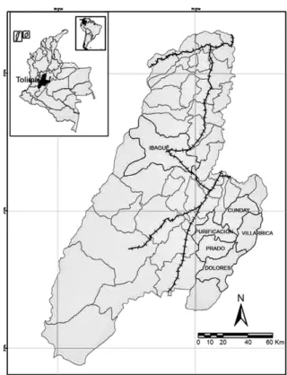
Study site - The malaria focal area of Cunday-Villarri-ca is composed of the municipalities of Cunday, VillarriCunday-Villarri-ca, Dolores, Prado and Purificación (03º57’N 074º36’W) (Fig. 1), which are situated on the western slopes of the eastern mountain chain and have an average temperature of 24ºC and an annual rainfall of between 2,000-4,000 mm. This area is approximately 100 Km2 and is characterised by
humid forests that have an abundance of epiphytes. This area has had stable malaria transmission for more than 25 years. Moreover, malaria cases reported in Tolima have been almost exclusively from that focal area.
Sampling strategy and adult mosquitoes - Collec-tions of biological material were made between Febru-ary 2009-June 2012. Adult females were captured using human landing collections inside and around houses and in the forest during periods of peak biting at various locations in the municipalities in this study. Captured females were individualised in vials and brought to the entomology laboratory of the National University of Co-lombia for preservation and taxonomic determination. Additionally, adult females of Kerteszia, which were collected between 1981-1983 in the same study area and belonged to the collection of the entomology laboratory of the Museum of Entomology, University of Valle, were analysed morphologically.
Entomological series - Mosquitoes in immature stages (larva and pupa) were captured directly from wa-ter at the base of bromeliads leaves by drawing out their
content with a larger pipette. Larvae of Anopheles were collected from 41 trees with 78 bromeliads and brought to the laboratory of the National University of Colombia for rearing to the adult stage. IV instar larvae were indi-vidualised and larval and pupal skins were obtained to achieve 37 full entomological series. Male genitalia and larval and pupal skins were mounted.
Morphological analysis - Diagnostic characters of females, IV instar larvae and male genitalia were stud-ied based on the descriptions and morphological keys of Zavortink (1973) and Harrison et al. (2012).
Molecular methods - Forty-four specimens were processed as belonging to An. (Kerteszia) sp. and their DNA was extracted using the DNeasy Blood & Tissue Kit (QIAgen®, USA). The rDNA ITS2 region was
ampli-fied using the primers of Collins and Paskewitz (1996) and the polymerase chain reaction (PCR) conditions de-scribed by Linton et al. (2001). A portion (710 bp) of the barcoding region of COI (mDNA) was amplified using the primers designed by Folmer et al. (1994) and the PCR conditions described by Ruiz et al. (2010). The products were visualised on 1% agarose gel containing 0.5 mg/ mL of ethidium bromide. The PCR product was purified using ExoSAP-IT® (USB Corporation, USA).
Sequencing reactions were carried out in both direc-tions using the Big Dye terminator Kit® (PE Applied
Bio-systems, England) on an ABI 3730 automated sequencer (PE Applied Biosystems). The sequences were edited with SequencherTM 4.10.1 (Gene Codes Corporation, USA) and
aligned manually and translated to amino acids in Mac-Clade v.4.06 (Maddison & Maddison 2003). Sequence similarities were compared with those available in Gen-Bank using Basic Local Alignment Search Tool (BLAST) (ncbi.nlm.nih.gov/genbank/) and sequence statistics were calculated in MEGA v.5 (Tamura et al. 2011).
Ethics - This study was approved by the Ethical Com-mittee of the Faculty of Medicine of the National Uni-versity of Colombia, according to consecutive number E-31, on 26 June 2008.
REsULts
Table shows the sampling locations in the study area and the sampling dates. A total of 1,737 anopheline fe-males were analysed morphologically. Of these fefe-males, 1,596 were collected landing on humans in the period between 2009-2012 and 141 females were sampled be-tween 1981-1983.
The first morphological analysis was conducted us-ing the dichotomous keys of Zavortink (1973). As a result of this analysis, 76% (1,218) of the collected specimens corresponded to either An. lepidotus or An. pholidotus
and 24% (378) were in agreement with an identification of An. boliviensis. According to Zavortink´s key (1973), the only difference existing between An. lepidotus and
An. pholidotus is restricted to the size of the scales on the female abdomen. As a result, it is difficult to deter-mine which of these two species is present in the study region using this morphological key. Considering this issue, a second morphological analysis was made using
the dichotomous key proposed by Harrison et al. (2012). This key can separate An. lepidotus females from An. pholidotus females according the following diagnostic features: (i) females with white scales on the proboscis, pedicel and palpomere I; hindtarsomeres I and 2 with-out apical pale band (from dorsal view) are classified as
An. lepidotus and(ii) females without white scales on the proboscis, pedicel and palpomere I; hindtarsomeres I and 2 with narrow apical pale band (from dorsal view) are classified as An. pholidotus.
All females captured in Tolima showed morphologi-cal characteristics that were similar to those assigned to
An. pholidotus. Additionally, the females of An. pholido-tus exhibited numerous scales with variable widths on tergites and sternites II-VII, hindtarsal segment 2 with white bands in the apical pale band between 0.1-0.2 as the total length of the tarsomere, mesanepimeron with large curved patch of scales extending from higher bris-tles to below the middle of the segment, scales in the proximal tergites and scales of the distal tergites form-ing transverse apical bands (Fig. 2).
IV instar larvae - Twenty-eight micro-preparations of IV instar larvae were analysed. The larvae showed a characteristic coloration pattern in the dorsal area of the thorax and abdominal segments I and V (Fig. 3). The morphological characteristics of this instar coincided with those described by Zavortink (1973) for An. pholi-dotus. Setae 5-7-C simple, not plumose, moderately long, characteristics of the Kerteszia subgenus; setae 6-VI moderately long, always different to 6-III-V, setae 1-III-VII small palmate with pointed leaflets, pecten teeth of similar size all long, with marginal spinules extended to the apex, setae 1-I not palmate, setae 4-C always less developed than 2-C, setae 1-VII filiform, not palmate.
According to Zavortink (1973), two morphological characteristics help differentiate An. lepidotus from An. pholidotus in this larval stage: An. pholidotus shows a se-tae 3-C that is moderately developed and approximately 1/2 the length of 2-C, while An. lepidotus shows a setae 3-C that is short and thick, fusiform and less than 1/2 the length of 2-C. The 28 specimens analysed in this study presented an average length of 0.19 ± 0.01 mm for the se-tae 2-C and of 0.11 ± 0.004 mm for the sese-tae 3-C, show-ing that in all cases the length of the setae 3-C is more than half the length of the setae 2-C (Fig. 4A), which corresponds to An. pholidotus. The clypeal index was 0.98 ± 0.15. The second discriminatory character is the setae 11-P, which is well developed for An. pholidotus. A very short 11-P would correspond to An. lepidotus. All larvae tested showed the setae 11-P to be well developed (length = 0.32 ± 0.03 mm), with an average of 66.7% of the length of the 12-P setae (0.48 ± 0.03 mm) (Fig. 4B).
Male genitalia - Twenty-five micro-preparations of male genitalia from a series were analysed. The mor-phological characteristics for this structure coincided with those described by Zavortink (1973) and González and Carrejo (2009) for An. pholidotus, but not for An. lepidotus. The specific characteristics were gonocoxite with long and sinusoid parabasal setae, always greater than 1/3 the total length of gonocoxite; one internal seta flattened at the apex and two accessory ones located api-cally, an aedeagus without leaflets (Fig. 5A), a lateral expansion of the ventral lobe of claspette, moderately spiculose except laterally and rounded (Fig. 5B, C). The 25 male genitalia analysed in this study showed a lateral expansion of the ventral lobe of claspette corresponding
to the typical shape of An. pholidotus described by Za-vortink (1973) and González and Carrejo (2009).
DNA sequences - Ninety-five specimens were se-quenced with the molecular markers COI and ITS2. Only one haplotype was observed for each marker, con-firming the lack of an intraspecific variation in all of the individuals analysed.
The COI barcode region (658 bp) consisted of a single open reading frame, discarding the presence of pseudo-genes. Its nucleotide frequencies (%) were A: 27.7%, T: 40.9%, G: 15.5% and C: 16% (Fig. 6). The ITS2 fragment showed no length variability (546 bp) and its nucleotide frequencies were A: 26.4%, T: 19.4%, G: 26.7% and C: 27.5% (Fig. 7).
TABLE
Sampling localities of Cunday-Villarrica focus in the Department of Tolima, Colombia
Municipality Coordinates Locality Sampling period
Cunday 04º47’59.1”N
74º34’ 50.21”W
San Francisco February 2009
Icononzo 04º51’02.1”N
74º09’003”W
Valencia Cuatimbal Alto Icononzo
November 1981 October 1982 October 1982
Villarrica 03º52’03.4”N
74º39’09.8”W
Puerto Lleras Totumal Guanacas Rio Lindo Rio Lindo Rio Lindo Rio Lindo
February 1981-December 1982 June 1981-February 1983 June 1982-February 1983 February-November 2009
August 2010 August-September 2011
April-July 2012
Purificación 03º51’58.11”N
74º55’59.83”W
Villa Esperanza May 2009
Prado 03º45’04.15”N
74º49’55.9”W
El Cruce May 2009
Fig. 2: lateral view of female of Anopheles pholidotus. Right arrow shows mesepimeron with one large and curved patch of scales that extends ventrally from upper setae. Left arrow shows palpomere 1 without scales.
The sequencesof ITS2 were submitted to BLAST, where high significant homology (100%) was found with the sequences of An. pholidotus (from Táchira, Venezu-ela) (accessions JN967769.1, JN967768.1) published by Harrison et al. (2012).
COI sequences, compared with sequences in the Bar-code of Life Data Systems (Bold Systems) (boldsystems. org), showed 100% similarity with An.pholidotus.
DisCUssiON
Until the study published by Harrison et al. (2012), the only morphological feature that had been proposed to differentiate females of An. pholidotus from An.
lepi-dotus were, according to Zavortink (1973), the size of the scales on proximal tergites. Those were described as moderately wide or broad for An. lepidotus and pre-dominantly narrow to moderately wide for An. pholido-tus, with those differences being rather subjective. This taxonomic characteristic is vague and difficult to inter-pret, so it would be inaccurate to separate female adults of these two species based only on these characteristics. Therefore, it was essential to analyse other morphologi-cal structures and associated stages, such as male geni-talia and to use the chaetotaxy of IV instar larvae. Even so, genitalia analysis presents a “weak spot” or a “bottle neck” effect in the separation of these two species be-Fig. 4: larvae IV instar of Anopheles pholidotus (400X). A: head. Clipeal setaes (2-C and 3-C); B: prothorax. Setae 11-P.
Fig. 7: internal transcribed spacer sequence of Anopheles pholidotus
from Villarrica, Department of Tolima, Colombia using the Collins and Paskewitz (1996) primers (n = 10, 546 bp). Only one haplotype was found. Primers sequences are underlined and in bold.
cause in Zavortink´s key (1973), the presence or absence of scales in VII tergite in the abdomen of adults must be verified at one point, which is an external feature of the genitalia. Thus, the dichotomous key of Harrison et al.
(2012) is useful for separating females of both species. As mentioned above, for several years, there was evi-dence of the presence of An. pholidotus rather than An. lepidotus in the Cunday-Villarrica focal region (González & Carrejo 2009). Even just three years after the report of Quiñones et al. (1984), the presence of An. lepidotus in that zone was questioned (Harrison et al. 2012).
The limited availability of specimens collected from the area for morphological analysis, the scarcity of asso-ciated stages, the poor condition of some of the samples and the difficulty of accessing the area to collect new biological information most likely delayed the opportu-nity to solve this problem by three decades.
The collections of mosquitoes for this study, which occurred between February 2009-October 2011, provid-ed updatprovid-ed information about the species of anophelines in these focal areas. According to the morphological fea-tures of females, the larvae chaetotaxy, the male genita-lia presented in the specimens of Cunday-Villarrica ma-laria focal area in Tolima and the analysis of the COI and ITS2 sequences, An. pholidotus was the most common
species found and was most likely misidentified as An. lepidotus in the previous report (Quiñones et al. 1984).
It is now possible to differentiate females of An. pholi-dotus from An. lepidotus using the dichotomous key. The fact that females of An. lepidotus were not found by analysing 1,737 individual mosquitoes captured indi-cates that the species that was previously reported as the vector of malaria in the area since 1984 (Quiñones et al. 1984) was An. pholidotus and not An. lepidotus.
The sequences obtained in this investigation, both
COI and ITS2, showed that the tested specimens cor-respond to An. pholidotus, thus confirming the results of the morphology analysis. Sequences from three locali-ties of Tolima exhibit no intraspecific variability, show-ing that An. pholidotus is present in the study area as a single species, although this may not be true in other parts of the country due to the high altitude of certain mountain ranges. Olano et al. (2001) and González and Carrejo (2009) provided records of supposed “An. lepi-dotus” in different departments of the country. For ex-ample, we have identified, by morphology, three females from Valle del Cauca (PNN Farallones: Cali, Pance, VI-6-1984, 1,500 m) as An. pholidotus. This result indicates that there is a possibility that An. pholidotus has a wider distribution than previously thought and that many of the specimens identified as An. lepidotus may correspond to An. pholidotus. Moreover, the sequences mentioned above show 100% homology with sequences of An. pholidotus from Táchira, Venezuela, recorded in Gen-Bank by Harrison et al. (2012). As a result, it is impor-tant to determine whether this homology is generalised to all countries where An. pholidotus is present.
Harrison et al. (2012) inadvertently forgot to include the precise location of the record for An. pholidotus in Costa Rica. A single female in the mosquito collection located at the NMNH at the Smithsonian Institution was collected resting on the wall of a highway tunnel. The collection data record was as follows: Costa Rica: Car-tago, Res. Tapanti; unnamed tribs., Ca. 9 Km (road NW tunnel, 9.72 N, 83.78 W, 8.9.vi.1988, elv. 1,400 m, CM & OS Flint, Molzenthal).
One of the key aspects for optimising control mea-sures is an appropriate taxonomic determination of the species that are present and those that are responsible for transmission. Taxonomic studies in Kerteszia species are relevant because the distribution of this subgenus along the continent is still poorly known, mainly due to the lack of research studies. Furthermore, the association of these species with areas where bromeliads are normally present means that environmental changes and human activity may restrict the distribution for some species in the subgenus (Marrelli et al. 2007) However, for other species, the same factors may stimulate the proliferation of breeding sites and possibly extend their geographical distribution (Solarte et al. 1994).
REFERENCEs
Benítez JA, Rodríguez A, Sojo M, Lobo H, Villegas C, Oviedo L, Brown E 2004. Descripción de un brote epidémico de malaria de altura en un área originalmente sin malaria del estado de Trujillo, Venezuela. Boletín de Malariología y Salud Ambiental 44: 93-99. Fig. 6: cytochrome oxidase I sequence of Anopheles pholidotus from
Carvajal H, de Herrera MA, Quintero J, Alzate A, Herrera S 1989.
Anopheles neivai: a vector of malaria in the Pacific low lands of Colombia. Trans R Soc Trop Med Hyg83: 609.
Collins FH, Paskewitz SM 1996. A review of the use of ribosomal DNA (rDNA) to differentiate among cryptic Anopheles species.
Insect Mol Biol 5: 1-9.
Collucci E, Sallum MAM 2003. Phylogenetic analysis of the subgenus
Kerteszia of Anopheles (Diptera: Culicidae: Anophelinae) based on morphological characters. Insect Syst Evol34: 361-372.
Cova-García P 1961. Notas sobre los anofelinos de Venezuela y su identificación, 2nd ed., Grafos, Caracas, 213 pp.
de Carvalho-Pinto CJ, Lourenço-de-Oliveira R 2004. Isoenzimatic analysis of four Anopheles (Kerteszia)cruzii (Diptera: Culicidae) populations of Brazil. Mem Inst Oswaldo Cruz 99: 471-475.
Escobar J, González R, Quiñones ML, Wilkerson RC, Harrison B 2010. Presence of Anopheles (Kerteszia) pholidotus in a malaria focus in Colombia. In G Clark, Y Rubio-Palis, Mosquito vector biology and control in Latin America - A 20th symposium. J Am Mosq Control Assoc26: 306-320.
Folmer O, Black M, Hoeh W, Lutz R, Vrijenhoek R 1994. DNA prim-ers for amplification of mitochondrial cytochrome c oxidase sub-unit I from diverse metazoan invertebrates. Mol Mar Biol Bio-technol 3: 294-299.
Forattini OP, Kakitani I, dos Santos RC, Ueno HM, Kobayashi KM 1999. Role of Anopheles (Kerteszia)bellator as malaria vector in southeastern Brazil (Diptera: Culicidae). Mem Inst Oswaldo Cruz 94: 715-718.
González R, Carrejo N 2009. Introducción al estudio taxonómico de
Anopheles de Colombia. Claves y notas de distribución, 2nd ed., Universidad del Valle, Cali, 248 pp.
Gutierrez LA, Nelson N, Jaramillo LM, Muskus C, Luckhart S, Conn JE, Correa M 2008. Natural infectivity of Anopheles species from the Pacific and Atlantic Regions of Colombia. Acta Trop 107: 99-105.
Harbach RE, Navarro JC 1996. A new species of Anopheles, subge-nus Kerteszia (Diptera: Culicidae) from Venezuela. Ent Scand 27: 207-216.
Harrison BA, Ruíz-López F, Calderón G, Savage HM, Pecor JE, Wilkerson RC 2012. Anopheles (Kerteszia) lepidotus (Diptera: Culicidae), not the malaria vector we thought it was: revised male and female morphology; larva, pupa and male genitalia charac-ters and molecular verification. Zootaxa3218: 1-17.
Komp WHW 1937. The species of the subgenus Kerteszia of Anophe-les. Ann Entomol Soc Am 30: 492-529.
Linton Y-M, Harbach RE, Anthony TG, Chang MS, Asmad M 2001. Morphological and molecular identity of Anopheles (Cellia) sundaicus (Diptera: Culicidae), the nominotypical member of a malaria vector species complex in Southeast Asia. Syst Entomol 26: 357-366.
Maddison DR, Maddison WR 2003. MacClade v.4.06: analysis of phylogeny and character evolution, Sinauer Associates, Sun-derland, MA.
Marrelli MT, Malafronte RS, Sallum MAM, Natal D 2007. Kerteszia
subgenus of Anopheles associated with the Brazilian Atlantic rain-forest: current knowledge and future challenges. Malar J 6: 127.
Montoya J, González R, Palma G, Solarte Y, Olano V 1994. Ma-laria. In BL Travi, J Montoya-Lerma, Centro Internacional de Entrenamiento e investigaciones Médicas (eds.), Manual de en-tomología médica para investigadores de América Latina, CI-DEIM, Cali, p. 163-209.
Montoya-Lerma J, Solarte YA, Giraldo-Calderón GI, Quiñones ML, Ruiz-López F, Wilkerson RC, González R 2011. Malaria vector species in Colombia - A review. Mem Inst Oswaldo Cruz106
(Suppl. I): 223-238.
Olano V, Brochero H, Sáenz R, Quiñones M, Molina J 2001. Mapas preliminares de la distribución de Anopheles vectores de malaria en Colombia. Biomédica 21: 402-403.
Quiñones M, Suárez M, Rodríguez A, Fleming G, Galvis L 1984. Comportamiento de Anopheles (Kerteszia) lepidotus Zavortink, 1973, y su incriminación como posible vector de malaria en el Departamento del Tolima, Colombia. Biomédica4: 5-13.
Rubio-Palis Y, Zimmerman RH 1997. Ecoregional classification of malaria vectors in the Neotropics. J Med Entomol 34: 499-510.
Ruiz F, Linton Y-M, Ponsonby DJ, Conn JE, Herrera M, Quiñones ML, Vélez ID, Wilkerson RC 2010. Molecular comparison of to-potypic specimens confirms Anopheles(Nyssorhynchus) dunha-mi Causey (Diptera: Culicidae) in the Colombian Amazon. Mem Inst Oswaldo Cruz 105: 899-903.
Sallum MAM, Schultz TR, Foster PG, Wirtz RA, Wilkerson RC 2002. Phylogeny of Anophelinae (Diptera: Culicidae) based on nuclear ribosomal and mitochondrial DNA sequences. Syst En-tomol27: 361-381.
Solarte Y, González R, Hurtado JC, Alzate A 1994. Influencia de la vegetación en la presencia, distribución y abundancia de criade-ros de tres especies de Anopheles (DIP.: Culicidae) en la parte baja del río Naya, costa pacífica de Colombia. Bol Mus Ent Univ Valle 2: 55-71.
Tamura K, Peterson D, Peterson N, Stecher G, Nei M, Kumar S 2011. MEGA 5: molecular evolutionary genetics analysis using maxi-mum likelihood, evolutionary distance and maximaxi-mum parsimony methods. Mol Biol Evol 28: 2731-2739.
Ueno HM, Forattini OP, Kakitani I 2007. Vertical and seasonal distri-bution of Anopheles (Kerteszia) in Ilha Comprida, southeastern Brazil. Rev Saude Publica41: 269-275.