Differential expression of the
pr
1A gene in
Metarhizium anisopliae
and
Metarhizium acridum
across different culture conditions
and during pathogenesis
Mariele Porto Carneiro Leão
1, Patricia Vieira Tiago
1, Fernando Dini Andreote
2,
Welington Luiz de Araújo
3and Neiva Tinti de Oliveira
11
Departamento de Micologia, Universidade Federal de Pernambuco, Recife, PE, Brazil.
2Departamento de Ciência do Solo, Escola Superior de Agricultura “Luiz de Queiroz”,
Universidade de São Paulo, Piracicaba, SP, Brazil.
3
Departamento de Microbiologia, Instituto de Ciências Biomédicas, Universidade de São Paulo,
São Paulo, SP, Brazil.
Abstract
The entomopathogenic fungi of the genusMetarhizium have several subtilisin-like proteases that are involved in pathogenesis and these have been used to investigate genes that are differentially expressed in response to differ-ent growth conditions. The iddiffer-entification and characterization of these proteases can provide insight into how the fun-gus is capable of infecting a wide variety of insects and adapt to different substrates. In addition, thepr1A gene has been used for the genetic improvement of strains used in pest control. In this study we used quantitative RT-PCR to assess the relative expression levels of thepr1A gene in M. anisopliae and M. acridum during growth in different cul-ture conditions and during infection of the sugar cane borer,Diatraea saccharalis Fabricius. We also carried out a pathogenicity test to assess the virulence of both species againstD. saccharalis and correlated the results with the pattern ofpr1A gene expression. This analysis revealed that, in both species, the pr1A gene was differentially ex-pressed under the growth conditions studied and during the pathogenic process.M. anisopliae showed higher ex-pression ofpr1A in all conditions examined, when compared to M. acridum. Furthermore, M. anisopliae showed a greater potential to controlD. saccharalis. Taken together, our results suggest that these species have developed different strategies to adapt to different growing conditions.
Keywords: entomopathogen,Diatraea saccharalis, quantitative RT-PCR, expression pattern.
Received: August 5, 2014; Accepted: October 30, 2014.
Introduction
In Brazil, sugarcane monoculture forms the basis of the sugar export and biofuel industries (Alveset al., 2008). The sugarcane borerDiatraea saccharalisFabricius is con-sidered one of the major sugarcane pests in the Americas. Due to its cryptic lifestyle, conventional control measures by deploying chemical insecticides targeting the larvae are ineffective.
Entomopathogenic fungi are used in biological pest control as a promising alternative to chemical pesticides due to a number of advantages, including lower environ-mental impact, lower costs, higher specificity and lesser risk of development of resistance (Samuels et al., 1989; Frazzon et al., 2000). Species that belong to the genus
Metarhiziumare collectively able to infect a broad range of insects (Samuelset al., 1989) and are adapted to life in the root rhizosphere (Roberts and St. Leger, 2004). However, little is known about the molecular and physiological mecha-nisms involved in their adaptation to different growth con-ditions, and this lack of understanding has prevented the development of new strategies to improve their effective-ness in biological pest control (Zhanget al., 2011).
The taxonomy ofMetarhiziumwas reassessed based on multigenic phylogenetic analysis covering thetef-1 gene (translation elongation factor 1-a)rpb1 (large subunit of RNA polymerase II)rpb2 (second largest subunit of RNA polymerase II) andb-tub (b-tubulin). This revaluation led to the recognition of different Metarhizium species:
M. anisopliae (Mestchnikoff) Sorokin, M. guizhouense
Q.T. Chen & H.L. Guo,M. pingshaenseQ.T. Chen & H.L. Guo,M. acridum(Driver and Milner) Bischoff, Rehner & Humber, M. lepidiotae (Driver and Milner) Bischoff, DOI: http://dx.doi.org/10.1590/S1415-475738138120140236
Send correspondence to Mariele Porto Carneiro Leão. Departa-mento de Micologia, Universidade Federal de Pernambuco, Av. Prof. Nelson Chaves s/no, Cidade Universitária, 50670-420 Recife, PE, Brazil. E-mail: [email protected].
Rehner & Humber and M. majus (Johnston) Bischoff, Rehner & Humber (Bischoffet al., 2009).
A large and diverse range of enzymes and toxins have been identified in several studies that are thought to be criti-cal for the ability of the fungus to infect diverse groups of insects and ticks, and to grow in different types of substrate (Freimoseret al., 2003, 2005; Wanget al., 2005). In partic-ular, subtilisin protease PR1A is the predominant protein produced during degradation of insect cuticle (Baggaet al., 2004). Thus, the gene encoding this protein (pr1A) is inves-tigated for its use in the development of advanced engi-neered biopesticides (St. Legeret al., 1989).
Identifying genes that are up- or down-regulated in response to a given host insect or growth condition should increase our understanding of the genetic mechanisms in-volved in host specificity and adaptation (Pathan et al., 2007; Heet al., 2012; Luoet al., 2013; Jinet al., 2014).M. anisopliaehas been reported to have a large host range of over 200 insect species (Samuelset al., 1989). In contrast,
M. acridumhas a very limited host-range and is only known to attack orthopteran insects (Bischoffet al., 2009). A more thorough understanding of this behavioral flexibility may be obtained by comparatively studying the two species, and such studies should contribute to the development of more effective pest control. Thus, the objectives of this study were to analyze the expression of the pr1A gene in M. anisopliaeandM.acridumduring growth in different cul-ture conditions and during the pathogenic process of
D. saccharalisinfection. We then went on to correlate the expression ofpr1A with the pathogenicity ofM. anisopliae
andM. acridum, againstD. saccharalis.
Materials and Methods
Fungal culture and identification
Metarhizium anisopliaeURM 4921, originally iso-lated fromMahanarva pos-ticataStal from Brazil, andM. acridumURM 4412, originally isolated from Austracnis guttulosaWalker from Australia, were obtained from the mycological collection of the Department of Mycology, Federal University of Pernambuco (URM-UFPE). Cultures were grown on potato dextrose agar at 28 °C for 12 days to obtain conidia. For confirmation of the species identity the partial sequence oftef-1 gene (translation elongation factor 1-a) was amplified and sequenced. The primers used for amplification and sequencing were designed for the EF-1a intron region: EF1T (5’-ATGGGTAAGGARGACAAG AC) and EF2T (5’-GGAAGTACCAGTGATCATGTT) (Bischoffet al., 2009). Sequence data were manually ad-justed by Staden software (Stadenet al., 1998) and com-pared to the GenBank database using BLASTn (Altschulet al., 1990).
Growth conditions for expression analysis of the
pr1A gene
Analysis of the pr1A gene expression pattern was performed after growth of the two fungi species in YPD medium (0.2% yeast extract, 1% peptone, 2% dextrose), in liquid minimal medium (MM) (Pontecorvo et al., 1953) without glucose and supplemented with 1% casein (MM + casein), and in liquid MM without glucose and supple-mented with 1% (w/v) insect cuticle (MM + cuticle). To ob-tain the cuticle, we used third-instar larvae of
D. saccharalis. The insects were crushed to remove inter-nal material, dried to a constant weight in an oven at 80 °C, and then the exoskeleton was macerated. The resulting powder was sieved and stored frozen at -25 °C. To obtain a cuticle suspension (1%), we resuspended the cuticle pow-der in an aqueous solution of potassium tetraborate (1%) and subjected the mixture to flowing steam for 20 min (Andersen, 1980). The cuticle extract was then added to sterile liquid MM.
Conidia were harvested in 0.01% Tween 80 aqueous solution, and the conidia suspen-sion was filtered through glass wool to remove mycelia. Conidia (2 x 10⁸ coni-dia/mL) were inoculated in 20 mL of culture medium as de-scribed above and incubated as shake cultures at 150 rpm, 28 °C. The mycelia were collected at 24 h and 72 h after in-oculation, immediately frozen in liquid nitrogen, and main-tained for 24 h at -80 °C for subsequent extraction of RNA. The experimental design followed a 2 x 3 randomized fac-torial scheme (two fungal strains + three culture medium) for a total of six treatments with two replicates each.
Preparation of insects for expression analysis of the
pr1A gene
Third-instar larvae ofD. saccharaliswere infected by immersion for 1 min in a conidia suspension of 2 x 10⁸ conidia/mL. Each infected larva was then placed in a Petri dish containing a small piece of sugarcane stalk, and the plates were incubated at 28 °C. The process of fungal pathogenesis is commonly divided into the following five phases: uninfected insect, 20 h after infection, dead in-fected insect, emergent mycelia from the insect cadaver, and insect cadaver mummified with conidia. The insects collected in the phases described above were immediately frozen in liquid nitrogen and maintained for 24 h at -80 °C for subsequent RNA extraction. Two biological replicates were performed for each phase analyzed. Eight insects were used per biological replicate.
Total RNA isolation and cDNA synthesis
50mL DEPC-treated water. The purity of the total RNA was determined based on the absorbance at 260/280 nM, and RNA integrity was verified by electrophoresis in a 1% agarose gel. Residual DNA was removed by treating RNA with RNase-free DNase I according to the manufacturer’s instructions (Invitrogen). RNA was stored at -80 °C until further use. An aliquot of 2mg DNase-treated RNA was transcribed into cDNA using the SuperScriptTM First-Strand Synthesis System for RT-PCR (Invitrogen) and oligo-dT primers (Invitrogen).
Quantitative real-time reverse transcription polymerase chain reaction (RT-qPCR)
To obtain qPCR products, the Platinum® SYBR® Green qPCR SuperMix-UDG kit (Invitrogen) was used. Each 25-mL qPCR reaction contained 12.5 mL of qPCR SuperMix-UDG Kit (Invitrogen), 0.5 mL of MgCl2 (50 mM), 0.1mL each of the forward and reverse primers (each at 100mM) (Bioneer), 10.8mL nuclease-free water and 1mL cDNA (20 ng/mL cDNA in each sample). Nega-tive controls (no DNA template) for each primer set were included in each run to ensure that there was no contamina-tion in any of the qPCR reagents. Uninfected insect cDNA was also used to control for amplification of insect DNA. Two qPCR assays were performed per biological replicate. The asays were done using an iCycler system (Bio-rad). qPCR with the following protocol: a 1.5-min activation/de-naturation step at 95 °C, followed by 40 cycles of 15 s at 95 °C, 30 s at 60 °C, and 30 s at 72 °C. Subsequently, the specificity of the primers was verified by melting curve analyses done at 72 °C to 96 °C. All PCR assays were per-formed in duplicate, and the mean of these values was cal-culated for the final analysis.
Thetrygene, which encodes an enzyme involved in the tryptophan synthesis, was used as the reference gene for these studies due to its constitutive expression across tissue types (Fang and Bidochka, 2006). Indeed, we found that qPCR oftrytranscripts generated highly similar quantifica-tion cycle (Cq) values across all of the cDNA samples ana-lyzed. We then went on to assess the expression of thepr1A gene (encoding the subtilisin protease PR1A) in
M. anisopliaeandM. acridumduring growth in different culture conditions and duringD. saccharalispathogenesis.
Trytranscripts were amplified using the forward primer 5’-TGCAATGCATGTTTGATGTC-3’ and the reverse primer 5’-CAAAGAGTGGTATCGAGTTAC-3’. Pr1A transcripts were amplified using the forward primer 5’-GATTGGTGGCAGCACTAAC-3’ and the reverse primer 5’-TCCTGGATCTTCTTGCAAAG- 3’ (Fang and Bidochka, 2006).
Generation of quantitative data by real-time qPCR is based on the number of cycles required for the optimal am-plification fluorescence to reach a specific threshold of de-tection (Cq value) (Bustin et al., 2009). The relative
expression ratios were calculated using a mathematical model that includes an efficiency correction for real-time qPCR efficiency of the individual transcripts (Pfaffl, 2001). Real-time qPCR amplification efficiencies (E) were deter-mined for each set of primers using the slope of a linear re-gression model (Pfaffl, 2001). The cDNA samples were diluted to 50, 25, 5, 1, and 0.25 ng/mL and used as template for RT-qPCR reactions. Standard curves were generated by plotting the log of the cDNA values against Cq values ob-tained over the range of dilutions. The slope of the curves was used to determine the reaction efficiency (E), as E = 10[-1/slope].When assessing gene expression during fun-gal growth in different culture media, the average Cq value of conidia collected at 24 h was considered the control treatment, and the average Cq value of conidia collected at 72 h was considered the experimental treatment. Similarly, when assessing gene expression during pathogenesis, the average Cq value of an insect 20 h after infection was con-sidered the control treatment, and the average Cq value of dead infected insects, insects with emergent mycelia from insect cadavers and insect cadavers mummified with coni-dia were all considered experimental treatments.
After an R value was calculated for each biological replicate, the relative gene expression levels ofpr1A inM. anisopliaeandM. acridumwere expressed as Log2R. In this way, gene expression levels can be expressed as a nu-merical value that directly correlates with the induc-tion or repression of that gene’s expression. Thus, if zero is consid-ered to be “no expression”, the genes analyzed are “in-duced” if they are associated with positive values or “re-pressed” if they are associated with negative values. Subsequently, the data were subjected to analysis of vari-ance (ANOVA) and means compared by the Tukey test at 5% probability using the Assistat 7.4 software (Silva and Azevedo, 2002). For the analysis ofpr1A gene expression across all stages of pathogenesis inD. saccharalis, the data were normalized by transformation (x = 0.4053 + c) before analysis of variance (ANOVA), using ASSISTAT 7.4 Beta software (Silva and Azevedo, 2002).
Pathogenicity test
Obser-vations were done daily for 10 days. The larval cadavers were placed in Petri dishes containing moist filter paper to confirm the causal agent. The data were subjected to analy-sis of variance (ANOVA), and the means were compared by Tukey test at the 5% probability level using Assistat 7.4 Beta software (Silva and Azevedo, 2002). The Pearson cor-relation coefficient was used to analyze the degree of corre-lation between pathogenicity andpr1A gene expression in culture medium supplemented with cuticle and during pathogenesis (at the phase in which mycelium are emerging from the insect corpse) induced by the two species.
Results
Denaturation curve and efficiency of amplification
Analysis of the melting curves confirmed that all cor-responded to a unique amplification product. According to the slope obtained from the standard curve of dilutions evaluated for each gene, the PCR efficiency (E) varied from 98% to 106.2%, and regression coefficient values (R2) var-ied from 0.994 to 0.998 (Table 1).
Expression of thepr1A gene in different culture conditions
In bothM. anisopliaeandM. acridum, thepr1A gene was induced after 72 h of culture in the three types of media tested (saprophytic phase) when compared with 24 h of cul-ture. There was a significant interaction between the
Metarhiziumspecies and the different culture media factors on the relative expression of thepr1A gene. Specifically, there was significantly lesspr1Aexpression in YPD me-dium and higher expression ofpr1Ain MM supplemented withD. saccharaliscuticle (F = 1206.595, p < 0.001). A significantly higher expression ofpr1Awas also observed inM. anisopliae(F = 332.6593, p < 0.001) (Figure 1).
Expression of thepr1A gene during pathogenicity
For bothM. anisopliaeandM. acridum, pr1A expres-sion was repressed when the fungi were grown in dead in-sects compared to expression in infectious conditions (20 h post-infection of insects). Thepr1A gene was induced in insects covered with mycelia or conidia compared to in-sects 20 h post-infection. We found that fungal species and phases of fungal pathogenesis all had significant effects on the relative expression of pr1A. By comparing different pathogenesis phases we observed higherpr1A expression
in insects covered with mycelia than in insects covered with conidia (F = 2224.833, p < 0.001) (Figure 2). We also ob-served differences inpr1A expression among fungal spe-cies. Specifically, we found thatM. anisopliaecaused less
pr1A repression in the dead insect than didM. acridumand greaterpr1A induction in mycelia- and conidia-covered in-sects (F = 668.4208, p < 0.001) (Figure 2).
Pathogenicity
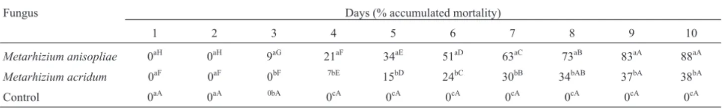
The pathogenicity test revealed that M. anisopliae
caused an 88% mortality rate in D. saccharalis larvae, whereasM. acridumcaused a mortality of 38%. We also observed that M. anisopliae caused an initial mortality (12%) on the third day after infection, whereasM.acridum
caused mortality (7%) only on the fourth day (Table 2). The Pearson correlation analysis showed a strong correlation between mortality andpr1A gene expression in medium containing cuticle (r = 0.9835, p < 0.0004) and during pathogenesis (r = 0.9987, p < 0.0001).
Discussion
Presumably, Metarhizium has different subsets of genes that are responsive to different growth conditions. Thus, identifying these genes should help to elucidate mechanisms of adaptation (Zhanget al., 2011; Liu et al., 2013; Jinet al., 2014). In this study, we found that expres-sion of thepr1A gene was affected by different growth con-ditions, indicating that pr1A expression may vary in the presence of different carbon sources. Previous work has shown that subtilisin PR1A protein is produced when grown on minimal medium, and this production is en-hanced by the addition of cuticle to the media; however, PR1A synthesis is repressed in rich medium (Freimoseret al., 2003, 2005). Our finding of a significant increase in
pr1A expression in medium supplemented with D. saccharalis cuticle compared to either medium supple-mented with casein or rich medium (YPD) corroborates these reports. The demonstration of differential expression of pr1A in different culture media suggests that the two fungal species can regulatepr1A gene expression during saprophytic growth. It is likely that differences inpr1A ex-pression are related to factors that allowM. anisopliaeand
M. acridumto flexibly respond to the conditions of the sur-rounding environment.
During the pathogenicity process, adhesion to the host and penetration through the cuticle are decisive stages
Table 1- E and R2 values of linear regression for dilutions of the reference and target gene ofMetarhizium anisopliaeandMetarhizium acridum.
Gene E value (%) R2value
Metarhizium anisopliae Metarhizium acridum Metarhizium anisopliae Metarhizium acridum
pr1 103% 104% 0.998 0.996
try 106.2% 98% 0.995 0.994
in establishing the infection, and these involve major chan-ges in gene expression patterns that result in dramatic changes in the amount of available nutrients from the host (Roberts and St. Leger, 2004; Heet al., 2012). In nutri-ent-deprived conditions, subtilisin PR1 is produced in the appressorium where it hydrolyzes cuticular proteins, thus facilitating hyphal penetration through the insect cuticle (Goettelet al., 1989; St. Legeret al., 1989).
Once the hyphae have breached the cuticle, the fun-gus grows in the hemolymph, which is rich in nutrients, and here PR1 is down-regulated by the presence of accessible
carbon and nitrogen sources (Small and Bidochka, 2005). This process explains whypr1A gene is repressed in dead insects infected by either of the two species. The death of the insect occurs after the fungus has penetrated the cuticle, formed the blastospores in the hemolymph and invaded the various internal organs that are rich in nutrients (Samuelset al., 1989). Metarhizium then utilizes and depletes the avail-able nutrients of the insect. Subsequently, PR1 is induced again and performs functions that facilitate the penetration of hyphae through the cuticle, allowing the fungus to emer-ge and produce conidia in laremer-ge masses on the surface of the insect cadaver, resulting in mummification (Scholteet al., 2007) Thus, induction ofpr1A expression in insects cov-ered with either mycelia or conidia suggests that PR1A may not only be directly involved in the formation of conidia but may also be required for the completion of the pathogenic cycle.
Regarding virulence factors, there is evidence that mutant strains ofM. anisopliaethat have increased expres-sion of the enzyme PR1A are more pathogenic to the host
Manduca sexta(St. Legeret al., 1989). Furthermore, spon-taneouspr1A-deficient mutants ofM. anisopliae demon-strated a reduction in their ability to infectTenebrio molitor
compared to wild-typeM. anisopliae(Wanget al., 2005). Gillespieet al. (1998) reported that differences in thein vivoproduction of proteases amongMetarhiziumisolates can influence the virulence of the individual isolates. In the current study we found a correlation between the level of
pr1A expression in MM supplemented with cuticle and during pathogenesis and observed pathogenesis. When comparing the two fungal species, we observed that
M. anisopliae, besides having higher expression ofpr1A in culture medium and in all phases of pathogenicity, showed better pathogenic action againstD. saccharalis, and herein we considered both the initial and final mortality caused by the infection. Taken together, our data suggest that the level ofpr1A gene expression may predict the ability of the fun-gus to cause disease.
Surface structure and the chemical composition of the host cuticle are believed to affect the adhesion of fungal spores and, consequently, pathogenicity.Metarhizium rec-ognizes specific host signals that induce the secretion of different host-specific proteins. As a consequence, this se-lectivity for host signals is most likely to define the
speci-Figure 1 - Relative expression levels of pr1A gene in Metarhizium anisopliaeandMetarhizium acridumafter 72 h of growth in different cul-ture media. Means followed by distinct letters differ by the Tukey test at the 5% probability level; lower-case letters represent differences between iso-lates, and upper-case letters represent differences between culture media.
Figure 2- Relative expression levels ofpr1A during pathogenesis of
Metarhizium anisopliaeandMetarhizium acridum. Means followed by distinct letters differ by the Tukey test at 5% probability; lower-case letters represent differences between isolates at each phase, and upper-case let-ters represent the differences between phases for each isolate. ID = dead infected insect; ME = emergent mycelia from insect cadavers; IC = insect cadavers completely covered with conidia.
Table 2- Accumulated mortality ofDiatraea saccharalisinfected byMetarhizium anisopliaeandMetarhizium acridumduring 10 days of evaluation.
Fungus Days (% accumulated mortality)
1 2 3 4 5 6 7 8 9 10
Metarhizium anisopliae 0aH 0aH 9aG 21aF 34aE 51aD 63aC 73aB 83aA 88aA
Metarhizium acridum 0aF 0aF 0bF 7bE 15bD 24bC 30bB 34bAB 37bA 38bA
Control 0aA 0aA 0bA 0cA 0cA 0cA 0cA 0cA 0cA 0cA
ficity for a particular isolate/host (Lazzariniet al., 2006; Pedrini et al., 2007; Santi et al., 2010). Thus, although
pr1A is expressed by two different species, we speculate that the higher level of expression detected inM. anisopliae
occurred in response to the host, indicating that induction of
pr1A gene expression is one of the factors that determines the ability of this pathogen to infectD. saccharalis. The dif-ference in pathogenicity factors between the species of
Metarhiziummay exist due to differential regulation of the same set of genes, rather than variations in the number and types of gene for each host species.
Although many studies involving genes related to pathogenicity have been reported in the literature (Fang and Bidochka, 2006; Scholteet al., 2007; Bischoffet al., 2009), much can still be learned about the functions of these genes. Gene functionality can be inferred from the differential ex-pression of these genes in response to different growing conditions and hosts. In support of this premise, Fanget al.
(2010) showed thatMetarhiziumcan induce genes differ-ently for each type of environment or host. We observed such differences in our assays withM. anisopliaeandM. acridum,species that have not previously been evaluated under these conditions. Our findings thatpr1A is differen-tially expressed inM. anisopliaeandM. acridumin differ-ent culture media, as well as during pathogenesis, indicates that these species have different strategies to adapt to dif-ferent growth conditions. This conclusion is consistent with the saprophytic and/or parasitic lifestyle of these pathogens and may be a sign of their versatility, which favors adapt-ability to environmental conditions.
Our results provide a better understanding of the roles that PR1A plays during the saprophytic and parasitic phases in the two species of Metarhizium studied. This study serves to emphasize the importance ofpr1A gene ex-pression in the biology of these fungi and also suggests that this gene is a potential virulence factor for the development of advanced engineered biopesticides.
Acknowledgments
This project received financial support by the Con-selho Nacional de Desenvolvimento Científico e Tecnoló-gico - CNPq (PhD fellowship to M.P. Carneiro-Leão).
References
Alves SB, Lopes RB, Vieira SA and Tamai MA (2008) Fungos entomopatogênicos usados no controle de pragas na Amé-rica Latina. In: Alves SB and Lopes RB (eds) Controle Microbiano de Pragas na América Latina: Avanços e Desa-fios. FEALQ, Piracicaba, pp 69-110.
Altschul SF, Gish W, Miller E, Myers EW and Lipton DJ (1990) Basic local alignment search tool. J Mol Biol 215:403-410. Andersen SO (1980) Cuticular sclerotization. In: Miller TA (ed)
Cuticle Techniques in Arthropods. Springer-Verlag, New York, pp 185-215.
Bagga S, Hu G, Screen SE and St. Leger RJ (2004) Recon-structing the diversification of subtilisins in the pathogenic fungusMetarhizium anisopliae. Gene 324:159-169. Bischoff JF, Rehner SA and Humber RA (2009) A multilocus
phylogeny of theMetarhizium anisopliae lineage. Myco-logia 101:512-530.
Bustin SA, Benes V, Garson JA, Hellemans J, Huggett J, Kubista M, Mueller R, Nolan T, Pfaffl MW, Shipley GL, et al.
(2009) The MIQE guidelines: Minimum information for publication of quantitative real-time PCR experiments. Clin Chem 4:611-622.
Fang W and Bidochka MJ (2006) Expression of genes involved in
germination, conidiogenesis and pathogenesis in
Metarhizium anisopliaeby quantitative real-time RT-PCR. Mycol Res 110:1165-1171.
Fang W, Fernandes EKK, Robert DW, Bidochka MJ and St. Leger RJ (2010) A laccase exclusively expressed byMetarhizium anisopliaeduring isotropic growth is involved in pigmenta-tion, tolerance to abiotic stresses and virulence. Fungal Genet Biol 47:602-607.
Frazzon APG, Da Silva V Jr I, Masuda A, Schrank A and Vainstein MH (2000) In vitroassessment ofMetarhizium anisopliae isolates to control the cattle tick Boophilus microplus. Vet Parasitol 94:117-125.
Freimoser FM, Hu G and St. Leger RJ (2005) Variation in gene expression patterns as the insect pathogen Metarhizium anisopliaeadapts to different host cuticles or nutrient depri-vation in vitro. Microbiology 151:361-371.
Freimoser FM, Screen S, Bagga S, HuG and St Leger RJ (2003) Expressed sequence tag (EST) analysis of two subspecies of
Metarhizium anisopliaereveals a plethora of secreted pro-teins with potential activity in insect hosts. Microbiology 149:239-247.
Gillespie JP, Bateman R and Charnley AK (1998) Role of cuti-cle-degrading proteases in the virulence ofMetarhiziumspp. for the desert locust, Schistocerca. J Invertebr Pathol 71:128-137.
Goettel MK, St Leger RJ, Rizzo NW, Staples RC and Roberts DW (1989) Ultrastructural localization of a cuticle degrading protease produced by the entomopathogenic fungus
Metarhizium anisopliae during penetration of host (Manduca sexta) cuticle. J Gen Microbiol 135:2233-2239. He M, Hu J and Xia Y (2012) Large scale expressed sequence tag
(EST) analysis ofMetarhizium acriduminfectingLocusta migratoriareveals multiple strategies for fungal adaptation to the host cuticle. Curr Genet 58:265-279.
Jin K, Han L and Xia Y (2014) MaMk1, a FUS3/KSS1-type mitogen-activated protein kinase gene, is required for appressorium formation, and insect cuticle penetration of
the entomopathogenic fungus Metarhizium acridum. J
Invertebr Pathol 115:68-75.
Lazzarini GMJ, Rocha LFN and Luz C (2006) Impact of moisture on in vitro germination of Metarhizium anisopliae and
Beauveria bassianaand their activity onTriatoma infestans. Mycol Res 110:485-492.
Liu Q, Ying SH, Li JG, Tian CG and Feng MG (2013) Insight into the transcriptional regulation of Msn2 required for coni-diation, multi-stress responses and virulence of two ento-mopathogenic fungi. Fungal Genet Biol 54:42-51.
Luo S, He M, Cao Y and Xia Y (2013) The tetraspanin gene
appressorial function and enzymes for cuticle degradation in the entomopathogenic fungus,Metarhizium acridum. Envi-ron Microbiol 15:2966-2979.
Pathan AAK, Devi KU, Vogel H and Reineke A (2007) Analysis of differential gene expression in the generalist entomo-pathogenic fungus Beauveria bassiana(Bals.) Vuillemin grown on different insect cuticular extracts and synthetic medium through cDNA-AFLPs. Fungal Genet Biol 44:1231-1241.
Pedrini N, Crespo R and Juárez MP (2007) Biochemistry of insect epicuticle degradation by entomopathogenic fungi. Comp
Biochem Physiol C Pharmacol Toxicol Endocrinol
146:124-137.
Pfaffl MW (2001) A new mathematical model for relative quanti-fication in real-time RT-PCR. Nucleic Acids Res 29:2002-2007.
Pontecorvo G, Roper JA, Hemons LM, MacDonald KD and Bufton AWJ (1953) The genetics ofAspergillus nidulans. Adv Genet 5:141-238.
Roberts DW and St Leger RJ (2004)Metarhiziumspp., cosmopol-itan insect-pathogenic fungi: Mycological aspects. Adv Appl Microbiol 54:1-70.
Samuels KDZ, Pinnock DE and Allsopp PG (1989) The potential of Metarhizium anisopliae (Metschnikoff) Sorokin (Deutermycotina, Hyphomycetes) as a biological
control-agent of Inopus rubriceps (Macquart) (Diptera,
Stratiomyidae). J Aust Entomol Soc 28:69-74.
Santi L, Silva WOB, Pinto AFM, Schrank A and Vainstein MH (2010) Metarhizium anisopliaehost-pathogen interaction: Differential immunoproteomics reveals proteins involved in the infection process of arthropods. Fungal Biol 114:312-319.
Scholte EJ, Takken W and Knols BGJ (2007) Infection of adult
Aedes aegyptiandAe. albopictusmosquitoes with the ento-mopathogenic fungus Metarhizium anisopliae. Acta Trop 102:151-158.
Silva FAS and Azevedo CAV (2002) Assistant computational program version for the windows operating system. Braz J Agro-Industr Prod 4:71-78.
Small CN and Bidochka MJ (2005) Up-regulation of Pr1, a subti-lisin-like protease, during conidiation in the insect pathogen
Metarhizium anisopliae. Mycol Res 3:301-313.
Staden R, Beal KF and Bonfield JK (1998) The Staden Package. Computer Methods in Molecular Biology. Bioinform Meth Prot 132:115-130.
St Leger RJ, Butt TM, Staples RC and Roberts DW (1989) Syn-thesis of proteins including a cuticle-degrading protease
dur-ing differentiation of the entomopathogenic fungus
Metarhizium anisopliae. Exp Mycol 13:253-262.
Wang C, Gang Hu and St. Leger RJ (2005) Differential gene ex-pression byMetarhizium anisopliaegrowing in root exudate and host (Manduca sexta) cuticle or hemolymph reveals mechanisms of physiological adaptation. Fungal Genet Biol 42:704-718.
Zhang C, Xia Y and Li Z (2011) Identification of genes differen-tially expressed byMetarhizium anisopliaegrowing on Lo-custa migratoria wings using suppression subtractive hy-bridization. Curr Microbiol 62:1649-1655.
Associate Editor: Célia Maria Soares