Brazilian Journal of Microbiology (2011) 42: 835-845 ISSN 1517-8382
TAXONOMIC STUDY AND PARTIAL CHARACTERIZATION OF ANTIMICROBIAL COMPOUNDS FROM A MODERATELY HALOPHILIC STRAIN OF THE GENUS ACTINOALLOTEICHUS
Farida Boudjelal1; Abdelghani Zitouni1; Florence Mathieu2; Ahmed Lebrihi2; Nasserdine Sabaou1*
1
Laboratoire de Recherche sur les Produits Bioactifs et la Valorisation de la Biomasse, Ecole Normale Supérieure de Kouba, BP
92, 16050 Kouba, Alger, Algérie; 2 Université de Toulouse, Laboratoire de Génie Chimique UMR 5503 (CNRS/INPT/UPS),
ENSAT/INP de Toulouse, 1 avenue de l’Agrobiopôle, Castanet-Tolosan Cedex, France.
Submitted: May 01, 2010; Returned to authors for corrections: September 29, 2010; Approved: January 31, 2011.
ABSTRACT
A moderately halophilic actinomycete strain designated AH97 was isolated from a saline Saharan soil, and
selected for its antimicrobial activities against bacteria and fungi. The AH97 strain was identified by
morphological, chemotaxonomic and phylogenetic analyses to the genus Actinoalloteichus. Analysis of the
16S rDNA sequence of strain AH97 showed a similarity level ranging between 95.8% and 98.4% within
Actinoalloteichus species, with A. hymeniacidonis the most closely related. The comparison of the
physiological characteristics of AH97 with those of known species of Actinoalloteichus showed significant
differences. Strain AH97 showed an antibacterial and antifungal activity against broad spectrum of
microorganisms known to be human and plant pathogens. The bioactive compounds were extracted from the
filtrate culture with n-butanol and purified using thin layer chromatography and high pressure liquid
chromatography procedures. Two active products were isolated, one hydrophilic fraction (F1) and another
hydrophobic (F2). Ultraviolet-visible, infrared, mass and 1H and 13C nuclear magnetic resonance
spectroscopy studies suggested that these molecules were the dioctyl phthalate (F2) and an aminoglycosidic
compound (F1).
Key words:Actinoalloteichus, taxonomy, biological activities, structure, dioctyl phthalate
INTRODUCTION
Actinomycetes are the most significant group of
microorganisms considered as an important producer of
antibiotics and other important bioactive substances (4). The
filamentous Actinomycetales species produces over 10000
bioactive compounds. The most frequent producers, the
Streptomyces species, produce 7600 compounds (74% of all
Actinomycetales), while other actinomycetes represent 26%,
altogether 2500 compounds (2). These organisms produce
perhaps the most diverse and most unique, unprecedented,
sometimes very complicated compounds exhibiting excellent
antibacterial potency and usually low toxicity (2).
In the course of screening for new bioactive compounds,
Boudjelal, F. et al. Taxonomic study of the genus Actinoalloteichus
several research studies are currently oriented towards isolation
of rare actinomycetes from different ecosystems (13). The
isolation of these microorganisms that produce bioactive
compounds is of great interest in the development of new
molecules to fight against many pathogens especially with the
emergence of antibiotic multi-resistance pathogenic bacteria.
Halophilic and salt-tolerant actinomycetes are
extremophilic microorganisms, which are used by several
research programs around the world to isolate new natural
compounds (4).
In fact, our laboratory takes a particular interest in extreme
environment, like saline soils and their microflora. The
ecological distribution of actinomycetes from Saharan soils has
been studied in Algeria and their biodiversity in those arid soils
has been demonstrated (23).
During a screening programme of the search for new
antibiotics from non-Streptomyces genera, new molecules were
obtained (12, 32, 33). As part of this program, we identified a
new halophilic actinomycete (strain AH97) having interesting
antimicrobial activity, which assigned to the genus
Actinoalloteichus.
In present study, we describe the polyphasic taxonomic
characterization of actinomycete strain AH97 isolated from an
Algerian Saharan soil having antimicrobial activity against
various bacteria and fungi. The production, extraction,
purification and partial characterization of active molecules of
this strain are reported as well.
MATERIALS AND METHODS
Origin of actinomycete
The strain AH97 was isolated from a Saharan soil
collected in Tamanrasset (Hoggar, south of Algeria; latitude,
22° 49’ N; longitude, 5° 28’ E).
Microbial strains and culture conditions
The strain AH97 was grown at 30ºC in tryptic soy broth
(TSB) (contents per liter: 17.0 g tryptose; 3.0 g soytone; 5.0 g
NaCl; 15 g agar) fortified with 5% NaCl. The pH was adjusted
to 7.3 prior to autoclaving.
For antibacterial and antifungal experiments, Bacillus
subtilis ATCC 6633 and Mucor ramannianus NRRL1829
(sensitive strains) were used as the indicator microorganisms.
Taxonomic study
Cultural characteristics of strain AH97 were studied on
yeast extract-malt extract agar (ISP2) (27), inorganic
salts-starch agar (ISP4) (27), tryptic soy agar (TSA) (27) and
complex medium agar (CMA) (26). All media were
supplemented with 5% of NaCl. Colors of aerial and substrate
mycelia were determined with the ISCC-NBS centroid color
charts (National Bureau of Standards, USA 1964). The
micromorphology of strain AH97 was examined under light
microscopy for the mycelial organization and sporulation.
For the physiological study, fifty nine tests were
considered, including the utilization of 20 carbohydrate
compounds, the degradation of adenine, guanine, xanthine,
hypoxanthine, milk casein, tyrosine, testosterone (6), tween 80,
gelatine, starch, esculin and arbutin (20), the decarboxylation
of nine organic acids (7) and the production of nitrate reductase
(20). The strain AH97 was checked for salt requirement or
tolerance by inoculation on TSA containing 0, 5, 7, 10, 15 and
20% of NaCl, as described by Kushner (11). The strain was
also examined for its ability to grow on TSA (with 5% of
NaCl), supplemented with 6 antibiotics or with lysozyme (8)
and to grow at pH 5 and 9, and at 42ºC.
For the chemotaxonomic analysis, biomass was obtained
from culture grown on shake TSB medium supplemented with
5% of NaCl, and incubated at 30ºC for 7 days. Diaminopimelic
acid (DAP) isomer, whole-cell sugars pattern and
phospholipids were analysed according to the methods of
Becker et al. (1), Lechevalier and Lechevalier (14) and
Boudjelal, F. et al. Taxonomic study of the genus Actinoalloteichus
DNA preparation, PCR amplification and sequence analysis
DNA was prepared according to the method of Liu et al.
(18). The strain AH97 was grown at 30ºC for 7 days in a shake
flask containing 100 mL of TSB medium supplemented with
5% of NaCl. PCR amplification of the 16S rDNA of the
halophilic strain was performed using two primers: 27f
(5'-AGTTTGATCCTGGCTCAG-3') and 1492r
(5'-GGTTACCTTGTTACGACTT-3'). The 16S rDNA was
amplified by PCR using Invitrogen kit. The final volume of
reaction mixture of 50 µ L contained 1X PCR buffer (10 mmol
of Tris-HCl, 50 mmol of KCl, pH 9.0 at 25ºC), 1.5 mmol of
MgC12, 200 mmol of each dNTP, 1 mmol of each primer, 1.25
U of Taq DNA polymerase and 1 µL (500 ng) of the purified
DNA. The amplification was performed on a STRATAGENE
RoboCycler Gradient 96, according to the following profile: an
initial denaturation step at 98ºC for 3 min, after which Taq
polymerase was added, followed by 30 amplification cycles of
94ºC for 1 min, 52ºC for 1 min, and 72ºC for 2 min and a final
extension step of 72ºC for 10 min. The PCR product was
detected by agarose gel electrophoresis and was visualized by
ultraviolet fluorescence after ethidium bromide staining.
The sequencing reaction was performed by MilleGen
Company (Toulouse, France). The same primers as before and
an automated sequencer were used for this purpose. The
obtained sequences were compared for similarity level with the
reference species of bacteria contained in genomic database
banks, using the ‘NCBI Blast’ available at
http://www.ncbi.nlm.nih.gov/
Phylogenetic analysis
Phylogenetic and molecular evolutionary analyses were
conducted using softwares included in MEGA version 3.0
package (10). The 16S rDNA sequence of the strain AH97 was
aligned using the CLUSTAL W program (30) against
corresponding nucleotide sequences of Actinoalloteichus
reference species retrieved from GenBank. Evolutionary
distance matrices were generated as described by Jukes and
Cantor (9) and a phylogenetic tree was inferred by the
neighbor-joining method (24). Tree topologies were evaluated
by bootstrap analysis (5) based on 1000 resamplings of the
neighbor-joining dataset.
Antimicrobial activity of the strain AH97
The antimicrobial spectrum was determined by
conventional agar method (agar cylinders method). The
microorganisms used are listed in Table 2.
Strain AH97 was grown on TSA agar plates supplemented
with 5% of NaCl for 7 days at 30°C, then calibrated agar
cylinders (5 mm in diameter) were cut out and placed onto the
agar surface (nutrient agar or Sabouraud agar, covered
respectively by 10 mL of culture containing 105 CFU mL-1 for
bacteria or 104 CFU mL-1 for fungi). A sterile cylinder of TSA
supplemented with 5% of NaCl was used as control. The plates
were incubated at 30°C for 24-48 h after a diffusion process for
4 h at 4°C. The diameters of any inhibition zones formed
around the cylinder of actinomycete isolate were then
measured.
Kinetic of growth and production of antimicrobial compounds
For the pre-culture, the strain AH97 was grown on TSA
supplemented with 5% of NaCl, for 7 days at 30ºC. The
mycelium was scraped and inoculated into a 250 mL
Erlenmeyer flask containing 50 mL of TSB supplemented with
5% of NaCl. Aliquots (5%, v/v) of this pre-culture were
transferred into 500-mL Erlenmeyer flasks containing 100 mL
of the same medium. The cultures were incubated on a rotary
shaker (250 rpm) at 30ºC for 7 days. The antibacterial and
antifungal activities were assayed daily against Bacillus subtilis
and Mucor ramannianus by the agar diffusion method (well
technique). Each well of 10 mm in diameter was filled with 0.2
mL of the supernatant. The growth (dry weight of mycelium)
Boudjelal, F. et al. Taxonomic study of the genus Actinoalloteichus
Production and extraction of antimicrobial compounds For the production of active compounds, fermentations
were carried out in 500 mL Erlenmeyer flasks containing 100
mL of TSB supplemented with 5% of NaCl (250 rpm, 30ºC
and 3 days).
The culture broth (10 L) was centrifuged at 5,000 × g for
10 min to remove the biomass. The cell-free supernatant was
extracted with an equal volume of n-butanol. The organic
extract was concentrated to dryness, recuperated in methanol
and bioassayed against B. subtilis and M. ramannianus by
paper disk diffusion method.
Biological and chemical detection of antimicrobial compounds
Crude extract samples were subsequently subjected to
thin-layer chromatography (TLC). Samples were spotted onto
20 × 20-cm2 silica gel plates (Merck Art 5735, kieselgel
60F254), and then developed with n-butanol-acetic acid-water
(B.A.W.) (3: 1: 1, v/v) as the solvent mixture. The active spot
was detected by bioautography (3) on silica gel plates seeded
with M. ramannianus or B. subtilis. Clear halos, due to growth
inhibition of the microorganisms, indicated the location of
antimicrobial compounds on the TLC plates, and the retention
factor (Rf) values were recorded. The antimicrobial compounds
were also detected under ultraviolet irradiations at 254 and 365
nm and by spraying with some chemical reagents such as
napthoresorcinol-H2SO4 and ninhydrin.
Purification of antimicrobial compounds
Concentrated extracts were purified on preparative TLC
silica gel plates (Merck 60 F-254; 0.5 mm thick). The plates
were developed with B.A.W. solvent system (3: 1: 1, v/v), and
then air dried. The active band was visualized in ultraviolet
light at 254 and 365 nm, scraped from the plates, eluted with
methanol and concentrated using the Rotavapor. The antibiotic
activity of the band against B. subtilis and M. ramannianus was
measured by the paper disk method. Final purification of the
antimicrobial compounds was carried out by a
high-performance liquid chromatography (HPLC) (Waters, Milford,
MA, USA) system equipped in first step (in order to separate
the hydrophilic fraction of the hydrophobic fraction) with a
C18 column (Uptisphere UP5ODB, 250 × 7.8 mm; Interchim)
and in second step with a C18 pyramid column (Nucleodur VP
250/10, 250 × 10 mm; Machery Nagel). The analysis
conditions were as follows: a mobile phase with a continuous
gradient (convex curve) solvent system from 0 to 100%
methanol in water (30 min) to separate the active fractions with
Uptisphere UP5ODB C18 column, then 80 to 100% methanol
in water (30 min) to purify the hydrophobic active fraction with
the same column, and 100% water (isocratic) (30 min) to
purify the hydrophilic active fraction with C18 pyramid
column; flow rate of 2 mL min-1 and UV detection at 220 nm.
The final purification was achieved after the third re-injection
in HPLC system.
Spectroscopic studies of antimicrobial compounds
The UV absorption spectra of the hydrophilic fraction F1
in distilled water and hydrophobic fraction F2 in methanol
were determined with a Shimadzu UV 1605
spectrophotometer. Mass spectrum of fraction F1 was recorded
on an ion-trap mass spectrometer (Finnigan MAT, San Jose,
CA), equipped with a nanospray ion electro-spray ionisation
(ESI) source (positif ion mode) and fraction F2 with electron
impact by using Nermag R-10-10C spectrometer. Infrared (IR)
spectrq of the fractions (F1) and (F2) were obtained with a
Perkin Elmer FT-IR 1760 spectrometer. 1H and 13C nuclear
magnetic resonance spectra of the hydrophobic compound F2
(in MeOD) and hydrophilic compound F1 (in D2O) were
recorded on a Bruker AMX 400 spectrometer operating at 500
MHz.
RESULTS AND DISCUSSION
Taxonomy
The strain AH97 grew well on TSA and CMA (7 days
Boudjelal, F. et al. Taxonomic study of the genus Actinoalloteichus
greenish blue to grayish blue (TSA) or pale yellowish (CMA)
aerial mycelium. The substrate mycelium was light yellow on
the two media used. Brown to black diffusible pigment was
observed on TSA. No growth was observed on ISP2 and ISP4
media with 5% of NaCl. The aerial mycelium was branched
and fragment into straight spore chains after 10 days of
incubation at 30°C. Endospores, sclerotia, sporangia,
synnemata and whirls were not observed.
Cell-wall hydrolysate of strain AH97 contained the
DL-diaminopimelic acid isomer but glycine was not found.
Galactose, mannose and glucose were the characteristic sugars
of the whole-cell extract in addition to ribose. Thus, strain
AH97 had cell-wall of type III and sugar pattern type C (14).
The phospholipid profile was of type PII, characterized by the
presence of phosphatidylethanolamine as major phospholipid
(15). Based on the morphological and chemical characteristics,
AH97 is classified in the genus Actinoalloteichus (19). The
genus currently comprises only three species, A. cyanogriseus
IFO14455T (29), A. spitiensis DSM44848T (28) and A.
hymeniacidonis HPA177T (29), and no one is halophilic.
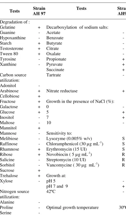
Physiological characteristics
The physiological properties of strain AH97 are shown in
Table 1. Optimal growth of strain was observed at 30°C at pH
7 and in presence of NaCl in range 5 to 7%. Strain was unable
to grow at concentration 0 and 10% of NaCl, at pH 5 and at
42°C, and able to grow at pH 9.
The strain was able to hydrolyse a great number of
compounds such as adenine, arbutin, casein, esculin, gelatine,
starch, testosterone, tween 80, arabinose, cellobiose, fructose,
galactose, glucose, mannitol, rhamnose, ribose, sucrose,
trehalose, sodium propionate, sodium pyruvate and sodium
succinate but did not utilize guanine, hypoxanthine, tyrosine,
xanthine, adonitol, inositol, maltose, mannose, melibiose,
raffinose, salicine, sorbitol, xylose, sodium acetate, sodium
benzoate, sodium butyrate, sodium citrate, sodium oxalate,
sodium tartrate, alanine, proline and serine. It reduced nitrate
and was resistant to novobiocin (5 µg mL-1), streptomycin (10
UI) and vancomycin (30 µg mL-1) and sensitive to
chloramphenicol (30 µ g mL-1), erythromycin (15 UI) and
lysozyme (0.005%).
Table 1. Physiological characters of strain AH97.
Tests Strain AH 97
Tests Strain
AH97
Degradation of :
Gelatine + Decarboxylation of sodium salts:
Guanine - Acetate -
Hypoxanthine - Benzoate -
Starch + Butyrate -
Testosterone + Citrate -
Tween 80 + Oxalate -
Tyrosine - Propionate +
Xanthine - Pyruvate +
Succinate +
Carbon source utilization:
Tartrate -
Adonitol -
Arabinose + Nitrate reductase +
Cellobiose +
Fructose + Growth in the presence of NaCl (%):
Galactose + 0 -
Glucose + 5 +
Inositol - 7 +
Maltose - 10 -
Mannitol +
Mannose - Sensitivity to:
Melibiose - Lysozyme (0.005% w/v) S
Raffinose - Chloramphenicol (30 µg mL-1) S
Rhamnose + Erythromycin (15 UI) S
Ribose + Novobiocin ( 5 µg mL-1) R
Salicine - Streptomycin (10 UI) R
Sorbitol - Vancomycine ( 30 µg mL-1) R
Sucrose +
Trehalose + Growth at:
Xylose - pH 5 -
pH 7 and 9 +
Nitrogen source utilization:
42ºC -
Alanine -
Proline - Optimal growth temperature 30ºC
Serine -
Boudjelal, F. et al. Taxonomic study of the genus Actinoalloteichus
Phylogenetic analysis
The 16S rDNA sequences of the strain AH97 (1248
nucleotides) was determined and deposited in GenBank under
the accession number FJ379336. This strain was first analysed
by a BLAST search and was aligned with those of
Actinoalloteichus reference species available in the GenBank
database, which confirmed its identification at the genus level.
The position of strain AH97 in the 16S rDNA
Actinoalloteichus tree is shown in Fig. 1
The neighbor-joining tree indicated that the novel strain
fall into one distinct clade with the type strains of
Actinoalloteichus spitiensis DSM44848T (96.2%), A.
cyanogriseus IFO14455T (95.8%), which were different from
strain AH97 (similarity level < 97%), and A. hymeniacidonis
HPA177T (98.4%), the most closely related species. The
results suggest that our strain represent a distinct phylogenetic
line.
Strain AH97 differed from A. hymeniacidonis (31) by
many characters. By contrast to our strain, A. hymeniacidonis
was not halophilic, grew well on ISP2, had a
brownish-purplish-grey aerial mycelium and a brownish-black substrate
mycelium, was able to degrade maltose, mannose, sorbitol,
xylose and sodium citrate, unable to degrade arabinose and
sodium succinate and reduction of nitrate is negative.
Figure 1. Neighbor-joining tree based on 16S rDNA sequences showing the relations between the moderately halophilic actinomycete isolate AH97 and type species of the genus Actinoalloteichus. The accession numbers of strain sequences are given
in parentheses.
The numbers at the nodes indicate the levels of bootstrap support based on neighbor-joining analyses of 1000 resampled datasets;
only values over 50% are given. Bar: 0.005 nucleotide substitution per nucleotide position. Amycolatopsis orientalis is given as
out group.
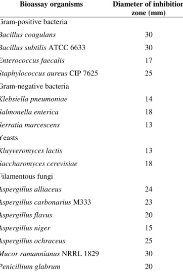
Antimicrobial activity on solid media
Strain AH97 showed a broad spectrum of antimicrobial
activity (Table 2). It was strongly active against Gram-positive
bacteria such as Bacillus coagulans, B. subtilis and
Staphylococcus aureus and against fungi such as Aspergillus
alliaceus, A. carbonarius, A. flavus, A. ochraceus, Mucor
ramannianus and Penicillium glabrum. It was moderately
active against Enterococcus faecalis, Klebsiella pneumoniae,
Salmonella enterica, Serratia marcescens, Kluyveromyces
lactis, Saccharomyces cerevisiae and Aspergillus niger.
However, no activity was observed against Pseudomonas
aeruginosa, P. syringae, Agrobacterium tumefaciens, Candida
Boudjelal, F. et al. Taxonomic study of the genus Actinoalloteichus
Table 2. Antimicrobial activity of strain AH97 against various microorganisms.
Bioassay organisms Diameter of inhibition zone (mm) Gram-positive bacteria
Bacillus coagulans 30
Bacillus subtilis ATCC 6633 30
Enterococcus faecalis 17
Staphylococcus aureus CIP 7625 25
Gram-negative bacteria
Klebsiella pneumoniae 14
Salmonella enterica 18
Serratia marcescens 13
Yeasts
Kluyveromyces lactis 13
Saccharomyces cerevisiae 18
Filamentous fungi
Aspergillus alliaceus 24
Aspergillus carbonarius M333 23
Aspergillus flavus 20
Aspergillus niger 15
Aspergillus ochraceus 25
Mucor ramannianus NRRL 1829 30
Penicillium glabrum 20
* Activity was performed using agar cylinder method.
The diameter of the cylinder agar does not include in diameters of inhibition zones.
The strain AH97 is non-active against Agrobacterium tumefaciens no 2410, Pseudomonas aeruginosa, P. syringae n° 2410, Candida albicans and Botrytis cinerea.
Growth kinetics and production of antimicrobial compounds
In TSB medium with 5% of NaCl, the biomass increased
and reached the maximum at 3 days, followed by a stationary
phase until 4 days, than decreased. The antifungal activity was
detected at 1 day after inoculation and reached a maximum at 3
days. The antibacterial activity began and reached a maximum
at 2 days of fermentation than decrease after this. The pH
varied between 7.2 and 8.6 during the incubation (Fig. 2).
Figure 2. Time course of growth and antibiotic production in TSB medium with 5% of NaCl: ( ) dry cell weight, ( )
antibacterial activity against Bacillus subtilis, ( ) antifungal
activity against Mucor ramannianus, ( ) pH.
Inhibition diameter values were measured without diameter of
well (10 mm).
Extraction, purification and partial characterization of antimicrobial compounds
Various separation steps were applied to 10 L culture
broth of strain AH97 grown in TSB medium supplemented
with 5% of NaCl.
Antimicrobial compounds were extracted from broth
culture with n-butanol. The active extract was concentrated to
dryness using the Rotavapor, recuperated in methanol and
analysed by TLC developed in B.A.W. solvent system. One
active band, named fraction TM2, was detected by
bioautography (at a Rf value of 0.44), which was active against
B. subtilis and M. ramannianus.
The active band was scraped from the plates, eluted with
methanol, concentrated and purified by HPLC in first with a
C18 column (Uptisphere UP5ODB). The chromatogram of the
Boudjelal, F. et al. Taxonomic study of the genus Actinoalloteichus
antibacterial and antifungal activity, which were designated F1
(retention time: RT, 5.00 min, hydrophilic and colorless
fraction), and F2 (RT, 17.00 min, hydrophobic and colorless
oil). Each fraction was collected separately and re-injected in
HPLC system with a C18 pyramid column (Nucleodur) to
purified F1 with 100% H2O eluted phase and C18 column
(Uptisphere UP5ODB) to purified F2 with 80 to 100%
methanol in water eluted phase.
Fraction 1 (F1)
Chromogenic reactions with napthoresorcinol-H2SO4 and
ninhydrin were positive (presence of sugar and amine). The
UV-visible spectrum in water (data not shown) of the pure
product exhibited a maximum at 200 nm in water. The IR
absorption spectrum (data not shown) exhibited the following
bands: 3390 cm-1 (O-H), 2933 and 673 cm-1 (C-H), 1675 cm-1
(C-OH) and 1543 cm-1 (NH2). The PM of the fraction F1 (data
not shown) is 850 with fragments at m/z 685 and 522. The
fragmentation of the majority peak in positive mode showed
differences between the ions at m/z 162-164 that could
correspond to C6 sugars.
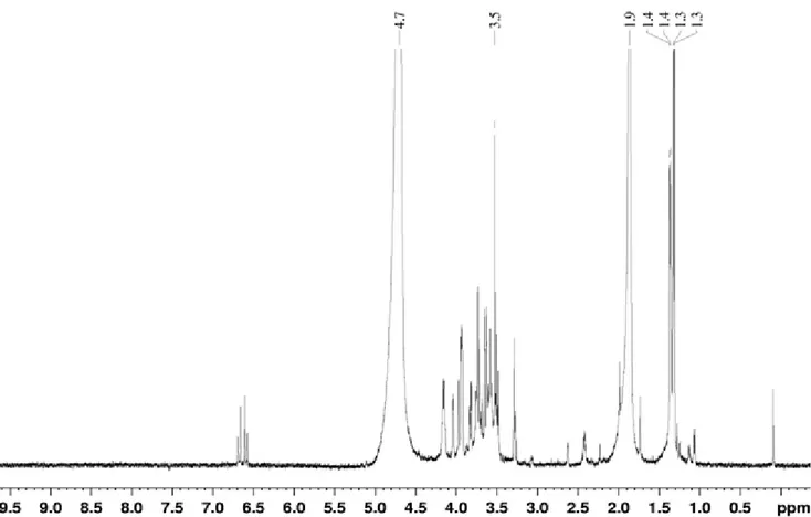
The 1H NMR (Figure 3) and 13C NMR spectra (data not
shown) seem to indicate the presence of sugars, the vast
majority of signals protons resonate between 3.3-4.2 ppm and
the majority of carbon signals resonate between 50-80 ppm.
These signals are characteristic of CH (OR), CH (OH) and CH2
(OR) groups. There are also the signals around 100 ppm carbon
(carbon bonded to two oxygens) and signals to 170-180 ppm
identified to a group O=CH-O-CH3. We also note a set of
proton signals (6.6-6.7 ppm) and carbon (130-140 ppm)
characteristic of C=C (alkene). These results suggest the
presence of an aminoglycosidic compound.
Boudjelal, F. et al. Taxonomic study of the genus Actinoalloteichus
Fraction 2 (F2)
Chromogenic reactions with napthoresorcinol-H2SO4 and
ninhydrin were negative for F2 compound. UV-visible
spectrum (data not shown) of the pure product F2 exhibited the
maxima at 225 and 275 nm in methanol suggesting the
presence of aromatic ring and the absence of polyenic
structure.
Compound F2 was isolated as colorless oil, which had an
IR absorption spectrum (data not shown): 3000-2800 cm-1
(C-H), 1800-1600 cm-1 (C-O), 1600-1580 cm-1 (aromatic ring) and
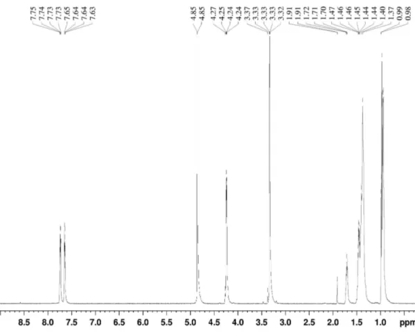
1300-1200 cm-1 (C-O).Molecular weight is 390. 1
H NMR spectrum of pure fraction F2 (Fig. 4) showed
some common main signals regions indicating the presence of
methyl group at 0.94-0.98 ppm, ethyl group at 1.37-1.47 ppm,
C-H at 1.70-1.71 ppm, O-CH2 at 4.24-4.25 ppm and aromatic
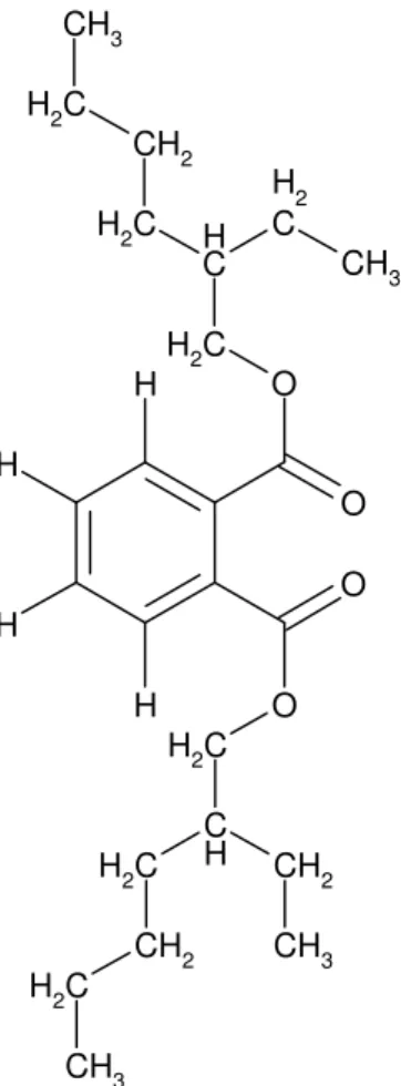
ring at 7.64-7.75 ppm. Based on the spectral data and search in
literature, fraction F2 was identified as dioctyl phthalate (Fig.
5), which has the same UV-visible and infrared spectrum, the
same molecular weight and the same 1H NMR spectrum.
Phthalate compounds are petrochemicals used as
plasticisers or solvents in a variety of industrial products.
Nevertheless, many phthalate derivatives have been isolated
from terrestrial and marine organisms including plants (16, 17),
marine algae (25), and fungal and bacterial culture broth,
particularly those belonging to the genus Streptomyces. Our
experiments were repeated 3 times at differentes periods to
confirm that the dioctyl phthalate produced by strain AH97
was a natural product and not an impurity.
Boudjelal, F. et al. Taxonomic study of the genus Actinoalloteichus O O O O C H 2 C H 2 C H C H C H 2 C H 2 CH 2 CH 3 CH 2 C H 2 CH 3 C H 2 CH 2 C H 2 CH 3 CH 3 H H H H
Figure 5. Chemical structure of the dioctyl phthalate.
CONCLUSION
A moderately halophilic actinomycete strain (AH97),
isolated from saline Saharan soil, was identified by a
polyphasic study to the genus Actinoalloteichus. Analysis of
the 16S rDNA sequence of this strain showed a similarity level
at 98.4% with A. hymeniacidonis the most closely related
which, in the other side, showed significant physiological
characteristic differences. Strain AH97 showed an antibacterial
and antifungal activity against broad spectrum of
microorganisms.
Our results indicate that Actinoalloteichus sp. AH97
produced two bioactive compounds, F1 (hydrophilic) and F2
(hydrophobic). The comparison of their characteristics with
those cited in the literature suggested that the product (F1) is an
aminoglycosidic compound and the product (F2) is the dioctyl
phthalate. Further studies will be necessary to determine the
structure of the F1 compound.
REFERENCES
1. Becker, B.; Lechevalier, M.P.; Gordon, R.E.; Lechevalier, H.A. (1964). Rapid differentiation between Nocardia and Streptomyces by paper chromatography of whole-cell hydrolysates. J. Appl. Microbiol., 12: 421-423.
2. Berdy, J. (2005). Bioactive microbial metabolites. J. Antibiot.,58: 1-26. 3. Betina, V. (1973). Bioautography in paper and thin layer
chromatography and its scope in the antibiotic field. J. Chromatogr., 78: 41-51.
4. Cai, Y. (2009). Classification and salt-tolerance of actinomycetes in the Qinghai lake water and lakeside saline soil. J. Sustain. Develop., 2: 107-110.
5. Felsenstein, J. (1985). Confidence limits on phylogenies: an approach using the bootstrap. Evolution., 39: 783-791.
6. Goodfellow, M. (1971). Numerical taxonomy of some nocardioform bacteria. J. Gen. Microbiol.,69: 33-90.
7. Gordon, R.E.; Barnett, D.A.; Handarhan, J.E.; Hor-Nay-Pang, C. (1974).
Nocardia coeliaca,Nocardia autotrophica and the nocardin strains. Int. J. Syst. Bacteriol., 24: 54-63.
8. Gordon, R.E.; Barnett, D.A. (1977). Resistance to rifampicin and lysozyme of strains of some species of Mycobacterium and Nocardia as a taxonomic tool. Int. J. Syst. Bacteriol.,27: 176-178.
9. Jukes, T.H.; Cantor, C.R. (1969). Evolution of protein molecules. In: Munro, H.N. (ed). Mammalian protein metabolism, vol. 3. Academic Press, New York, pp. 21-132.
10. Kumar, S.; Tamura, K.; Nei, M. (2004). MEGA3: Integrated software for molecular evolutionary genetic analysis and sequence alignment. Brief. Bioinfo., 5: 150-163.
11. Kushner, D. J. (1993). Growth and nutrition of halophilic bacteria. In:
Vreeland, L.I. (ed). The biology of halophilic bacteria, pp. 87-103. 12. Lamari, L.; Zitouni, A.; Boudjella, H.; Badji, B.; Sabaou, N.; Lebrihi, A.;
Lefebvre, G.; Seguin, E. (2002). New dithiolopyrrolone antibiotics from
Saccharothrix sp. Sa 233. I. Taxonomy, fermentation, isolation and biological activities. J. Antibiot., 55: 696-701.
13. Lazzarini, A.; Cavaletti, L.; Toppo, G.; Marinelli, F. (2001). Rare genera of actinomycetes as potential producers of new antibiotics. Ant. v. Leeuw., 78: 399-405.
14. Lechevalier, M.P.; Lechevalier, H.A. (1970). Composition of whole-cell hydrolysates as a criterion in the classification of aerobic actinomycetes.
Boudjelal, F. et al. Taxonomic study of the genus Actinoalloteichus
15. Lechevalier, M.P.; De Bièvre, C.; Lechevalier, H.A. (1977). Chemotaxonomy of aerobic actinomycetes: phospholipids composition.
Biochem. Syst. Ecol., 5: 249-260.
16. Lee, D.S., (2000). Dibutyl phthalate, a glucosidase inhibitor from
Streptomyces melanosporofaciens. J. Biosci. Bioeng., 89: 271-273.
17. Lee, K.H.; Kim, J.H.; Lim, D.S.; Kim C.S.; Kim C.H. (2000).
Anti-leukaemic and antimutagenic effects of di(2.ethylhexyl) phthalate
isolated from aloe linne. J. pharm. Pharmacol.,52: 593-598.
18. Liu, D.; Coloe, S.; Baird, R.; Pedersen, J. (2000). Rapid mini-preparation of fungal DNA for PCR. J. Clinical. Microbiol., 38: 471. 19. Liu, Z.; Zhang, Y.; Yan, X. (1984). A new genus of the order
Actinomycetales. Acta Microbiol. Sin.,24: 295-298.
20. Marchal, N.; Bourdon, J.L.; Richard, C.L. (1987). Milieux de culture pour l'isolement et l’identification biochimique des bactéries. Doin Press, Paris.
21. Minnikin, D.E.; Patel, P.V.; Alshamaony, L.; Goodfellow, M. (1977). Polar lipid composition in the classification of Nocardia and related bacteria. Int. J.Syst. Bacteriol., 27: 104-117.
22. Pfefferle, C.; Theobald, U.; Gürtler, H.; Fiedler, H-P. (2000). Improved secondary metabolite production in the genus Streptosporangium by optimization of the fermentation conditions. J. Biotechnol., 80: 135-142.
23. Sabaou, N.; Boudjella, H.; Bennadji, A.; Mostefaoui, A.; Zitouni, A.; Lamari, L.; Bennadji, H., Lefebvre G.; Germain, P. (1998). Les sols des oasis du Sahara algérien, source d’actinomycetes rares producteurs d’antibiotiques. Sécheresse,9: 147-153.
24. Saitou, N.; Nei, M. (1987). The neighbour-joining method: a new method for reconstructing phylogenetic trees. Mol. Biol. Evol., 4: 406-425.
25. Sastry, V.M.V.S.; Rao, G.R.K. (1995). Dioctyl phthalate and antibacterial compound from marine brown alga-Sargassum wightii. J. Appl. Phycology,7: 185-186.
26. Sehgal, S.N.; Gibbons, N.E. (1960). Effect of some metal ions on the growth of Halobacterium cutirubrum. Can. J. Microbiol., 6: 165-169. 27. Shirling, E.B.; Gottlieb, D. (1966). Methods for characterization of
Streptomyces species. Int. Syst. Bacteriol., 16: 3313-3340.
28. Singla, A. K.; Mayilraj, S.; Kudo, T.; Krishnamurthi, S.; Prasad, G. S.; Vohra, R. M.(2005). Actinoalloteichus spitiensis sp. nov., a novel
Actinobacterium isolated from a cold desert of the Indian Himalayas. Int. J.Syst. Evol. Microbiol., 55: 2561-2564.
29. Tamura, T.; Zhiheng, L.; Yamei, Z.; Hanato, K. (2000).
Actinoalloteichus cyanogriseus gen. nov., sp. nov. Int. J. Syst. Evol. Microbiol., 50: 1035-1040.
30. Thompson, J.D.; Higgins, D.G.; Gibson, T.J. (1994). CLUSTAL W: improving the sensitivity of progressive multiple sequence alignment through sequence weighing, position specific gap penalties and weight matrix choice. Nucl. Acids. Res., 22: 4673-4680.
31. Zhang, H.; Zheng, W.; Huang, J.; Luo, H.; Jin, Y.; Zhang, W.; Liu, Z.; Huang, Y. (2006). Actinoalloteichus hymeniacidonis sp. nov., an actinomycete isolated from the marine sponge Hymeniacidon perleve.
Int. J. Syst. Evol. Microbiol., 56: 2309.
32. Zitouni, A.; Boudjella, H.; Mathieu, F.; Sabaou, N.; Lebrihi, A. (2004a). Mutactimycin PR, a new anthracycline antibiotic from Saccharothrix sp. Sa 103. Taxonomy, fermentation, isolation and biological activities. J. Antibiot., 57: 367-372.
33. Zitouni, A.; Lamari, L.; Boudjella, H.; Badji, B.; Sabaou, N.; Gaouar, A.; Mathieu, F.; Lebrihi, A. (2004b). Saccharothrix algeriensis sp. nov., isolated from Saharan soil. Int. J. Syst. Evol. Microbiol., 54: 1377-1381.