J. Braz. Chem. Soc., Vol. 16, No. 6B, 1454-1457, 2005. Printed in Brazil - ©2005 Sociedade Brasileira de Química 0103 - 5053 $6.00+0.00
S
ho
rt
R
e
p
o
rt
* e-mail: [email protected]
Diploflavone, a New Flavonoid from
Diplotropis ferruginea
Benth. (Fabaceae)
Jackson Roberto G. S. Almeidaa,b, José Maria Barbosa-Filho*,a, Analúcia G. S. Cabral a, Maria de Fátima Agraa, Emidio V. Leitão da-Cunhaa,c, Marcelo S. da Silvaa,
Silene C. do Nascimentod and Raimundo Braz-Filhoe
a
Laboratório de Tecnologia Farmacêutica, Universidade Federal da Paraíba, CP 5009, 58051-970 João Pessoa - PB, Brazil
b
Universidade Federal do Vale do São Francisco, CP 252, 56306-410 Petrolina - PE, Brazil
c
Departamento de Farmácia, CCBS, Universidade Estadual da Paraíba, 58100-000 Campina Grande - PB, Brazil
d
Departamento de Antibióticos, Universidade Federal de Pernambuco, 50740-521 Recife - PE, Brazil
e
Setor de Química de Produtos Naturais- LCQUI-CCT - Universidade Estadual do Norte Fluminense, 28015-620 Campos, Rio de Janeiro - RJ, Brazil
A análise química de Diplotropis ferruginea Benth. resultou no isolamento da 3-metoxiflavona, 3-metoxi-6-O-prenil-6”,6”-dimetilcromeno-(7,8,2”,3”)-flavona, à qual foi dado o nome trivial de diploflavona (1), bem como da 3,6-dimetoxi-6”,6”-dimetilcromeno-(7,8,2”,3”)-flavona (2). A estrutura
do novo composto foi estabelecida por análises espectrais. A atividade citotóxica dos compostos isolados foi testada contra células NCI-H292 (carcinoma de pulmão), HEp-2 (carcinoma de laringe) e KB (carcinoma epidermóide oral). As células HEp-2 foram as mais afetadas pelas substâncias testadas.
The chemical examination of Diplotropis ferruginea Benth. resulted in the isolation of a new 3-methoxyflavone, 3-methoxy-6-O-prenyl-6”,6”-dimethylchromene-(7,8,2”,3”)-flavone, to which was given the trivial name diploflavone (1); as well as the known
3,6-dimethoxy-6”,6”-dimethylchromene-(7,8,2”,3”)-flavone (2). The structure of the new compound was established by spectral analyses. Cytotoxic activity of the isolated compounds was tested against the cells NCI-H292 (lung carcinoma), HEp-2 (larynx carcinoma) and KB (oral epidermoid carcinoma). The cells HEp-2 were the most affected by the substances tested.
Keywords:Diplotropis ferruginea, Fabaceae, flavonoids, cytotoxicity
Introduction
The Fabaceae have a cosmopolitan distribution, consisting of ca 700 genera and more than 17000 species.1
The genus, Diplotropis consists of approximately 22 species, including, Diplotropis ferruginea Benth. Investigations of only two species have been reported in the literature: the isolation of quinolizidine alkaloids from Diplotropis martiusii,2 and flavonoids from Diplotropis
purpurea.3
Diplotropis ferruginea is a tree native to Northeastern Brazil, where it is popularly known as “sucupira”. It is used in folk medicine for the treatment of rheumatism, arthritis and diabetes.4 Recently, a chemical investigation
of this species resulted in the isolation of lupeol, ethyl 2-hydroxy-4-methoxy-6-propyl benzoate5 and of the
flavonoid 3,4,5,8-tetramethoxy-6,7,2”,3”-furanoflavan.6
Spasmolytic activity was reported for the crude EtOH extract of this plant.7
This paper describes the isolation of two more flavonoids, whose structures were established by spectroscopic techniques, mainly EIMS and 1D and 2D NMR.
Experimental
General experimental procedures
1455 Diploflavone, a New Flavonoid from Diplotropis ferruginea Benth. (Fabaceae)
Vol. 16, No. 6B, 2005
spectrophotometer. 1H and 13C NMR spectra were run on a
Jeol Eclipse+ 400 spectrometer operating at 400 MHz for
1H and 100 MHz for 13C, using CDCl
3 as solvent
(approximately 10 mg of sample were dissolved in 0.5 mL of solvent and transferred into a 5 mm NMR tube) and solvent signals were used as internal reference for the chemical shifts
δH 7.26 (CHCl3) and δC 77.00 (CDCl3). The one-dimensional (1D) 1H and 13C NMR spectra were acquired under standard
conditions (5 mm multinuclear probe). The two-dimensional (2D) experiments were acquired and processed with the Delta software provided by Jeol. Standard pulse sequences were used for all experiments. 1H-1H-COSY spectra were obtained
with X-points 512/Y-points 256, X-resolution 11.7 Hz/Y-resolution 23.4 Hz, X-acquisition time 85.4 ms/Y-acquisition time 42.7 ms, pulse 90o, relaxation delay 1.5 s, zerofill: 4.
For homonuclear 2D 1H-1H-NOESY experiments were used
mixing time 0.5 s, X-points 512/Y-points 256, X-resolution 11. 7Hz/Y-resolution 23.4 Hz, X-acquisition time 85.4 ms/ Y-acquisition time 42.7 ms, pulse 90o relaxation delay 1.5 s,
zerofill: 4. Two-dimensional inverse hydrogen detected heteronuclear shift correlation 1H-13C-HMQC-1J
CH spectra
were obtained with 1J
CH = 140 Hz, X-points 1024/Y-points
128, X-resolution 5.86 Hz/Y-resolution 196 Hz, X-pulse 90o/
Y-pulse 90o, X-acquisition time 0.17 s/Y-acquisition time
5.09 ms, pulse 90o, relaxation delay 2.0 s, gradient 1/3 1 ms
square, zerofill: 4. Two-dimensional inverse hydrogen detected heteronuclear long-range correlation 1H-13
C-HMBC-nJ
CH (n=2 and 3) experiments were carried out by
using nJ
CH = J constant 140 Hz/J long range 8 Hz, X-points
1024/Y-points 128, X-resolution 5.86 Hz/Y-resolution 196 Hz, X-pulse 90o/Y-pulse 90o, X-acquisition time 0.17
s/Y-acquisition time 5.09 ms, pulse 90o, relaxation delay 2.0 s,
gradient 1/3 1 ms square, zerofill: 4. EIMS were measured at 70 eV on a GC/MS System Shimadzu QP-5050.
Plant material
The stem bark of Diplotropis ferruginea was collected in the municipality of Caraúbas, State of Rio Grande do Norte, Northeastern Brazil in May 2002. Botanic material was identified by Prof. Maria de Fátima Agra, of the Laboratório de Tecnologia Farmacêutica. A voucher specimen (AGRA & D. ALMEIDA 5559) is deposited at the Herbario Prof. Lauro Pires Xavier (JPB), of the Universidade Federal da Paraíba.
Extraction and isolation
The dried and powdered stem bark of D. ferruginea (3 kg) was exhaustively extracted with 95% EtOH at room temperature. The extract was concentrated under vacuum
yielding 95 g of the crude product. This was suspended in a MeOH:H2O (3:7 v/v) mixture and partitioned with hexane, CHCl3 and EtOAc. The hexane fraction was then subjected to silica gel column chromatography and eluted with hexane, CHCl3 and MeOH in an increasing polarity gradient to give 152 fractions. The fractions were monitored by TLC and classified into 25 groups. Fraction 97-102 was purified by preparative TLC over silica gel using CHCl3:MeOH (9:1) to afford flavonoid 1 (61 mg)
and the fraction 89-96 was purified in the same way using hexane:EtOAc (2:1) to afford flavonoid 2 (123 mg).
Biological assay
The cytotoxic activity assays were based on the methylazoetetrazolium (MTT) method or the 3-(4,5-dimethylazol-2-yl)-3,5-diphenyltetrazolium bromide method.8 For the evaluation of cytotoxity the cellular strain
HEp2 (larynx carcinoma) NCIH-292 (lung carcinoma) KB (mouth carcinoma)9 with proven viability were used. The
cells were grown in MEM- Minimal Essential Medium10
with 10% bovine fetal serum containing 1% antibiotics solution (penicillin 1000 UI mL-1 + streptomycin 250 mg
mL-1) and 1% glutamine (200 μM). A cellular suspension
of 5°104 cells mL-1 was used and distributed in plates of
96 wells. The test samples of 0.15 mL were added into each well. The plates were incubated for 72 h at 37 oC in a
humid atmosphere enriched with 5% CO2. After incubation 15 mL MTT in phosphate buffered saline (BPS) solution at (5 mg mL-1) was added into each well. After 2h the culture
medium was removed and 100 μL of DMSOwere added in each well for quantitation of blue formazan. The readings were performed with the aid of a MultskanELX 800 cell reader (Bio-Tec Instruments – USA) at 540 nm.
3-Methoxy-6-O-prenyl-6”,6”-dimethylchromene-(7,8,2”,3”)-flavone or diploflavone, (1)
It was obtained as amorphous powder, mp 163-165 oC.
IR (KBr) νmax/cm-1: 3062, 2971, 2847, 1620, 1404, 1379,
1300, 1100. EI-MS: m/z (%): 418 (8, [M+]), 364 (19), 349
(100, [M+ - prenyl]), 335 (38) (Calc. for C
26H26O5). 1H NMR
(CDCl3, 400 MHz) and 13C NMR (CDCl
3, 100 MHz) (Table
1).
3,6-Dimethoxy-6”,6”-dimethylchromene-(7,8,2”,3”)-flavone, (2)
It was obtained as amorphous powder, mp 203-204 oC.
IR (KBr) νmax/cm-1: 2995, 2844, 1615, 1402, 1382, 1300,
1456 Almeida et al. J. Braz. Chem. Soc.
[M+ - CH
3]), 319 (4) (Calc. for C22H20O5).
1H NMR (CDCl
3,
400 MHz) and 13C NMR (CDCl
3, 100 MHz).
Results and Discussion
Flavonoid 1 was obtained as a colorless amorphous solid.
Its molecular formula was deduced as C26H26O5 (14 degrees of unsaturation), supported by the occurrence of the molecular ion at m/z 418 in the MS, in combination with 1H
and 13C-APT-NMR spectral data. The IR spectrum showed
absorptions at 1620 cm-1, attributed to an α-β unsaturated
carbonyl group; 3062 cm-1 attributed to unsaturated C-H
and absorptions in the region 1379-1404 cm-1, suggesting
the presence of a gem-dimethyl group. 1H NMR of 1 showed
signals at δH 8.07 (2H, br, d J = 7.7 Hz) and 7.56-7.46 (3H, m) which indicates the possibility of a mono-substituted ring B in a flavonoid. The presence of a 2,2-dimethyl-chromene moiety was indicated by the characteristic signals of its two vinyl hydrogens forming an AB system11 at δ
H
5.74 (1H, d, J = 9.9 Hz) and 6.87 (1H, d, J = 9.9 Hz) and a signal at δH 1.54 (6H, s) attributed to the two methyl groups. A signal at δH 1.79 (6H, s) was also observed and signals at
δH 4.68 (1H, d, J = 6.2 Hz) and 5.53 (1H, t, J = 6.2 Hz), suggesting the presence of a prenyl group in the molecule. This suggestion is confirmed by the 13C-APT NMR spectra
which shows signals at δC 18.25 and 25.72, for 2 methyl carbons and a methylene carbon at δC 66.29. The chemical shift of the methylene carbon in the 13C NMR indicates that
the prenyl group is bound to an oxygen atom. The HMBC experiment showed the location of the O-prenyl group at C-6, due to the 3J
CH correlation between the signal at δH
4.68 (prenyl’s methylene hydrogens) with the signal at δC
146.06 (C-6). The analysis of all the spectral data for 1 led
to the elucidation of its structure as 3-methoxy-6-O-prenyl-6”,6”-dimethylchromene-(7,8,2”,3”)-flavone. This substance is described here for the first time and was given the trivial name diploflavone.
Flavonoid 2 was isolated as a colorless amorphous
solid. Its molecular formula deduced as C22H20O5 (13 degrees of unsaturation), was confirmed by the molecular ion at m/z 364 in the MS in combination with 1H-NMR
(1D and 2D 1H-1H-COSY) and 13C-APT-NMR spectral data.
IR and 1H and 13C-NMR spectra showed the similarity with
substance 1. The only difference between the two
substances was the absence of the prenyl moiety in 2,
having a methoxy in the same position. The presence of the methoxy group was indicated by the signal at δH 3.97 (3H, s). The substance was thus characterized as the flavonoid 3,6-dimethoxy-6”,6”-dimethylchromene-(7,8,2”,3”)-flavone (2), previously isolated from
Bowdichia virgilioides and the NMR data are in accordance with the literature. 12
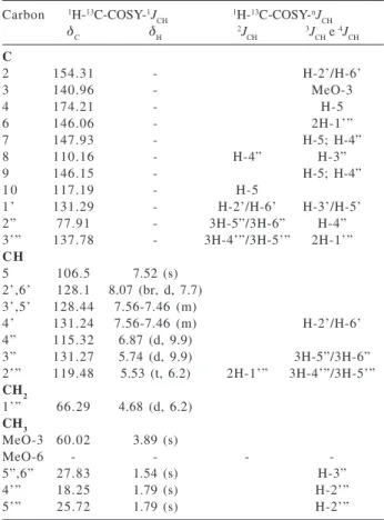
The 2D experiments HMQC and HMBC were used to confirm the 1H and 13C chemical shifts of 1 (Table 1) and
2.12
Figure 1. Flavonoids from Diplotropis ferruginea.
Table 1. 1H (400 MHz) and 13C (100 MHz) NMR data for 1 inclu-ding results obtained by heteronuclear 2D shift-correlated HMQC and HMBC spectra, in CDCl3 as solvent and TMS as internal refe-rence. Chemical shifts in δ (ppm) and coupling constants (J, in parenthesis) in Hz*
Carbon 1H-13C-COSY-1J
CH 1H-13C-COSY-nJCH
δC δH 2JCH 3JCH e 4JCH
C
2 154.31 - H-2’/H-6’
3 140.96 - MeO-3
4 174.21 - H-5
6 146.06 - 2H-1’”
7 147.93 - H-5; H-4”
8 110.16 - H-4” H-3”
9 146.15 - H-5; H-4”
1 0 117.19 - H-5
1’ 131.29 - H-2’/H-6’ H-3’/H-5’
2” 77.91 - 3H-5”/3H-6” H-4”
3’” 137.78 - 3H-4’”/3H-5’” 2H-1’”
C H
5 106.5 7.52 (s) 2’,6’ 128.1 8.07 (br, d, 7.7) 3’,5’ 128.44 7.56-7.46 (m)
4’ 131.24 7.56-7.46 (m) H-2’/H-6’ 4” 115.32 6.87 (d, 9.9)
3” 131.27 5.74 (d, 9.9) 3H-5”/3H-6” 2’” 119.48 5.53 (t, 6.2) 2H-1’” 3H-4’”/3H-5’”
CH2
1’” 66.29 4.68 (d, 6.2)
CH3
MeO-3 60.02 3.89 (s)
MeO-6 - - -
-5”,6” 27.83 1.54 (s) H-3”
4’” 18.25 1.79 (s) H-2’”
5’” 25.72 1.79 (s) H-2’”
* Homonuclear 1H-1H-COSY spectra were also used for these assignments. Chemical shifts of hydrogen atoms obtained from 1D 1H NMR spectrum. Carbon atoms corresponding to C, CH, CH
1457 Diploflavone, a New Flavonoid from Diplotropis ferruginea Benth. (Fabaceae)
Vol. 16, No. 6B, 2005
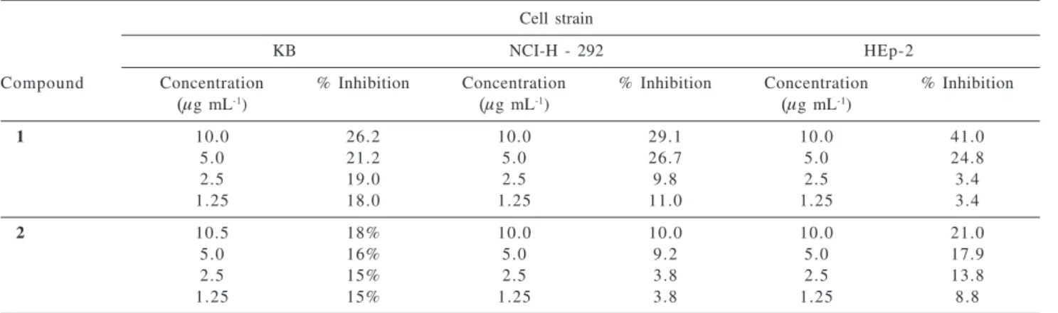
The cytotoxic activity of cells NCI-H292 and KB were not affected by flavonoids 1 and 2, however, cells HEp-2 were affected by diploflavone (1). At the concentration of
10 μg mL-1 it showed an inhibition of proliferation of 41%
(Table 2).
Acknowledgements
The authors are grateful to the Instituto do Milênio do Semi-Árido (IMSEAR/CNPq), CAPES and FAPERJ by financial support. Sincere thanks are also due to NAPRALERT.
References
1. Heywood, V. H.; Flowering Plants of the World, B. T. Batsford LTD: London, 1996, p.149.
2. Kinghorn, A. D.; Balandrin, M. F.; Lin, L. J.; Phytochemistry
1982, 21, 2269.
3. Braz-Filho, R.; Gottlieb, O. R.; Pinho, S. L. V.; Monte, F. J. Q.; Rocha, A. I.; Phytochemistry 1973, 12, 1184.
Table 2. Cytotoxic activity of 1 and 2 against the cells KB, NCI-H 292 and HEp-2
Cell strain
KB NCI-H - 292 HEp-2
Compound Concentration % Inhibition Concentration % Inhibition Concentration % Inhibition
(μg mL-1) (μg mL-1) (μg mL-1)
1 10.0 26.2 10.0 29.1 10.0 41.0
5.0 21.2 5.0 26.7 5.0 24.8
2.5 19.0 2.5 9.8 2.5 3.4
1.25 18.0 1.25 11.0 1.25 3.4
2 10.5 18% 10.0 10.0 10.0 21.0
5.0 16% 5.0 9.2 5.0 17.9
2.5 15% 2.5 3.8 2.5 13.8
1.25 15% 1.25 3.8 1.25 8.8
KB (oral epidermoid carcinoma), NCI-H - 292 (lung carcinoma), HEp-2 (larynx carcinoma).
4. Pio-Correia, M.; Dicionário das Plantas Úteis do Brasil e das Exóticas Cultivadas, Ministério da Agricultura: Brasil, 1984, p. 149.
5. Almeida, J. R. G. S.; Cunha, E. V. L.; Silva, M. S.; Athayde-Filho, P. F.; Braz-Athayde-Filho, R.; Barbosa-Athayde-Filho, J. M.; Rev. Bras. Farmacogn. 2003,13, Suppl. 2, 44.
6. Almeida, J. R. G. S.; Cunha, E. V. L.; Silva, M. S.; Braz-Filho, R.; Marques, A. S.; Zheng, C.; Barbosa-Filho, J. M.; Ann. Magn. Reson. 2003,1, 33.
7. Lima, J. T.; Claudino, F. S.; Cavalcante, F. A.; Almeida, J. R. G. S.; Barbosa-Filho, J. M.; Silva, B. A.; Rev. Bras. Ci. Farm.
2003,39, Suppl. 2, 158.
8. Alley, M. C.; Scudiero, D. A.; Monks, A.; Hursey, M. L.; Czerwinski, M. J.; Fine, D. L.; Abbot, B. J.; Mayo, J. G.; Shoemaker, R. H.; Boyd, M. R.; Cancer Res. 1988, 48, 589.
9. Eagle, H.; Proc. Soc. Exper. Biol. Med. 1955,89, 362. 10. Eagle, H.; Science 1959, 130, 432.
11. Campos, A. M.; Khac, D. D.; Fetizon, M.; Phytochemistry
1987, 26, 2819.
12. Arriaga, A. M. C.; Gomes, G. A.; Braz-Filho, R.; Fitoterapia
2000,71, 211.