BIOMEDICAL SCIENCES
AND
CLINICAL INVESTIGATION
www.bjournal.com.br
www.bjournal.com.br
Campus
Institutional Sponsors
The Brazilian Journal of Medical and Biological Research is partially financed by
Hotsite of proteomics metabolomics developped by:
Braz J Med Biol Res, December 2010, Volume 43(12) 1203-1214
doi: 10.1590/S0100-879X2010007500125
Proteomic analysis of cytosolic proteins associated with petite
mutations in Candida glabrata
Proteomic analysis of cytosolic proteins
associated with petite mutations in
Candida glabrata
C.V. Loureiro y Penha
1, P.H.B. Kubitschek
1, G. Larcher
2, J. Perales
3,
I. Rodriguez León
3, L.M. Lopes-Bezerra
1and J.P. Bouchara
21Laboratório de Micologia Celular e Proteômica, Instituto de Biologia Roberto Alcântara Gomes,
Universidade do Estado do Rio de Janeiro, Rio de Janeiro, RJ, Brasil
2Host-Parasite Interaction Study Group, UPRES-EA 3142, Laboratory of Parasitology-Mycology,
Angers University Hospital, Angers, France
3Laboratório de Toxinologia, Instituto Oswaldo Cruz, Fiocruz, Rio de Janeiro, RJ, Brasil
Abstract
The incidence of superficial or deep-seated infections due to Candida glabrata has increased markedly, probably because of the low intrinsic susceptibility of this microorganism to azole antifungals and its relatively high propensity to acquire azole resistance. To determine changes in the C. glabrata proteome associated with petite mutations, cytosolic extracts from an azole-resistant petite mutant of C. glabrata induced by exposure to ethidium bromide, and from its azole-susceptible parent isolate
were compared by two-dimensional polyacrylamide gel electrophoresis. Proteins of interest were identified by peptide mass fingerprinting or sequence tagging using a matrix-assisted laser desorption/ionization tandem time-of-flight mass spectrom
-eter. Tryptic peptides from a total of 160 Coomassie-positive spots were analyzed for each strain. Sixty-five different proteins were identified in the cytosolic extracts of the parent strain and 58 in the petite mutant. Among the proteins identified, 10 were
higher in the mutant strain, whereas 23 were lower compared to the parent strain. The results revealed a significant decrease
in the enzymes associated with the metabolic rate of mutant cells such as aconitase, transaldolase, and pyruvate kinase,and
changes in the levels of specific heat shock proteins. Moreover, transketolase, aconitase and catalase activity measurements decreased significantly in the ethidium bromide-induced petite mutant. These data may be useful for designing experiments
to obtain a better understanding of the nuclear response to impairment of mitochondrial function associated with this mutation in C. glabrata.
Key words: Candida glabrata; Petite mutations; Cytosolic extracts; Proteomic analysis; Azole resistance
Introduction
The incidence of life-threatening fungal infections that are mainly caused by the Candida species has increased dramatically in the past decades along with the development of antibiotic treatments, the widespread use of immunosup-pressive therapy, and the emergence of the AIDS epidemic (1). Among the causative agents of these infections, Candida albicans remains by far the most frequent, but infections due to other Candida species are being reported increasingly (2,3). For instance, C. glabrata has recently emerged as a
significant pathogen in various hospital settings, where it
is responsible for an increasing number of systemic
infec-tions and candiduria (4,5). In a recent study conducted in
the United States, C. glabrata ranked second among the
causative agents of fungemia, accounting for 21% of all
Candida bloodstream isolates (6,7). The rise in the number of C. glabrata systemic infections is due to the poor intrinsic susceptibility of this yeast to azole antifungals and to its
propensity to acquire azole resistance (5,8-12).
The mechanisms of resistance to azole antifungals have been studied mainly in C. albicans and can be categorized as i) changes in the cell wall or plasma membrane, which
lead to impaired azole uptake; ii) alterations in the affinity of azoles for their target Erg11p (lanosterol 14α-demethylase)
or increase in the cellular content of Erg11p due to muta-tions in or overexpression of the ERG11 gene, respectively,
and iii) increased efflux of the azole drugs mediated by
Correspondence: C.V. Loureiro y Penha, LMCProt, IBRAG, UERJ, Rua São Francisco Xavier, 524, PHLC 501-D, 20550-013 Rio de Janeiro, RJ, Brasil. Fax: +55-21-2587-7377. E-mail: [email protected]
membrane transport proteins belonging to the ATP-binding cassette (ABC) transporter family (CDR1 and CDR2) or to the major facilitator superfamily (MDR1 and FLU1). For instance, the CDR1, CDR2 and MDR1 genes have been shown to be overexpressed in many azole-resistant isolates, and deletion of these genes resulted in hypersensitivity to azoles (13). However, different mechanisms including overexpression of
genes encoding the efflux pumps and overexpression or point
mutations in the ERG11 gene frequently combine, resulting in a stepwise development of azole resistance over time (14). In addition, compensatory pathways that involve alterations
of specific steps in ergosterol biosynthesis have been docu -mented as resistance mechanisms to both azoles and polyene
antifungals (15).
More recently, increased levels of expression of the ABC transporter genes C. glabrata CDR1 (CgCDR1) and CDR2
(CgCDR2) were detected in azole-resistant isolates of C. glabrata (16-18). Furthermore, the azole resistance of C. glabrata petite mutants obtained by exposure to fluconazole or
induced by ethidium bromide (ETB) was shown to be associ-ated with the up-regulation of the nuclear genes CgCDR1 and
CgCDR2 (19,20). However, due to the numerous cross-talks between the nucleus and mitochondria, petite mutations may also lead to the deregulation of the expression of other nuclear genes (21). For instance, an increased cellular content of free
ergosterol due to a defect in sterol esterification has been
reported in petite mutants (19). Similarly, changes in the bio-chemical composition of the cell wall associated with a lower cell surface hydrophobicity were also observed in mutant cells, as well as an increased expression of the CgEPA1 gene encoding a lectin involved in adherence to epithelial cells (22).
In the present study, changes in the C. glabrata pro-teome associated with petite mutations and azole resistance were examined by investigation of an azole-susceptible wild-type isolate and an ETB-induced azole-resistant petite mutant.
Material and Methods
Yeast strains and culture conditions
The study was carried out using a C. glabrata clinical isolate
designated 90.1085, which was obtained at the Laboratory
of Parasitology and Mycology of Angers University Hospital, Angers, France, from a urine sample collected in 1990, and a derived petite mutant induced by exposure to the intercalat-ing agent ETB (Sigma-Aldrich, USA). The mutant presented cross-resistance to azoles due to overexpression of the CDR1
and CDR2 genes.The parent isolate and its ETB-induced petite mutant were maintained by biweekly passages in yeast
extract-peptone-glucose (YEPD) agar containing 5 g/L yeast extract, 10 g/L peptone, 20 g/L glucose, 0.5 g/L chlorampheni
-col, and 20 g/L agar. Mutant cells were subcultured on yeast
extract-peptone agar containing 2% glycerol as the sole carbon
source to ascertain their respiratory deficiency. Both isolates
were preserved in 20% glycerol at -80°C.
Cytosolic protein extraction
For the isolation of cytosolic proteins, blastoconidia were washed in deionized distilled water and resuspended in 10 mM Tris-HCl, pH 7.4, containing 300 U RNase and a mix of
protease inhibitors (1 mM phenylmethylsulfonyl fluoride, 1 mM
ethylenediamine tetraacetic acid and 1 µM pepstatin). Cells were disrupted using glass beads in a Braun homogenizer with cooling CO2 and the suspensions obtained were centrifuged at
12,000 g for 30 min at 4°C. The pellets were discarded, and the
supernatants were centrifuged for 1 h at 75,000 g. The result-ing supernatants, which correspond to the cytosolic extracts, were desalted by dialysis and protein content was measured using the Bradford (Bio-Rad Laboratories, USA) and the BCA
protein assay kit (Thermo Scientific, USA).
2-D SDS-PAGE
Samples containing 150 µg (analytical gels) or 500 µg (pre -parative gels) protein were solubilized in a lysis buffer consisting of 7 M urea, 2 M thiourea, 4% CHAPS, 64 mM dithioerythritol,
0.5% Pharmalyte, pH 3-11 (Amersham Biosciences, Sweden)
and bromophenol blue and then applied onto Immobiline, pH 3-11, nonlinear DryStrips (18 cm long; Amersham Biosciences). Isoelectric focusing was performed using an IPGphor system (Amersham Biosciences) at 20°C and the following program:
30 V (active rehydration) for 12 h, 200 V for 1 h, 500 V for 1 h, 500-10,000 V for 3 h, and 10,000 V for 1 h. Immobilized pH
gradient strips were then reduced (1% dithioerythritol) and
al-kylated (1.5% iodoacetamide) in equilibration buffer (6 M urea, 75 mM Tris-HCl, pH 8.8, 29.3% glycerol, 2% SDS) (23). The
second dimension run was performed in homogeneous 12%
polyacrylamide slab gels (1.5 mm thick) at 5 mA for 30 min, 8
mA for 1 h and 60 mA for 4 h using a Protean II electrophoresis apparatus (Bio-Rad).
For the determination of the total number of spots, analytical gels were silver-stained as described by Bjellqvist et al.(24),
fixed first in methanol-acetic acid-distilled water (4:1:5) for 1 h and then in ethanol-acetic acid-distilled water (0.5:0.5:9) over
-night. The gels were rinsed with 7.5% acetic acid and incubated in 1% glutaraldehyde containing 0.5 M sodium acetate for 30
min. The gels were then extensively washed with water and stained with an ammoniacal silver nitrate solution for 30 min. Gels were washed and color was developed in 0.01% citric acid containing 0.1% formaldehyde. Staining was stopped with
5% Tris in 2% acetic acid.
Preparative gels for further analysis of proteins by mass
spectrometry were stained with colloidal Coomassie blue R-250
(Bio-Rad). The gels were destained using several changes of distilled water over a 2-h period. Finally, spots were excised and in gel-digested for analysis by matrix-assisted laser
des-orption/ionization tandem time-of-flight mass spectrometry (MALDI-TOF/TOF MS).
Image analysis
images were processed for detection, volumetric quantification,
matching, and editing of molecular masses and pI of spots, using ImageMaster 2-D Platinum software (Amersham Biosci-ences). Proteins were considered to be differentially increased or decreased if the protein spot in the ETB-induced mutant
showed statistically significant differences of 2-fold or more in
their mean spot volume on at least three of four gels (P < 0.05,
t-test) compared to its parent isolate.
In-gel tryptic digestion
Digestion was performed by the method of Pitarch et al.
(25). Protein spots were excised from Coomassie-stained 2-D gels and transferred to 0.5-mL tubes. Gel fragments were destained with acetonitrile (ACN), washed twice with 50% ACN in 25 mM ammonium bicarbonate (AmBic), and vacuum-dried. The proteins were then reduced with 10 mM dithioerythritol in 25 mM AmBic for 30 min at 56°C and subsequently alkylated with 55 mM iodoacetamide in 25 mM AmBic for 20 min in the dark. Next, the gel fragments were washed with 25 mM AmBic and
ACN and dried under vacuum. All gel fragments were incubated
with 12.5 ng/µL sequencing grade trypsin (Promega, USA) in 25 mM AmBic overnight at 37°C. Peptides were then extracted from the gel fragments with 50% ACN, 1% trifluoroacetic acid in 25 mM AmBic, and finally with 100% ACN. The extracts were
pooled, and concentrated by evaporation of the solvent with a
SpeedVac apparatus (Thermo Fisher Scientific, USA).
MALDI-TOF/TOF MS
Peptides were applied onto a MALDI plate after
co-crys-tallization with and α-cyano 4-hydroxycinnamic acid (CHCH)
matrix (Sigma-Aldrich). One microliter of each sample with 0.4
µL 3 mg/mL CHCH matrix in 50% ACN and 0.01% trifluoroacetic acid was spotted onto a MALDI plate. MS/MS sequencing analyses were carried out using a MALDI-TOF/TOF MS 4700
Proteomics Analyzer (Applied Biosystems,USA).
Database search
Peak listsfrom all MS/MS spectra were submitted to a database searchusing an in-house copy of MASCOT, version 3.1 (Matrix ScienceInc., USA). The following criteria were used for alldatabase searches: a minimum signal-to-noise ratio thresholdof 5-10; mass values in the 0-60 Da range and masses within 20Da of the precursor ion mass were excluded. A maximum of 60 peaksper spectrum were included as product ions. The mass tolerancewas ±75 ppm for MS data, ±200 ppm
for MS/MS precursorions, and ±250 ppm for MS/MS product
ions. The samplewas searched against the NCBInr database (October 4, 2007).The ExPASy (Expert Protein Analysis Sys-tem) proteomics server of the Swiss Institute of Bioinformatics (SIB) was used for the analysis of protein sequences.
Measurements of transketolase and aconitase activity To estimate the transketolase activity, samples were added
to a cuvette containing buffer (50 mM Tris-HCl, pH 7.6), 2 mM ribose 5-phosphate, 1 mM xylulose 5 phosphate, 5 mM MgCl2,
0.2 U/mL triosephosphate isomerase, 0.2 mM NADH, and 0.1
mM thiamine pyrophosphate. Reactions were initiated by the addition of cytosolic extracts at 37°C. The aconitase activity was determined by addition of the samples to a cuvette
con-taining buffer (50 mM Tris-HCl, pH 7.4), 0.6 mM MnCl2, and
0.5 mM ferrous ammonium sulfate. Reactions were initiated
by the addition of cytosolic extracts at 37°C. Transketolase and aconitase activities were measured by spectrophotometry (340 nm) (Thermo Spectronic Genesys 10 uv), and data are reported as ng product·min-1·mg total protein-1. Total protein
content of cytosolic extracts was determined by the method of Bradford. Each experiment was repeated three times.
Measurements of catalase activity
Catalase activity was measured by the method of Aebi (26), which is based on the principle that the absorbance will decrease due to dismutation of H2O2 at 240 nm (UV-visible
spectrophotometry). The amount of H2O2 converted into H2O
and oxygen in 1 min under standard conditions is accepted as enzyme reaction velocity. Data are reported as µmol H2O2
metabolized·min-1·mg total protein-1.
Statistical analysis
Statistical analysis for the enzyme activity data was per-formed using the Student t-test, with the level of significance set
at P < 0.05. For identification of protein, the Mascot protein score
reports a match as significant if it has a match with less than 5%
chance of being a random hit. Protein scores are derived from ion scores as a non-probabilistic basis for ranking protein hits. Protein score is -10*Log(P), where P is the probability that the observed match is a random event. Individual protein scores
>50 indicate identity or extensive homology (P < 0.05).
Results
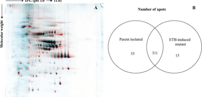
In order to identify differences in protein expression between the parent isolate and its derived petite mutant, differences in
protein profiles between cytosolic protein extracts from cultures
of the two isolates were examined. Equal amounts of each protein extract (parent isolate and ETB-induced petite mutant) were submitted to 2-D electrophoresis. Multiple gels from three independent experiments were run for each sample to ascer-tain reproducibility. One representative gel of each extract was used as a reference 2-D map (Figure 1). Analysis of the virtual image generated using the ImageMaster Platinum software and based on the overlay of the reference 2-D
maps permitted the observation of significant differences
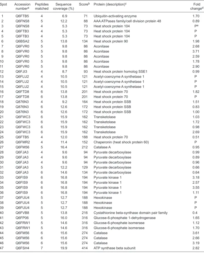
For protein identification, the spots were digested in-gel and analyzed by MALDI-TOF/TOF MS. Bioinformatic analysis of MALDI-TOF spectra permitted the identification of a total of 65 different proteins in the parent isolate and
58 in the ETB-induced petite mutant corresponding to the
140 and 121 spots, respectively, numbered in Figure 1. A
list of identified proteins is presented in Table 1, together
with their accession numbers, peptides matched, sequence
Figure 1. 2-D gels of cytoplasmic protein extracts of the Candida glabrata parent isolate and its ethidium bromide (ETB)-induced petite mutant separatedon immobilized pH gradient (IPG) strips covering the pH range of 3 to 11. The gels werestained withcolloidal Coomassie blue. A, Parent isolate; B, ETB-induced petite mutant. The protein spots that were identified are numbered and listed in
Table 1.
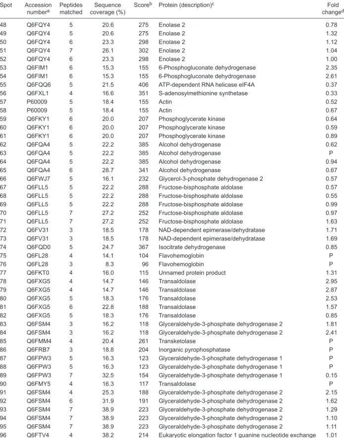
Table 1. Proteins identified by two-dimensional sodium dodecyl sulfate-polyacrylamide gel electrophoresis and matrix-assisted laser desorption/ionization tandem time-of-flight mass spectrometry.
Spot Accession numbera
Peptides matched
Sequence coverage (%)
Scoreb Protein (description)c Fold
changed
1 Q6FTB5 4 6.9 71 Ubiquitin-activating enzyme 1.70
2 Q6FNS8 5 12.2 88 AAA ATPases family/cell division protein 48 0.89 3 Q6FNS8 4 5.3 73 Heat shock protein 104 Pe
4 Q6FTB3 4 5.3 73 Heat shock protein 104 P
5 Q6FTB3 4 5.3 73 Heat shock protein 104 P 6 Q6BSA2 5 13.8 134 Heat shock protein 90 1.98 7 Q6FVR0 5 9.8 86 Aconitase 2.68 8 Q6FVR0 5 9.8 86 Aconitase 3.71 9 Q6FVR0 5 9.8 86 Aconitase 2.59 10 Q6FVR0 5 9.8 86 Aconitase 1.78 11 Q6FVR0 5 9.8 86 Aconitase 2.90 12 Q6FJI3 4 8.7 93 Heat shock protein homolog SSE1 0.99 13 Q6FLU2 4 10.5 121 Acetyl-coenzyme A synthetase 1 P 14 Q6FLU2 4 10.5 121 Acetyl-coenzyme A synthetase 1 P
15 Q6FLU2 4 10.5 121 Acetyl-coenzyme A synthetase 1 P 16 Q6FTD8 6 13.8 201 Heat shock protein 70 1.82 17 Q6FTD8 6 13.8 201 Heat shock protein 70 P 18 Q876N3 4 9.2 164 Heat shock protein SSB 1.51 19 Q876N3 6 12.6 172 Heat shock protein SSB 0.83 20 Q876N3 6 12.6 172 Heat shock protein SSB 1.00 21 Q6FWC3 6 15.9 162 Transketolase 1.03 22 Q6FWC3 6 15.9 162 Transketolase 1.72 23 Q6FWC3 6 15.9 162 Transketolase 0.98 24 Q6FWC3 6 15.9 162 Transketolase 2.69
25 Q6FTB5 4 12.0 188 Heat shock protein 70 0.51
26 Q6FMR2 4 11.4 152 Chaperonin (heat shock protein 60) P
27 Q6FM56 5 16.4 212 Catalase A 0.95
28 Q6FJA3 4 9.6 94 Pyruvate decarboxylase 0.99 29 Q6FJA3 4 9.6 94 Pyruvate decarboxylase 0.89 30 Q6FJA3 4 9.6 94 Pyruvate decarboxylase 0.96 31 Q6FJA3 5 12.2 129 Pyruvate decarboxylase 0.66 32 Q6FJA3 6 14.6 134 Pyruvate decarboxylase 0.64 33 Q6FIS9 6 16.8 194 Pyruvate kinase 1 3.18 34 Q6FIS9 6 16.8 194 Pyruvate kinase 1 2.57
35 Q6FIS9 6 16.8 194 Pyruvate kinase 1 3.55 36 Q6FIS9 6 16.8 194 Pyruvate kinase 1 1.11 37 Q6FUU4 5 12.7 188 Hexokinase P 38 Q6FUU4 5 12.7 188 Hexokinase P 39 Q6FUU4 5 12.7 188 Hexokinase P 40 Q6FVB8 5 13.8 216 Cystathionine beta-synthase domain pair family 0.4 41 Q6FP06 5 16.0 316 Glucose-6-phosphate 1-dehydrogenase 1.65 42 Q6FRW1 5 14.6 316 Glucose-6-phosphate isomerase 1.12 43 Q6FRW1 5 14.6 316 Glucose-6-phosphate isomerase 1.70
44 Q6FM56 6 15.6 274 Catalase 3.61
45 Q6FM56 6 15.6 274 Catalase 2.66
46 Q6FM56 6 15.6 274 Catalase 3.19
Table 1 continued Spot Accession
numbera
Peptides matched
Sequence coverage (%)
Scoreb Protein (description)c Fold
changed
48 Q6FQY4 5 20.6 275 Enolase 2 0.78 49 Q6FQY4 5 20.6 275 Enolase 2 1.32
50 Q6FQY4 6 23.3 298 Enolase 2 1.12
51 Q6FQY4 7 26.1 302 Enolase 2 1.04
52 Q6FQY4 6 23.3 298 Enolase 2 1.00
53 Q6FIM1 6 15.3 155 6-Phosphogluconate dehydrogenase 2.35
54 Q6FIM1 6 15.3 155 6-Phosphogluconate dehydrogenase 2.61
55 Q6FQQ6 5 21.5 406 ATP-dependent RNA helicase eIF4A 0.37
56 Q6FXL1 4 16.6 351 S-adenosylmethionine synthetase 0.33
57 P60009 5 18.4 155 Actin 0.52
58 P60009 5 18.4 155 Actin 0.67
59 Q6FKY1 6 20.0 207 Phosphoglycerate kinase 0.64 60 Q6FKY1 6 20.0 207 Phosphoglycerate kinase 0.59 61 Q6FKY1 6 20.0 207 Phosphoglycerate kinase 0.89 62 Q6FQA4 5 22.2 385 Alcohol dehydrogenase 0.62 63 Q6FQA4 5 22.2 385 Alcohol dehydrogenase P 64 Q6FQA4 5 22.2 385 Alcohol dehydrogenase 0.94
65 Q6FQA4 6 28.7 341 Alcohol dehydrogenase 0.67 66 Q6FWJ7 5 16.1 232 Glycerol-3-phosphate dehydrogenase 2 0.57
67 Q6FLL5 5 22.2 288 Fructose-bisphosphate aldolase 0.57
68 Q6FLL5 5 22.2 288 Fructose-bisphosphate aldolase 0.55
69 Q6FLL5 5 22.2 288 Fructose-bisphosphate aldolase 0.99
70 Q6FLL5 7 27.2 252 Fructose-bisphosphate aldolase 0.97
71 Q6FLL5 7 27.2 252 Fructose-bisphosphate aldolase 1.63
72 Q6FV31 3 18.5 178 NAD-dependent epimerase/dehydratase 1.71 73 Q6FV31 3 18.5 178 NAD-dependent epimerase/dehydratase 1.69 74 Q6FQD0 5 24.7 367 Isocitrate dehydrogenase 0.85
75 Q6FL28 4 14.1 104 Flavohemoglobin P 76 Q6FL28 3 8.3 96 Flavohemoglobin P 77 Q6FKT0 4 16.0 115 Unnamed protein product 1.31
78 Q6FXG5 4 14.7 146 Transaldolase 2.95
79 Q6FXG5 4 14.7 146 Transaldolase 2.87
80 Q6FXG5 5 18.3 176 Transaldolase 2.53
81 Q6FXG5 6 22.8 188 Transaldolase 1.57
82 Q6FXG5 5 18.3 176 Transaldolase 0.85
83 Q6FSM4 3 16.2 118 Glyceraldehyde-3-phosphate dehydrogenase 2 1.81 84 Q6FSM4 3 16.2 118 Glyceraldehyde-3-phosphate dehydrogenase 2 2.41
85 Q6FMM4 4 20.4 261 Transketolase P 86 Q6FRB7 3 18.8 204 Inorganic pyrophosphatase P 87 Q6FPW3 5 16.3 123 Glyceraldehyde-3-phosphate dehydrogenase 1 P 88 Q6FPW3 5 16.3 123 Glyceraldehyde-3-phosphate dehydrogenase 1 P 89 Q6FPW3 7 32.5 154 Glyceraldehyde-3-phosphate dehydrogenase 1 0.15
90 Q6FMY5 4 16.3 117 Transaldolase P
91 Q6FSM4 4 25.3 188 Glyceraldehyde-3-phosphate dehydrogenase 2 2.15 92 Q6FSM4 6 31.9 191 Glyceraldehyde-3-phosphate dehydrogenase 2 1.62 93 Q6FSM4 7 38.9 223 Glyceraldehyde-3-phosphate dehydrogenase 2 1.29 94 Q6FSM4 7 38.9 223 Glyceraldehyde-3-phosphate dehydrogenase 2 1.10
95 Q6FSM4 7 38.9 223 Glyceraldehyde-3-phosphate dehydrogenase 2 1.11 96 Q6FTV4 4 38.2 214 Eukaryotic elongation factor 1 guanine nucleotide exchange 1.01
Table 1 continued Spot Accession
numbera
Peptides matched
Sequence coverage (%)
Scoreb Protein (description)c Fold
changed
97 Q6FJX4 5 25.5 351 Ribosomal protein S2 0.51 98 Q6FN26 4 14.8 112 Aldose 1-epimerase 0.72 99 Q6FX79 3 17.6 123 Phosphatidylethanolamine-binding protein 1.99 100 Q6FN26 4 23.6 186 Aldose 1-epimerase 1.06 101 Q6FMG7 4 11.5 89 Cytochrome C peroxidase 1.29 102 Q6FW89 4 27.8 314 WD domain, G-beta repeat family 0.31 103 Q6FW89 4 27.8 314 WD domain, G-beta repeat family 0.39 104 Q6FWB8 4 20.8 251 Spermine/spermidine synthase family 1.44
105 Q6FQI9 5 27.5 255 Haloacid dehalogenase-like hydrolase 1.42 106 Q6FQI9 6 34.0 416 Haloacid dehalogenase-like hydrolase 1.04 107 Q6FXR3 2 16.2 233 Flavodoxin 0.14 108 Q6FXR3 2 16.2 233 Flavodoxin 0.22 109 Q6FUX8 4 23.5 154 Phosphoglycerate mutase 0.12 110 Q6FKF2 5 28.8 233 14-3-3 protein family 0.15 111 Q6FKF2 5 28.8 233 14-3-3 protein family 0.31 112 Q6FUX8 4 24.7 298 Phosphoglycerate mutase 3.00 113 Q6FUX8 4 24.7 298 Phosphoglycerate mutase 2.52 114 Q6FUX8 5 32.4 404 Phosphoglycerate mutase 2.76
115 Q6FUZ5 4 50.0 333 Unnamed protein product P
116 Q6FLR5 3 14.7 184 Haloacid dehalogenase-like hydrolase 0.89
117 Q6FLR5 4 24.7 197 Haloacid dehalogenase-like hydrolase 0.79
118 Q6FRI3 5 23.4 155 Triosephosphate isomerase 1.08 119 Q6FRI3 5 23.4 155 Triosephosphate isomerase 1.16 120 Q6FRI3 6 29.4 276 Triosephosphate isomerase 1.23 121 Q6FM32 4 23.4 271 Adenylate kinase 1.51
122 Q6FX51 4 25.0 112 DJ-1/PfpI family 4.91
123 Q6FX51 5 33.9 123 DJ-1/PfpI family 1.51
124 Q6FX51 4 25.0 112 DJ-1/PfpI family 1.71
125 Q6FX51 6 41.1 298 DJ-1/PfpI family 1.22
126 Q6FSW7 5 39.0 367 NADH dehydrogenase 0.84 127 Q6FSW7 5 39.0 367 NADH dehydrogenase 0.57 128 Q6FM13 2 27.3 223 Flavodoxin 2.71 129 Q6FM13 2 27.3 223 Flavodoxin 2.80 130 Q6FM13 3 42.9 316 Flavodoxin 1.03 131 Q6FM13 4 47.5 388 Flavodoxin 0.94 132 Q6FIU4 3 33.7 221 Redoxin 0.76 133 Q6FIU4 4 42.9 288 Redoxin 0.76 134 Q6FIU4 3 33.7 221 Redoxin 0.89
135 Q6FV81 4 39.9 415 Cofilin/tropomyosin-type actin-binding protein 1.13
136 Q6FWL5 4 24.8 191 Copper/zinc superoxide dismutase 0.94
137 Q6FVK5 5 37.7 267 Peptidyl-prolyl cis-trans isomerase 1.08
138 Q6FVK5 5 37.7 267 Peptidyl-prolyl cis-trans isomerase 1.04
139 Q6FYB0 3 33.0 199 60S acidic ribosomal protein 0.57 140 Q6FPF6 5 57.3 424 Heat shock protein 9/12 0.23 1M Q6FUU4 6 17.4 145 Hexokinase Mf aPrimary accession numbers from the Candida glabrata NCBI blast database (http://www.expasy.org/tools/blast/). bMascot protein
score obtained for the protein indicated. Individual protein scores >50 indicate identity or extensive homology (P < 0.05). cDescription of
C. glabrata or Saccharomyces cerevisiae homologous proteins as denoted in the NCBI blast database. dRelative amount of the protein
spot in the ethidium bromide (ETB)-induced petite mutant compared to its parent isolate (parent isolate/ETB-induced petite mutant).
coverage, peptide score, and assignment to the strains. Although most proteins (N = 120) were detected in 2-D gels of both extracts from strains, quantitative changes were shown for some of them. Quadruplicate gel images for each biological sample (at least three protein extracts for each cell type) were obtained and quantitatively analyzed using the ImageMaster 2-D Platinum software to select statistically
significant changes (P < 0.05, t-test). We only considered differences of at least 2-fold in protein expression levels. At least 12 matched spots were higher in the ETB-induced petite mutant (Table 2) and 24 were diminished (Table 2), corresponding to 9 and 11 distinct proteins, respectively. Additionally, some qualitative differences in protein pat-terns between the two strains were also observed, and the corresponding proteins are listed in Table 3. For example,
five proteins involved in carbohydrate degradation, three
hexokinases and two glyceraldehyde-3-phosphate dehy-drogenases 1 (spots 37-39, 87, 88) were not detected in the cytosolic extract from the ETB-induced petite mutant.
Transketolase (spot 85), transaldolase (spot 90), alcohol
dehydrogenase (spot 63), inorganic pyrophosphatase (spot
86), and acetyl-coenzyme A synthetase 1 (spots 13-15), also involved in the carbohydrate pathway, were identified
only in the parent isolate. The heat shock proteins, Hsp104,
Hsp70 and Hsp60 (spots 3-5, 17, and 26, respectively),
were exclusive to the parental strain. In contrast, a single hexokinase (spot 1M), which was not detected in the parent
isolate, was identified in the ETB-induced petite mutant. In order to confirm whether some enzymes were essen -tially decreased in ETB-induced petite mutant, we measured total transketolase, aconitase and catalase activity in both
extracts. As expected, we found a significant decrease in
enzymatic activity of these three enzymes in the cytosolic
extract from the ETB-induced petite mutant (P < 0.0001,
t-test; Table 4).
Discussion
Mechanisms of antifungal resistance in C. glabrata are being elucidated at the molecular level. Azole resistance,
Table 2. Spots identified by MS/MS that were higher or lower in the ethidium bromide (ETB)-induced petite mutant compared to parent
cells.
Spot Protein (description) Function Abundance changea
7,8,9,11 Aconitase Carbohydrate degradation (citric acid cycle) ↓ 24 Transketolase Pentose-phosphate pathway ↓
33-35 Pyruvate kinase 1 Carbohydrate degradation (glycolysis pathway) ↓
40 Cystathionine beta-synthase domain pair family Biosynthesis of amino acids ↑ 44-46 Catalase Catalyzes the conversion of hydrogen peroxide to water
and molecular oxygen, antioxidant activity
↓
47 ATP synthase beta subunit ATP synthesis and/or hydrolysis ↓
53 6-Phosphogluconate dehydrogenase Pentose-phosphate pathway ↓
55 ATP-dependent RNA helicase eIF4A Translation initiation factor ↑
56 S-adenosylmethionine synthetase Methionine metabolism ↑ 78-80 Transaldolase Pentose-phosphate pathway ↓ 84,91 Glyceraldehyde-3-phosphate dehydrogenase 2 Carbohydrate degradation (glycolysis pathway) ↓ 89 Glyceraldehyde-3-phosphate dehydrogenase 1 Carbohydrate degradation (glycolysis pathway) ↑ 102,103 WD domain, G-beta repeat family Involved in diverse functions such as RNA-procession,
signal transduction, vesicular trafficking, cytoskeletal
assembly, and cell cycle control
↑
107,108 Flavodoxin Flavin mononucleotide binding ↑ 109 Phosphoglycerate mutase Carbohydrate degradation (glycolysis pathway) ↑ 110,111 14-3-3 protein family Modulates the activity of transcription factors, vesicular
transport and cortical actin network organization
↑
112-114 Phosphoglycerate mutase Carbohydrate degradation (glycolysis pathway) ↓ 122 DJ-1/PfpI family Regulation of RNA-protein interaction, thiamine biosynthesis,
Ras-related signal transduction and protease activity
↓
128,129 Flavodoxin Flavin mononucleotide binding ↓
140 Heat shock protein 9/12 Glucose and lipid-regulated protein ↑
which commonly occurs in patients receiving fluconazole
for prophylaxis or therapy, is usually associated with increased mRNA levels of the ATP binding cassette trans-porters, CgCDR1, CgCDR2 and PDH1 (16). However, the number of molecular events analyzed in previous studies was usually limited to the overexpression of genes
encod-ing lanosterol demethylase or some efflux pumps and
to mutations in the ERG11 gene. Furthermore, putative changes in other enzymes of the ergosterol pathway were not investigated in most of these studies. The sequencing of the C. glabrata genome and recent refinements in pro
-tein resolution and identification techniques have greatly
enhanced the application of proteomics to the study of this fungal pathogen. Proteome analysis has been applied to studies of virulence, drug response and antifungal resis-tance in the yeast C. albicans. At present, few data are available regarding protein levels in resistant strains of C. glabrata. In a laboratory-derived azole-resistant C. glabrata
isolate, Rogers et al. (27) demonstrated increased levels of the products of the genes CgCDR1 and ERG11 using
proteome analysis, confirming the up-regulation of these
genes. Previous studies from our group and others have
revealed that petite mutations, which are caused by the partial or total loss of mitochondrial DNA, are associated with a cross-resistance to almost all the azole drugs due to an increased expression of CgCDR1 and CgCDR2 (17,20). Although CgERG11 expression was not affected in petite mutants, mutant cells showed a marked increase in free ergosterol content (20). The present study was designed to provide additional data using a new methodological approach regarding changes in the expression of nuclear genes induced by the impairment of mitochondrial func-tion. Here, the proteomic analysis of cytosolic proteins of an ETB-induced petite mutant is described and compared to that of the parent strain.
The experiments revealed a huge variety of proteins in the azole-susceptible parent isolate and its derived petite mutant. Changes were observed in the protein pat-tern in association with the petite mutation, including the low expression of some proteins involved in carbohydrate
metabolism. Some of these proteins were identified as aco -nitase, phosphoglycerate mutase, glyceraldehyde-3-phosphate dehydrogenase 2, and pyruvate kinase 1. Moreover, three isoforms of hexokinase and two of glyceraldehyde-3-phosphate Table 3. Summary of spots detected only in the parent isolate or its derived ethidium bromide (ETB)-induced petite mutant.
Spot Protein (description) Strain Function
3-5 Heat shock protein 104 Parent isolate ATPase family associated with various cellular activities
13-15 Acetyl-coenzyme A synthetase 1 Parent isolate Propanoate and pyruvate metabolism
17 Heat shock protein 70 Parent isolate Controls signal transducers, promotes regulation of a transcription activator (Hap1)
26 Chaperonin (heat shock protein 60) Parent isolate Assembly and disassembly of protein to mitochondria 37-39 Hexokinase Parent isolate Carbohydrate degradation (glycolysis pathway) 63 Alcohol dehydrogenase Parent isolate Alcohol fermentation pathway
75,76 Flavohemoglobin Parent isolate Protect from nitrosative stress
85 Transketolase Parent isolate Pentose phosphate pathway 86 Inorganic pyrophosphatase Parent isolate Glycogen biosynthetic pathway
87,88 Glyceraldehyde-3-phosphate dehydrogenase 1 Parent isolate Carbohydrate degradation (glycolysis pathway) 90 Transaldolase Parent isolate Link between the glycolytic and pentose-phosphate
pathways
115 Unnamed protein product Parent isolate
-1M Hexokinase ETB-induced mutant Carbohydrate degradation (glycolysis pathway)
Table 4. Transketolase, aconitase and catalase activities in cytosolic preparations obtained from the parent isolate and the ethidium bromide (ETB)-induced petite mutant.
Transketolase Aconitase Catalase Parent isolate 13.88 ± 1.4 15.44 ± 1.1 3.13 ± 0.9 ETB-induced petite mutant 7.21 ± 1.53* 7.01 ± 1.1* 1.01 ± 0.35* Data for transketolase and aconitase are reported as means ± SD and are expressed as ng·min-1·mg
pro-tein-1. Data for catalase are reported as means ± SD and are expressed as µmol·min-1·mg protein-1. *P <
dehydrogenase were not detected in the ETB-induced mutant. Similarly, a transketolase, a transaldolase and a 6-phosphogluconate dehydrogenase, which play a key role in the regulation of the pentose-phosphate pathway, were down-regulated or not detected in mutant cells. These results
suggest a significant decrease in enzymes associated with
the metabolic rate of mutant cells aside from the mitochondrial loss, and are therefore in agreement with the limited growth of petite mutants on YEPD agar plates and the increase in the generation time observed in a previous study (22). Although the amount of numerous glycolytic enzymes was lower in mutant cells, an up-regulated isoform of hexokinase was
identified, suggesting a possible mechanism of compensa -tion for energy produc-tion from glucose.
Sulfur amino acid biosynthesis is peripherally linked to ergosterol biosynthesis. Homocysteine is required for the biosynthesis of S-adenosylmethionine, which is necessary for the ability of sterol C-24 methyltransferase to convert zymosterol to fecosterol (28). The up-regulation of cystathio-nine beta-synthase and S-adenosylmethionine synthetase detected in the ETB-mutant may therefore impact the er-gosterol biosynthesis pathway in azole resistance, since these enzymes are involved in homocysteine metabolism and S-adenosylmethionine synthesis, respectively.
Certain heat shock proteins including Hsp60, Hsp70 and Hsp104, were also shown to be down-regulated or not detected in the ETB-induced mutant. Hsp104, a cytosolic chaperone system member of the AAA+ protein family, is directly involved in the refolding of heat-denatured proteins (29), but full activity of this protein requires cooperation with the Hsp70 chaperone system (30). In 2006, Matsumoto et al. (31) demonstrated that mutant yeasts lacking both the cytosolic Hsp70 genes SSA1 and SSA2 presented numer-ous changes in gene expression with the up-regulation of genes involved in the stress response, protein synthesis and ubiquitin-proteasome protein degradation. A proteome analysis of the yeast strain ssa1/2 was also carried out and
revealed up-regulation of elongation factor eIF-5A proteins
and stress-inducible proteins (31). These stress proteins have been shown to play a direct role in the repair of mac-romolecular complexes involved in the RNA metabolism of yeast cells conditioned to environmental stresses (32).
In Saccharomyces cerevisiae, heme, a molecule that indicates oxygen level, binds to and activates Hap1 (33). Hsp70 also promotes regulation of Hap1, required for the activation of aerobic genes (expressed only in the pres-ence of oxygen), including those required for respiration and for the control of oxidative damage, and also indirectly represses hypoxic genes (low-oxygen conditions) by ac-tivating ROX1 (34). Interestingly, among hypoxic genes
that are down-regulated in the Hap1-deficient mutant are genes encoding flavohemoglobin and catalase, which are
both down-regulated in the petite mutant together with low
levels of Hsp70 (35).
Down-regulation of cytosolic Hsp70 in mutant cells
is not surprising since they were shown by transmission electron microscopy to be devoid of mitochondria.Indeed, this protein isimportant for the maintenance of compe-tence for the importation of some mitochondrialprecursor proteins (36). The absence of Hsp104 and cytosolic Hsp70 might also lead to a decrease in thermotolerance in petite mutants, which could contribute to their reduced growth rate. Conversely, another heat shock protein, Hsp12, was shown to be up-regulated in mutant cells. Interestingly, in
C. albicans, the transcriptional factor Tac1p was shown to be responsible not only for the overexpression of the genes
CDR1 and CDR2,but also for the up-regulation of other genes including HSP12 (37). The gene HSP12 encodes a protein responsible for a shift from the carbohydrate to the lipid metabolism. The present results suggest that HSP12
overexpression in C. glabrata petite mutants, their resis-tance to azoles and possibly the down-regulation of their carbohydrate metabolism could be regulated by a common
transcriptional factor similar to Tac1p. Recently, a zinc-finger
protein homologous to Tac1p, called Pdr1p, has been
identi-fied in C. glabrata, acting as a transcriptional regulator of a pleiotropic drug resistance network (38,39). Disruption of the gene PDR1 largely reversed azole resistance in azole-resistant isolates with high expression levels of CDR1
and microarray analysis demonstrated an overexpression of several genes including HSP12 in a laboratory mutant
resistant to fluconazole (40).
Although quite descriptive by having as its main objec-tive the determination of the protein changes induced by petite mutations, the present study provides new insights into the current understanding of the relationships between the impairment of mitochondrial function and azole resis-tance of petite mutants. The results suggest that regulation
of the expression of genes encoding the efflux pumps by
certain by-products of mitochondrial metabolism could occur through the regulation of the transcription factors of these genes. Further experiments will be performed to
confirm and extend our results by quantifying the expres -sion level of genes encoding up-regulated proteins by PCR, particularly the HSP12 gene, and to establish the role of the transcription factor Pdr1p in the protein changes observed in mutant cells.
Acknowledgments
The authors are most grateful to Jorge Nascimento Car-doso for excellent technical assistance. We would also like
to thank Dr. Gilberto Domont for scientific suggestions and
discussions. We are thankful for the use of the MS Platform from the Program for Technological Development of Health
Products (PDTIS/FIOCRUZ) and the Proteomic Network
of Rio de Janeiro. Research supported by FAPERJ
(#E-26/171521/04 and #E-26/171557/06). L.M. Lopes-Bezerra
1. Carrillo-Munoz AJ, Giusiano G, Ezkurra PA, Quindos G. Antifungal agents: mode of action in yeast cells. Rev Esp Quimioter 2006; 19: 130-139.
2. Garcia-Ruiz JC, Amutio E, Ponton J. [Invasive fungal infec-tion in immunocompromised patients]. Rev Iberoam Micol 2004; 21: 55-62.
3. Walsh TJ, Groll A, Hiemenz J, Fleming R, Roilides E, Anais-sie E. Infections due to emerging and uncommon medically important fungal pathogens. Clin Microbiol Infect 2004; 10 (Suppl 1): 48-66.
4. Ruhnke M. Epidemiology of Candida albicans infections and role of non-Candida albicans yeasts. Curr Drug Targets 2006; 7: 495-504.
5. Moran GP, Sullivan DJ, Coleman DC. Emergence of non-Candida albicans non-Candida species as pathogens. In: Cal-derone RA (Editor), Candida and candidiasis. Washington:
ASM Press; 2002. p 37-53.
6. Pfaller MA, Diekema DJ, Jones RN, Sader HS, Fluit AC, Hollis RJ, et al. International surveillance of bloodstream infections due to Candida species: frequency of occurrence and in vitro susceptibilities to fluconazole, ravuconazole, and
voriconazole of isolates collected from 1997 through 1999 in the SENTRY antimicrobial surveillance program. J Clin Microbiol 2001; 39: 3254-3259.
7. Malani A, Hmoud J, Chiu L, Carver PL, Bielaczyc A, Kauff-man CA. Candida glabrata fungemia: experience in a tertiary care center. Clin Infect Dis 2005; 41: 975-981.
8. Pfaller MA, Diekema DJ. Twelve years of fluconazole in clinical practice: global trends in species distribution and flu -conazole susceptibility of bloodstream isolates of Candida.
Clin Microbiol Infect 2004; 10 (Suppl 1): 11-23.
9. Panackal AA, Gribskov JL, Staab JF, Kirby KA, Rinaldi M,
Marr KA. Clinical significance of azole antifungal drug
cross-resistance in Candida glabrata. J Clin Microbiol 2006; 44: 1740-1743.
10. Sanguinetti M, Posteraro B, Fiori B, Ranno S, Torelli R, Fadda G. Mechanisms of azole resistance in clinical isolates of Candida glabrata collected during a hospital survey of antifungal resistance. Antimicrob Agents Chemother 2005;
49: 668-679.
11. Khan ZU, Ahmad S, Al-Obaid I, Al-Sweih NA, Joseph L, Farhat D. Emergence of resistance to amphotericin B and triazoles in Candida glabrata vaginal isolates in a case of recurrent vaginitis. J Chemother 2008; 20: 488-491. 12. Charlier C, Hart E, Lefort A, Ribaud P, Dromer F, Denning
DW, et al. Fluconazole for the management of invasive
candidiasis: where do we stand after 15 years? J Antimicrob Chemother 2006; 57: 384-410.
13. Sanglard D, Bille J. Current understanding of the modes of action and resistance mechanisms to conventional and emerging antifungal agents for treatment of Candida infec-tions. In: Calderone RA (Editor), Candida and candidiasis. Washington: ASM Press; 2002. p 349-383.
14. Franz R, Kelly SL, Lamb DC, Kelly DE, Ruhnke M, Morschhauser J. Multiple molecular mechanisms contribute
to a stepwise development of fluconazole resistance in clini -cal Candida albicans strains. Antimicrob Agents Chemother 1998; 42: 3065-3072.
15. Sanglard D, Ischer F, Parkinson T, Falconer D, Bille J.
Can-References
dida albicans mutations in the ergosterol biosynthetic path-way and resistance to several antifungal agents. Antimicrob Agents Chemother 2003; 47: 2404-2412.
16. Bennett JE, Izumikawa K, Marr KA. Mechanism of increased
fluconazole resistance in Candida glabrata during prophy-laxis. Antimicrob Agents Chemother 2004; 48: 1773-1777. 17. Sanglard D, Ischer F, Calabrese D, Majcherczyk PA, Bille
J. The ATP binding cassette transporter gene CgCDR1 from Candida glabrata is involved in the resistance of clini-cal isolates to azole antifungal agents. Antimicrob Agents Chemother 1999; 43: 2753-2765.
18. Sanglard D, Ischer F, Bille J. Role of ATP-binding-cassette transporter genes in high-frequency acquisition of resistance to azole antifungals in Candida glabrata. Antimicrob Agents Chemother 2001; 45: 1174-1183.
19. Brun S, Aubry C, Lima O, Filmon R, Berges T, Chabasse D, et al. Relationships between respiration and susceptibility to azole antifungals in Candida glabrata. Antimicrob Agents Chemother 2003; 47: 847-853.
20. Brun S, Berges T, Poupard P, Vauzelle-Moreau C, Renier G, Chabasse D, et al. Mechanisms of azole resistance in petite mutants of Candida glabrata. Antimicrob Agents Chemother
2004; 48: 1788-1796.
21. Traven A, Wong JM, Xu D, Sopta M, Ingles CJ.
Interorganel-lar communication. Altered nuclear gene expression profiles
in a yeast mitochondrial DNA mutant. J Biol Chem 2001; 276: 4020-4027.
22. Brun S, Dalle F, Saulnier P, Renier G, Bonnin A, Chabasse D, et al. Biological consequences of petite mutations in Can-dida glabrata. J Antimicrob Chemother 2005; 56: 307-314.
23. Rabilloud T. Use of thiourea to increase the solubility of membrane proteins in two-dimensional electrophoresis.
Electrophoresis 1998; 19: 758-760.
24. Bjellqvist B, Sanchez JC, Pasquali C, Ravier F, Paquet N, Frutiger S, et al. Micropreparative two-dimensional elec-trophoresis allowing the separation of samples containing milligram amounts of proteins. Electrophoresis 1993; 14:
1375-1378.
25. Pitarch A, Sanchez M, Nombela C, Gil C. Sequential frac -tionation and two-dimensional gel analysis unravels the complexity of the dimorphic fungus Candida albicans cell wall proteome. Mol Cell Proteomics 2002; 1: 967-982. 26. Aebi H. Catalase in vitro. Methods Enzymol 1984; 105:
121-126.
27. Rogers PD, Vermitsky JP, Edlind TD, Hilliard GM. Proteomic analysis of experimentally induced azole resistance in Can-dida glabrata. J Antimicrob Chemother 2006; 58: 434-438.
28. De Backer MD, Ilyina T, Ma XJ, Vandoninck S, Luyten WH,
Vanden Bossche H. Genomic profiling of the response of Candida albicans to itraconazole treatment using a DNA microarray. Antimicrob Agents Chemother 2001; 45:
1660-1670.
29. Seppa L, Makarow M. Regulation and recovery of functions of Saccharomyces cerevisiae chaperone BiP/Kar2p after
thermal insult. Eukaryot Cell 2005; 4: 2008-2016.
30. Bosl B, Grimminger V, Walter S. The molecular chaperone Hsp104 - a molecular machine for protein disaggregation. J Struct Biol 2006; 156: 139-148.
Yonekura M, et al. Search for novel stress-responsive pro-tein components using a yeast mutant lacking two cytosolic Hsp70 genes, SSA1 and SSA2. Mol Cells 2006; 21: 381-388.
32. Bond U. Stressed out! Effects of environmental stress on mRNA metabolism. FEMS Yeast Res 2006; 6: 160-170. 33. Hon T, Dodd A, Dirmeier R, Gorman N, Sinclair PR, Zhang
L, et al. A mechanism of oxygen sensing in yeast. Multiple oxygen-responsive steps in the heme biosynthetic pathway affect Hap1 activity. J Biol Chem 2003; 278: 50771-50780.
34. Hickman MJ, Winston F. Heme levels switch the function of Hap1 of Saccharomyces cerevisiae between transcriptional activator and transcriptional repressor. Mol Cell Biol 2007; 27: 7414-7424.
35. Ter Linde JJ, Steensma HY. A microarray-assisted screen
for potential Hap1 and Rox1 target genes in Saccharomyces cerevisiae. Yeast 2002; 19: 825-840.
36. Asai T, Takahashi T, Esaki M, Nishikawa S, Ohtsuka K, Nakai M, et al. Reinvestigation of the requirement of cytosolic ATP for mitochondrial protein import. J Biol Chem 2004; 279:
19464-19470.
37. Coste AT, Karababa M, Ischer F, Bille J, Sanglard D. TAC1, transcriptional activator of CDR genes, is a new transcrip-tion factor involved in the regulatranscrip-tion of Candida albicans
ABC transporters CDR1 and CDR2. Eukaryot Cell 2004; 3:
1639-1652.
38. Vermitsky JP, Edlind TD. Azole resistance in Candida glabrata: coordinate upregulation of multidrug transporters and evidence for a Pdr1-like transcription factor. Antimicrob Agents Chemother 2004; 48: 3773-3781.
39. Tsai HF, Krol AA, Sarti KE, Bennett JE. Candida glabrata
PDR1, a transcriptional regulator of a pleiotropic drug resis-tance network, mediates azole resisresis-tance in clinical isolates and petite mutants. Antimicrob Agents Chemother 2006; 50:
1384-1392.