Evaluatio n o f an anti-carcino e m bryo nic
m o no clo nal antibo dy suitable fo r
im m uno scintigraphy
1Instituto Ludwig de Pesquisa sobre o Câncer, São Paulo, SP, Brasil 2Hospital A.C. Camargo, Fundação Antonio Prudente, São Paulo, SP, Brasil 3Departamento de Microbiologia, Imunologia e Parasitologia,
Escola Paulista de Medicina, Universidade Federal de São Paulo, São Paulo, SP, Brasil
4Departamento deGastroenterologia, Faculdade de Medicina,
Universidade de São Paulo, São Paulo, SP, Brasil D.W. Moraes1,
E.N.P. Lima2,
I.B. Prado1,4 and
C.R.W. Carneiro1,3
Abstract
An anti-carcinoembryonic antigen (CEA) monoclonal antibody (mAb 6D1.1) was evaluated in vitro and in vivo to determine its suitability as
a tracer for immunoscintigraphy of colorectal carcinomas. Determina-tion of mAb affinity for CEA showed a constant of associaDetermina-tion of 0.63 ± 0.11 x 10⁹ M-1. Binding of technetium-99m (99mTc)-6D1.1, labeled by a direct method, to human cultured lineages was highly specific. Binding to only CEA-positive LS-174T cells resulted in a saturable curve inhibited by pre-incubation with unlabeled mAb. No binding at all was observed for the human lineages MeWo (melanoma) or ZR75-30 (breast carcinoma), neither of them expressing CEA cells. Intrave-nous injection of 99mTc-6D1.1 into nude mice xenografted with human LS-174T tumors resulted in planar images of excellent quality. Local-ization of an irrelevant mAb labeled with either 99mTc or iodine-125 was never observed in tumor masses. Biodistribution studies on excised tumoral tissue showed retention of 28.48% of the injected dose per gram of LS-174T tumor. The tumor-to-blood ratio was 3.46. The same analysis performed on the other three human xenografted tumors studied demonstrated that only the CEA-producing HT-29 (colorectal adenocarcinoma) retained 99mTc-6D1.1 while the other two (ZR75-30 and MeWo) did not. These data demonstrate that this mAb is an adequate tool for targeting CEA-expressing tumors in experimental models.
Co rre spo nde nce
C.R.W. Carneiro
Departamento de Microbiologia, Imunologia e Parasitologia EPM/UNIFESP
Rua Botucatu, 862 - 4º andar 04023-900 São Paulo, SP Brasil
Fax: + 55-11-572-3328 E-mail: crwcarneiro.dmip@ epm.br Publication supported by FAPESP.
Received January 29, 1999 Accepted May 18, 1999
Ke y wo rds
·Monoclonal antibody
·Carcinoembryonic antigen
·Colorectal carcinoma
·Immunoscintigraphy
·Technetium-99m
Intro ductio n
Immunoscintigraphy is a valuable and useful approach in clinical oncology. The use of radiolabeled antibodies directed against tumor antigens can be of practical use, mainly for detection of occult, metastatic or recur-rent disease. Also, there are several clinical trials with unmodified monoclonal
antibod-ies (mAbs) or immunoconjugates being used as drugs to treat cancer (1-4).
first human tumor antigen detected in CRC xenografted into nude mice (6) and thereaf-ter in patients (7) using radiolabeled poly-clonal antibodies. Soon after, mAbs replaced polyclonal antibodies (8) and are still being used today (9,10).
Imaging protocols have been performed with different radioactive isotopes but, in the nineties, technetium-99m (99m
Tc) became an optimal radionuclide for camera scintigra-phy and single photon emission computed tomography (SPECT). The main reasons were low cost, ready availability, reduced radiation dose and better imaging (11,12).
The efficacy of mAbs in targeting tumors is dependent on the particularities of the tumor mass such as volume (13), vascular-ization and heterogeneity of antigen expres-sion (14) as well as on some characteristics of the reagent, such as antigen specificity and association constant, in addition to the radiochemical purity of the mAb solution. As a consequence, the use of labeled mAbs for clinical purposes requires extensive evalu-ation. Here we report in vitro as well as in vivo studies that demonstrate the suitability of an anti-CEA mAb obtained in our labora-tory as a tracer for immunodetection of hu-man CRC.
Mate rial and Me tho ds
Anim als
Swiss nu/nu mice were purchased from Taconic Quality Lab. Animals and Service for Research (Germantown, NY, USA) and maintained in an adequate room in the ani-mal house of the Ludwig Institute for Cancer Research, São Paulo, Brazil. Animals were fed germ-free food and water ad libitum.
Ce ll line s
Human colorectal carcinoma cell lines LS-174T and HT-29 as well as the breast lineage ZR75-30 were purchased from the
American Type Culture Collection (ATCC; Rockville, MD, USA). The human mela-noma MeWo was kindly provided by Dr. Lloyd Old (Ludwig Institute for Cancer Re-search, New York, USA). LS-174T and HT-29 were cultivated in MEM, and the other two lineages were cultivated in RPMI-1640, both from Gibco (Grand Island, NY, USA). Media were supplemented with 10% fetal calf serum (Cultilab, Campinas, SP, Brazil) and buffered with 24 mM sodium bicarbo-nate and 10 mM HEPES. Gentamicin (40 mg/l) and L-glutamine (to a final concentra-tion of 2 mM) were always added. All four human cell lines were xenografted subcuta-neously in nude mice and the animals were used for biodistribution studies.
Mo no clo nal antibo die s
The cell line 6D1.1 was obtained by recloning a hybridoma denoted 6D1 (15). Bulk production of the mAb was achieved by ascites induction in BALB/c mice. This IgG1 antibody was purified by affinity chro-matography using the Affi-gel Protein A MAPS II kit (Bio-Rad Laboratories, Her-cules, CA, USA) according to manufacturers instructions. mAb 8F9, an anti- Staphylococ-cus aureus laminin receptor mAb of the same isotype, was used as control in the immunolocalization experiments.
Immuno staining o f human CRC
USA) in the presence of 0.05% H2O2.
1 2 5I-labe ling
Carcinoembryonic antigen (kindly provid-ed by Dr. Jean-Pierre Mach, Institute of Bio-chemistry, University of Lausanne, Switzer-land) and the irrelevant mAb 8F9 were labeled with iodine-125 (125I) by the chloramine T method using iodo-beads (Pierce Chemical Co., Rockford, IL, USA). Labeled proteins were isolated from the free isotope by gel filtration on Sephadex G-25 columns. The specific activity was measured after precipita-tion with trichloroacetic acid.
D e te rminatio n o f mAb affinity fo r CEA
The anti-CEA mAb 6D1.1 affinity con-stant was measured using CEA radiolabeled with 125
I to a specific activity of 1.86 µCi/µg. Flat-bottom well strips were coated with purified mAb in increasing amounts (0.03 to 1 µg/well) diluted in 50 µl phosphate-buff-ered saline (PBS), for 1 h at 37o
C. Twelve nanograms of 125
I-CEA diluted in 50 µl PBS was added to each well and incubated for 16 h at 37o
C. The immunoreactive fraction was determined by linear extrapolation of the bound fraction at infinite antibody excess (17). A binding assay was then carried out by adding increasing amounts of 125
I-CEA (1.9 hg to 244 hg/well) diluted in 50 µl PBS to multi-well strips previously coated with 500 hg of purified mAb 6D1.1. After an incuba-tion step of 16 h at 37oC, the amount of free and bound 125
I-CEA was determined. Scatchard plot analysis was done after cor-recting the data for nonspecific binding and for the nonimmunoreactive fraction.
99mTc labe ling
Anti-CEA mAb 6D1.1 and the irrelevant mAb 8F9 were labeled with 99m
Tc using a direct method (18). The radioisotope in the form of sodium pertechnetate was obtained
by elution with saline from molybdenium-technetium generators provided by the Nuclear Energy Research Institute (IPEN, University of São Paulo, São Paulo, Brazil). Before labeling, purified mAbs were par-tially reduced by treatment with 14.26 M 2-mercaptoethanol diluted 1:10 in PBS, for 30 min at room temperature. The reducing agent was removed by gel filtration on a Sephadex G-50 column (Pharmacia Fine Chemicals, Uppsala, Sweden).
For labeling, 13 µl of a mixture com-posed of 5 mg of methylenediphosphonic acid plus 0.75 mg of stannous chloride di-luted in 5 ml saline was added to 100 µg of partially reduced mAb. Immediately after, 3 to 4 mCi of sodium pertechnetate in saline was added to a final volume of 500 µl. Labeling efficiency was always analyzed by measuring the radiochemical purity on thin layer chromatography-silica gel (ITLC-SG) (Gelman Sciences Inc., Ann Arbor, MI, USA) using saline and acetone separately as sol-vents.
99mTc 6D 1.1 binding to ce lls
Binding of labeled mAb to CEA-pro-ducer and CEA-non-proCEA-pro-ducer cell lines in vitro was done as previously described (17). Briefly, 2 x 10⁵
LS-174T cells/ml were pre-incubated or not for 30 min at 4oC with unlabeled 6D1.1 (50 µg/ml). Cells were then incubated with 50 µl/ml of 99m
Tc-6D1.1 in serial dilutions starting from 5 µg/ml. After 2-h incubation at 4o
C cells were washed with PBS-0.1% BSA (w/v) and bound radioactiv-ity was measured in a gamma counter (Mini-gamma; LKB-Pharmacia, Bromma, Sweden). As controls, MeWo and ZR75-30 lines were similarly tested with omission of the unla-beled mAb preincubation step.
Tumo r lo calizatio n and tissue distributio n
studie s with radio labe le d m Abs
subcutaneous injection of human cell lines in the dorsal region were analyzed. The num-ber of injected cells was 1 x 10⁵
per animal for LS-174T and 1 x 10⁷
for the other three lines (HT-29, ZR75-30 and MeWo) since the former exhibits a higher rate of tumor growth. All experiments were performed when tumors reached about 1 cm in their largest diameter.
Animals were injected with 20 µg of 99mTc-6D1.1 (500 to 700 µCi) into the tail
vein. As controls, animals bearing LS-174T tumors were injected either with the irrel-evant mAb 99mTc-8F9 or with 99mTc-6D1.1 plus 125
I-8F9. Two micrograms (10 µCi) of the iodine-labeled mAb was used per ani-mal. Animals that received 125I-8F9 had their thyroid blocked by the addition of Lugols iodine (5%) solution to their drinking water for 3 days before mAb administration.
For the imaging studies animals were anesthetized immediately prior to scintigra-phy with an intramuscular injection of 0.1 ml ketamine chlorhydrate (Ketalar®
; Aché, Guarulhos, SP, Brazil) and fixed with adhe-sive tape to a styrofoam board in dorsal decubitus. Radiotracers were administered intravenously through the tail vein in a
vol-ume of 100 µl. Planar images were obtained 24 h after mAb administration using a Digi-tal SPECT gamma camera coupled to a com-puter (Starcam 4000 XR/T, General Elec-tric, Milwaukee, WI, USA). Image acquisi-tion time was 10 min and a high resoluacquisi-tion, low energy collimator was used. The photopeak was centered at 140 KeV with a 15% window. Images were enclosed with a zoom factor of 2.0 in 512 x 512 pixel word matrix. All data were stored on hard and flexible disks for further visual analyses.
Biodistribution experiments on animals bearing CEA-producing or -nonproducing lines were evaluated by counting the radia-tion retained in excised organs and tissues. For numerical quantification of radiotracer distribution animals were exsanguinated be-fore removal of tissues and organs. The tu-mor plus heart, lungs, liver, spleen, kidneys, stomach, small and large bowels, thyroid, sternum, tail and thigh muscle were removed, washed in PBS and weighed. The amount of radioactivity in each sample was measured using a gamma-counter (Mini-gamma; LKB-Pharmacia). Results are reported as the per-centage of injected dose retained per gram of tissue. The ratio between the radioactivity retained in tumors and the radioactivity in normal tissues and organs was calculated individually.
Re sults and D iscussio n
The literature is rich in reports concern-ing the utilization of anti-CEA mAbs in ex-perimental models and clinical trials to lo-calize or treat cancer. With the same objec-tive in mind we have generated anti-CEA mAbs that have already proved to be ad-equate for serological diagnostic purposes (15,19).
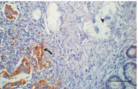
One of our mAbs called 6D1 is an IgG1 antibody with the property of specific recog-nition of CEA-expressing neoplastic cells in CRC tissue sections by the immunoperoxi-dase method. Thus, in the present study we Figure 1 - Immunoperoxidase staining of a colorectal carcinoma tissue section w ith mAb
performed a complete evaluation of mAb 6D1 for in vivo tumor targeting. For this purpose, the hybridoma was recloned to as-sure monoclonality and a subclone was des-ignated 6D1.1.
IgG1 mAb 6D1.1 was also shown to be CEA specific in immunoperoxidase reac-tions on CRC tissue secreac-tions (Figure 1) since it binds only to tumor tissue and not to neutrophils, rich in nonspecific cross react-ing antigen (NCA) (20).
Before starting the in vivo experimental studies, we measured mAb 6D1.1 affinity for CEA. The association constant (Ka) was 0.63 ± 0.11 x 10⁹
M-1
derived from a linear Scatchard plot as depicted in Figure 2. The percentage of 125I-CEA binding to mAb-coated strips was 40%, indicating a non-immunoreactive fraction of 60% radiolabeled CEA, while nonspecific binding was 2%. The affinity constant order of magnitude suggested adequacy for immunolocalization when compared to literature data (21).
For further study of mAb 6D1.1, 99m Tc labeling was standardized using a direct method (18). Labeling mAb 6D1.1 with 99mTc was always highly efficient, resulting in 97% radiochemical purity as evaluated by ITLC-SG. This result is in absolute accordance with others (22) and reflects an excellent labeling efficiency.
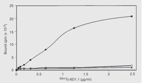
In vitro assays of 99mTc-6D1.1 binding to the CEA-producing CRC cell line LS-174T demonstrated that immunoreactivity was maintained after mAb labeling, as demon-strated in Figure 3, where a saturable binding curve of 99m
Tc-6D1.1 to LS-174T cells is shown. Specificity was confirmed by the binding inhibition obtained by pre-incuba-tion with unlabeled 6D1.1. No binding was observed to cell lines that do not produce CEA, i.e.,the melanomaMeWo line and the breast carcinoma ZR75-30 line. These re-sults were important for planning further 6D1.1 studies in vivo.
Imaging experiments were then per-formed on nude mice individually
xe-1.6
B
/F
1.4
1.2
1.0
0.6
0.4
0.2
0 0.8
0 0.2 0.4 0.6 0.8 1.0
B (nM ) nografted with either LS-174T or ZR75-30 cell lines as shown in Figure 4. 99mTc-6D1.1 was retained in the LS-174T tumor, the CEA-producing cell line (animal B). Also, the unrelated mAb 8F9, identically labeled, did not bind to the LS-174T tumor (animal A), showing that radioactive retention was not a consequence of tumor vascularization. This is one of the possible factors affecting tumor targeting by radiolabeled mAbs. The confir-mation of uptake specificity of mAb 6D1.1
B
o
u
n
d
c
p
m
(
x
1
0
-3) 25
20
10
5
0 0
0.5 1 1.5 2 2.5
99mTc-6D1.1 (µg/ml)
15
0
Figure 3 - 99mTc-6D1.1 binding curves to tumor cell lines: LS-174T (CEA positive), w ithout
(squares) and w ith (circles) prior to incubation w ith unlabeled antibody, and M eWo (CEA negative; triangles).
Figure 2 - Determination of mAb 6D1.1 affinity for CEA. Scatchard plot analysis obtained from binding of increasing amounts of 125I-CEA to mAb 6D1.1-coated w ells. The calculated
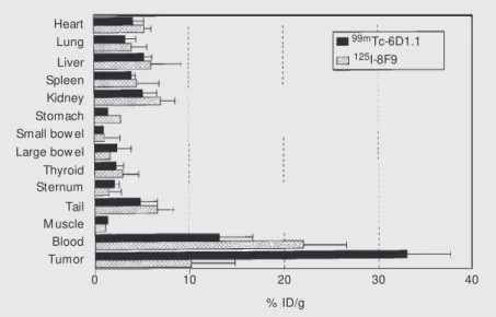
mAb was concentrated in the CEA-express-ing tumor as demonstrated in Figure 5. Taken together, these in vivo data are compatible with the in vitro results, indicating specific-ity and adequate labeling yield. Concerning the latter, it is worth mentioning that thyroid and/or stomach uptake, which was not ob-served, would indicate an excess of free 99m
Tc.
To quantitate antibody capture in differ-ent tumor masses in comparison to normal tissues and organs and to further evaluate the efficacy of 99m
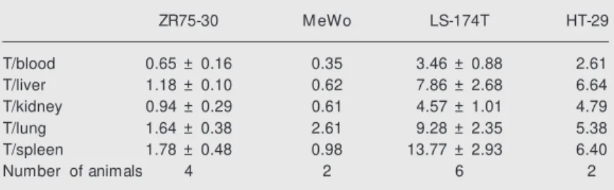
Tc-6D1.1 biodistribution, ex-periments were performed in animals xe-nografted with four cell lines expressing or not CEA. For comparison we used two lines that express CEA in different amounts and two lines that do not. Table 1 shows that the obtained tumor-to-blood ratio for the lines that do not express CEA (MeWo and ZR75-30) was lower than 1.0, while the CRC pre-sented ratios of 3.46 for LS-174T and 2.6 for HT-29. These results once again confirm CEA recognition by 99m
Tc-6D1.1 and also demonstrate that different CEA-producing tumors can be targeted. It is necessary to mention that we have shown elsewhere (23) that the HT-29 line expresses approximately three and a half times less CEA than LS-174T when CEA is measured in membrane-enriched cell extracts. The tumor-to-blood ratios obtained are as good as those de-scribed by others who studied anti-CEA mAbs (21,24).
Taken together, the presented results dem-onstrate that mAb 6D1.1, apart from being a good reagent for detecting CEA in neoplas-tic tissue sections, is a highly suitable tool for experimental immunoscintigraphy in hu-man CRC tumor models.
The obvious next step should be the utili-zation of this mAb in CRC patients to locate metastatic lesions. Nevertheless, more strin-gent tests to confirm CEA specificity are required before human trials are started. Thus, using binding assays to CEA-family trans-fectants and to isolated human granulocytes Heart
Lung
Liver Spleen Kidney Stomach Small bow el Large bow el Thyroid Sternum Tail M uscle Blood Tumor
0 10 20 30 40
% ID/g
99mTc-6D1.1 125I-8F9
Figure 5 - Biodistribution of radiolabeled mAbs injected simultaneously in mice xenografted w ith the LS-174T line and analyzed 24 h after injection. Black bars represent 99mTc-6D1.1
anti-CEA mAb and grid bars, unrelated 125I-labeled mAb 8F9. Results are reported as % of
injected dose per gram of tissue (% ID/g) obtained as the mean values ± SD from three animals.
by CEA-producing tumors is the non-visual-ization of non-CEA-producing ZR75-30 tu-mor (animal C).
These results were further corroborated by experiments where both 99mTc-6D1.1 and 125
I-8F9 mAbs were injected simultaneously into the same animals. Only the anti-CEA
Figure 4 - Images of nude mice bearing tumors of 1 cm in their largest diameter treated w ith
99mTc-labeled mAbs 24 h after injection. Animals: A) isotype control 8F9 mAb in a
CEA-positive tumor (LS-174T); B) anti-CEA 6D1.1 mAb in a CEA-CEA-positive tumor (LS-174T) indi-cated by the arrow , and C) anti-CEA 6D1.1 mAb in a CEA-negative tumor (ZR75-30).
it was demonstrated that mAb 6D1.1 prob-ably recognizes non-specific cross-reacting antigen, NCA-95 (personal communication, Dr. Fritz Grunert, Institute of Immunobiol-ogy, Albert-Ludwigs University, Freiburg, Germany). The apparent contradiction be-tween this last result and the immunoperoxi-dase reaction (Figure 1) may be explained either by the possibility that the epitope rec-ognized by this mAb was damaged by the tissue section fixation procedure which re-sulted in neutrophil nonstaining or by the lower sensitivity of this reaction. In view of these last results, we are not yet authorized to use this mAb in patients.
On the other hand, the importance of this report is also that it provides the complete sequence of experiments necessary for pre-vious evaluation of any mAb potentially use-ful for targeting human CRC in vivo.
Table 1 – Tumor (T)-to-normal-tissue ratios in mouse tissues obtained 24 h after 99m
Tc-6D1.1 mAb injection.
Tumor-to-normal tissue ratios w ere obtained by dividing the concentration of radioac-tivity in tumors by those of each normal tissue indicated. LS-174T and HT-29 are CEA-producing cells w hile ZR75-30 and M eWo are not. Results for the tw o groups composed of more than tw o animals are presented as means ± SD.
ZR75-30 M eWo LS-174T HT-29
T/blood 0.65 ± 0.16 0.35 3.46 ± 0.88 2.61
T/liver 1.18 ± 0.10 0.62 7.86 ± 2.68 6.64
T/kidney 0.94 ± 0.29 0.61 4.57 ± 1.01 4.79
T/lung 1.64 ± 0.38 2.61 9.28 ± 2.35 5.38
T/spleen 1.78 ± 0.48 0.98 13.77 ± 2.93 6.40
Number of animals 4 2 6 2
Ackno wle dgm e nts
We are greatly indebted to Prof. Ricardo Renzo Brentani for his invaluable support and for kindly revising the manuscript.
Re fe re nce s
1. Behr TM , Sharkey RM , Juw eid M E, Dunn RM , Vagg RC, Ying Z, Zhang C-U, Sw ayne LC, Vardi Y, Siegel JA & Goldenberg DM (1997). Phase I/II clinical radioimmuno-therapy w ith an iodine-131-labeled anti-carcinoembryonic antigen murine mono-clonal antibody IgG. Journal of Nuclear M edicine,38: 858-870.
2. W ersäll P, Boet hius J, Ohlsson I, Biberfeldt P, Larsson S, Krusenstjerna S, Lindström V, Collins P & M ellsted H (1997). Intratumoral infusions of mono-clonal antibody (M ab 425) to the epider-mal grow th factor receptor in epider-malignant
glioma. Cancer Immunology,
Immuno-therapy,44:157-164.
3. Juw eid M , Sharkey RM , Sw ayne LC, Griffiths GL, Dunn R & Goldenberg DM (1998). Pharmacokinetics, dosimetry and t oxicit y of rhenium -188-labeled ant i-carcinoembryonic antigen monoclonal an-tibody, M N-14, in gastrointestinal cancer.
Journal of Nuclear M edicine, 39: 34-42. 4. Ychou M , Pelegrin A, Faurous P, Robert
B, Saccavini J-C, Guerreau D, Rossi J-F, Fabbro M , M ach J-P & Artus J-C (1998). Phase-I/II radio-im m unot herapy st udy w ith iodine-131-labeled anti-CEA mono-clonal antibody F6 F(ab’)2 in patients w ith
non-resectable liver metastases from
co-lorectal cancer. International Journal of Cancer,75: 615-619.
5. Landerson JH, M cDonald JM , Landt M & Schw artz M K (1980). Colorectal carcino-ma and carcinoembryonic antigen. Clini-cal Chemistry, 26: 1213-1220.
6. M ach J-P, Carrel S, M erenda C, Sordat B & Cerottini J-C (1974). In vivo localisation of radiolabelled antibodies to carcinoem-bryonic ant igen in hum an carcinom a grafted into nude mice. Nature, 248: 704-706.
7. Goldenberg DM , DeLand F, Euishin K, Kim E, Bennett S, Primus FJ, Van Nagell Jr JR, Estes N, DeSimone P & Rayburn P (1978). Use of radiolabeled antibodies to carcinoembryonic antigen for the detec-tion and localizadetec-tion of diverse cancers by external photoscanning. New England Journal of M edicine,298: 1384-1388. 8. M ach J-P, Buchegger F, Forni M ,
Ritschard J, Berche C, Lumbroso J-D, Schreyer M , Girardet C, Accolla RS & Car-rel S (1981). Use of radiolabelled mono-clonal anti-CEA antibodies for the detec-tion of human carcinomas by external photoscanning and tomoscintigraphy. Im-munology Today, 2: 239-249.
9. Granow ska M , Britton KE, M ather SJ, M orris G, Soobramoney S, Talbot IC &
Northover JM A (1993). Radioimmunoscin-tigraphy w ith technetium-99m labelled monoclonal antibody, 1A3, in colorectal cancer. European Journal of Nuclear M edicine, 20: 690-698.
10. Juw eid M , Sharkey RM , Sw ayne LC & Goldenberg DM (1997). Improved selec-tion of patients for reoperaselec-tion for medul-lary thyroid cancer by imaging w ith radio-labeled anticarcinoembryonic antigen an-tibodies. Surgery, 122: 1156-1165. 11. Goldenberg DM , Goldenberg H, Sharkey
RM , Higginbot ham -Ford E, Lee RE, Sw ayne LC, Burger KA, Tsai D, Horow itz JA, Hall TC, Pinsky CM & Hansen HJ (1990). Clinical studies of cancer radioim-munodetection w ith carcinoembryonic an-tigen monoclonal antibody fragments la-beled w ith 123I or 99mTc. Cancer
Re-search,50 (Suppl): 909s-921s.
12. Hertel A, Baum RP, Lorenz M , Baew -Christow T, Encke A & Hör G (1990). Immunoscintigraphy using a technetium-99m labelled monoclonal antibody in the follow -up of colorectal cancer and other tumors producing CEA. European Journal of Cancer, 62 (Suppl X): 34-36.
antibodies in human colorectal primary tu-mors as a function of tumor mass. Euro-pean Journal of Nuclear M edicine, 20: 345-347.
14. Boxer GM , Begent RHJ, Kelly AM B, Southall PJ, Blair SB, Theodorou NA, Daw son PM & Ledermann JA (1992). Fac-tors influencing variability of localization of antibodies to carcinoembryonic antigen (CEA) in patients w ith colorectal carcino-ma - implications for radioimmunotherapy.
British Journal of Cancer,65: 825-831. 15. Carneiro CRW, Lopes JD & Brentani M M
(1987). Carcinoembryonic antigen (CEA): production of immunoprecipitating mono-clonal antibodies and development of an
enzyme immunoassay. Hybridom a, 6:
689-692.
16. Hsu SM , Raine L & Fanger H (1981). Use of avidin-biotin-peroxidase complex (ABC) in immunoperoxidase techniques. Jour-nal of Histochemistry and Cytochemistry, 29: 577-580.
17. Lindmo T, Boven E, Cuttita F, Fedorko J &
Bunn-Jr PA (1984). Determination of the immunoreactive fraction of radiolabeled monoclonal antibodies by linear extrapo-lation to binding at infinite antigen ex-cess. Journal of Immunological M ethods, 72: 77-89.
18. M ather SJ & Ellison D (1990). Reduction-mediated Technetium-99m labeling of monoclonal antibodies. Journal of Nuclear M edicine, 31: 692-697.
19. Carneiro CRW, Campos ACE, Pacheco M M & Brentani M M (1994). M onoclonal-antibody-based immunoassay for carcino-embryonic (CEA) measurement in human serum. Ciência e Cultura, 46: 111-114. 20. Buchegger F, Schreyer M , Carrel S &
M ach J-P (1984). M onoclonal antibodies identify a CEA crossreacting antigen of 95 kD (NCA-95) distinct in antigenicity and tissue distribution from the previously de-scribed NCA of 55 kD. International Jour-nal of Cancer,33: 643-649.
21. M oraes JZ, Gesztesi JL, Westermann P, Le Doussal JM , Lopes JD & M ach J-P
(1994). Anti-idiotypic monoclonal antibody AB3, reacting w ith the primary antigen (CEA), can localize in human colon-carci-noma xenografts as efficiently as AB1.
International Journal of Cancer, 57: 1-6. 22. Castiglia SG, Duran A, Fiszm an G &
Horenstein A (1995). A 99mTc direct
label-ing of anti-CEA monoclonal antibodies: quality control and preclinical studies.
Nuclear M edicine and Biology, 22: 367-372.
23. Prado IB, Laudanna AA & Carneiro CRW (1995). Susceptibility of colorectal carci-noma cells to natural-killer-mediated lysis: relationship to CEA and degree of differ-entiation. International Journal of Cancer, 61: 854-860.