ISSN 0100-879X
BIOMEDICAL SCIENCES
AND
CLINICAL INVESTIGATION
www.bjournal.com.br
www.bjournal.com.br
Volume 45 (2) 93-178 February 2012
Braz J Med Biol Res, February 2012, Volume 45(2) 104-112
doi: 10.1590/S0100-879X2012007500003
Distribution of human immunodeficiency virus type 1 subtypes in
the State of Amazonas, Brazil, and subtype C identification
L.K.H. Cunha, S. Kashima, M.F.C. Amarante, R. Haddad, E.S. Rodrigues, K.L.T. Silva, T.A. Lima,
D.B. Castro, F.C. Brito, E.G. Almeida, D.T. Covas and A. Malheiro
Institutional Sponsors
The Brazilian Journal of Medical and Biological Research is partially financed by
Faculdade de Medicina de Ribeirão Preto Campus
Ribeirão Preto
Ex plor e H igh - Pe r for m a n ce M S Or bit r a p Te ch n ology I n Pr ot e om ics & M e t a bolom ics
Distribution of human immunodeficiency virus
type 1 subtypes in the State of Amazonas,
Brazil, and subtype C identification
L.K.H. Cunha
1, S. Kashima
3, M.F.C. Amarante
3,
R. Haddad
3, E.S. Rodrigues
3,
K.L.T. Silva
2, T.A. Lima
2, D.B. Castro
2, F.C. Brito
2, E.G. Almeida
2,
D.T. Covas
3and A. Malheiro
1,21Departamento de Parasitologia, Universidade Federal do Amazonas, Manaus, AM, Brasil 2Diretoria de Ensino e Pesquisa, Fundação de Hematologia e Hemoterapia do Amazonas, Manaus, AM, Brasil
3Laboratório de Biologia Molecular, Hemocentro de Ribeirão Preto, Faculdade de Medicina de Ribeirão Preto,
Universidade de São Paulo, Ribeirão Preto, SP, Brasil
Abstract
Few studies have reported the molecular epidemiological characterization of HIV-1 in the Northern region of Brazil. The pres
-ent study reports the molecular and epidemiological characterization of 31 HIV-1 isolates from blood donors from the State of Amazonas who donated blood between April 2006 and March 2007. Serum/plasma samples from all donors were screened for HIV antibodies by ELISA and the results confirmed by Western blot analysis. Genomic DNA was extracted from the buffy coat using the Super Quik-Gene-DNA Isolation kit. Nested PCR was performed on the env, gag, and pol regions of HIV-1 using the Gene Amp PCR System 9700. Sequencing reactions were performed using the inner PCR primers and the DYEnamic™ ET Dye Terminator Kit, and phylogenetic analysis was performed using the gag, pol, and env gene sequences. We collected samples from 31 blood donors who tested positive for HIV-1 in confirmatory experiments. The male:female ratio of blood donors was 3.4:1, and the mean age was 32.4 years (range: 19 to 61 years). Phylogenetic analysis showed that subtype B is the most prevalent among Northern Brazilian HIV-1-seropositive blood donors. One HIV-1 subtype C and one circulating recombinant form (CRF_BF) of HIV-1 were identified in the State of Amazonas. This is the first study showing the occurrence of a possible “homogenous” subtype C in this region of Brazil. This finding could contribute to a better characterization of the HIV-1 strains that circulate in the country.
Key words: HIV-1; Subtypes; Phylogenetic analysis; Blood donors; Molecular and epidemiological characterization
Introduction
Correspondence: A. Malheiro, Diretoria de Ensino e Pesquisa, Fundação de Hematologia e Hemoterapia do Amazonas, Av. Constantino Nery, 2943, 69040-002 Manaus, AM, Brasil. Fax: +55-92-3655-0112. E-mail: [email protected]
Received July 16, 2011. Accepted January 4, 2012. Available online January 20, 2012. Published February 17, 2012. Two major reasons for the extensive genetic diversity
of HIV-1 are high levels of viral replication and error-prone reverse transcription, which incorporates mutations into the viral genome. Investigating the genetic diversity of HIV-1 can lead to further understanding of HIV-1 evolution and
dissemination and vaccinedevelopment.
The HIV-1 genome is composed of approximately 9.0
kb distributed among structural (gag, env, and pol), trans
-activation (tat and rev) and accessory (nef, vpu, vif, and
vpr) genes. Based on phylogeneticanalyses of env and
gag gene sequences, HIV-1 is classified into three main
groups: the major (M), outlier(O), and new (N) groups (1-4).
Nine genetic subtypes have been identified in the M group:
A-D, F-H, J, and K; at least 34 circulating recombinant forms (CRFs) have been recognized (5-7). These genetic subtypes
may have an impact on drugsusceptibility, the emergence
of new drug resistancemutations, and the performance
of laboratory tests for diagnostics and measurements of viral loads (8).
HIV subtypes in the State of Amazonas, Brazil 105
(CRF_BC, BF) (14,15). Subtype F1 was identified primarily in female prostitutes and drug users, and was introduced in Brazil in the early 1980s (10). Although most HIV-1 infections in Brazil are subtype B, the southern region of the country represents the main subtype C focus (16). Isolated cases of this subtype have also been reported in the States of Espírito Santo, Goiás, and Rio de Janeiro (17-20).
The Northern region of Brazil contains 7 States
(Rondônia, Acre, Amazonas, Roraima, Pará, Amapá, and Tocantins). Amazonas is located in the central region, comprises the largest territorial area of Brazil and shares borders with Venezuela, Colombia, and Peru. According to Boletim Epidemiológico DST/AIDS (2006), the Northern
region reports approximately 13,000 AIDS cases, which
corresponds to 3% of all cases in Brazil. The State of Ama
-zonas has around 5000 notified AIDS cases, and Manaus (the capital of Amazonas) is responsible for 90% of these cases. However, few genotyping studies of HIV-1-infected people in the Northern region of Brazil have been reported. Recently, a study describing the presence of subtypes D and C in the cities of Belém (Pará) and Macapá (Amapá) has been reported (21), but the epidemiology of HIV in the State of Amazonas is unknown. The only study that
describes HIV-1 subtypes in Manaus, the largest com
-munity within the central rain forest of Brazil’s Amazon Basin, showed that subtypes B and F are represented equally (22).
In the present study, we performed the
molecular and epidemiological characteriza
-tion of 31 HIV-1 isolates from the Northern region of Brazil. We also reported, for the first time, the presence of subtype C in the State of Amazonas.
Material and Methods
Patient samples
The samples were obtained from
HIV-1-seropositive blood donors at Fundação Hemo
-centro de Manaus (HEMOAM), AM, Brazil, and presented positive serology to HIV-1. The study
was approved by the Institutional Ethics Com
-mittee and all blood donors signed an informed
consent form and were informed about the procedures of sample collection and analysis. Between April 2006 and March 2007, 10-mL intravenous blood samples were collected from
each individual into tubes containing the anti
-coagulant EDTA. Serum/plasma samples from all donors were screened for HIV antibodies by ELISA Axsym HIV I/II gO (Abbott, Germany) and combined HIV Ag/Ac (Murex Biotech, UK). Confirmatory screening tests were performed by Western blot analysis using HIV Blot 2.2 (Genelabs Diagnostics, Singapore).
Proviral DNA extraction
DNA was extracted from the buffy coat (EIA-positive samples) using the Super Quik-Gene-DNA Isolation kit (Promega, USA) according to manufacturer instructions.
Polymerase chain reaction (PCR)
Nested PCR of HIV-1 was performed on the env, gag, and pol regions using a Gene Amp PCR System 9700 (Applied Biosystems, USA). To amplify a fragment of the genes, the first round of PCR was performed on samples using the primers ED5 and ED12 for the env gene, H1G777 and H1P202 or C1 and C2 for the gag gene, and K1 and
K2 for the pol gene (18). A second round of PCR was car
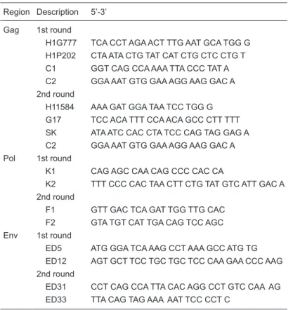
-ried out using the nested primers ED31 and ED33 for env, H11584 and G17 or SK and C2 for gag, and F1 and F2 for pol amplification. The primer sequences of HIV-1 genomic regions are presented in Table 1. Reactions were performed in 50-µL mixtures containing 500 ng DNA, 1.0 U Taq DNA polymerase, 50 mM KCl, 10 mM Tris-HCl, pH 8.3, 1.5 mM
MgCl2, 0.8 mM each deoxynucleotide triphosphate (dNTPs),
and 10 ρmol of each primer. PCR cycling conditions for the first and second rounds consisted of one cycle at 94°C for 5 min, 35 cycles at 94°C for 90 s, 55 at 60°C for 90 s, and 72°C for 30 s. Five microliters of the initially amplified product
Table 1. Primer sequences of HIV genomic regions (18).
Region Description 5’-3’
Gag 1st round
H1G777 TCA CCT AGA ACT TTG AAT GCA TGG G H1P202 CTA ATA CTG TAT CAT CTG CTC CTG T C1 GGT CAG CCA AAA TTA CCC TAT A C2 GGA AAT GTG GAA AGG AAG GAC A
2nd round
H11584 AAA GAT GGA TAA TCC TGG G
G17 TCC ACA TTT CCA ACA GCC CTT TTT
SK ATA ATC CAC CTA TCC CAG TAG GAG A
C2 GGA AAT GTG GAA AGG AAG GAC A Pol 1st round
K1 CAG AGC CAA CAG CCC CAC CA
K2 TTT CCC CAC TAA CTT CTG TAT GTC ATT GAC A 2nd round
F1 GTT GAC TCA GAT TGG TTG CAC F2 GTA TGT CAT TGA CAG TCC AGC Env 1st round
ED5 ATG GGA TCA AAG CCT AAA GCC ATG TG ED12 AGT GCT TCC TGC TGC TCC CAA GAA CCC AAG
2nd round
(first round) was used in the second round. The amplified products were analyzed by 2% agarose gel electrophoresis followed by ethidium bromide staining.
Nucleotide sequencing and phylogenetic analysis
Amplified products (gag, pol, and env) were purified using the Wizard PCR Preps DNA Purification System
Kit (Promega). Sequencing reactions were performed us
-ing the inner PCR primers and the DYEnamic™ ET Dye Terminator Kit according to manufacturer instructions, and products were sequenced using the MegaBace 1000 DNA Sequencing System. Electropherograms were analyzed with ChromasPro Version 1.41 (Technelysium Pty Ltda., Australia).
The HIV-1 sequences of Brazilian isolates (gag, pol, and env) were aligned and edited using the BioEdit 7.0.5.3 program. The sequences were compared to those in the Los Alamos database, which includes 40 prototypes for the genotypes A-D, F-H, J, and K, 14 CRFs and the out group sequence (O group). Neighbor joining (NJ) trees were constructed using the Phylip package (version 3.6)
and PAUP* 4.0b10 program (Sinauer Associates, USA).
The NJ trees were constructed using the (GTR+I+G) and (TMV+I+G) nucleotide substitution models, as selected by the Modeltest 3.06 program. The reliability of the NJ trees was assessed by analyzing 1000 bootstrap replicates. The likelihood ratio test was used to calculate statistical support (reported as P values) for the branches. Trees were drawn with the TreeView X 0.5.0 program. Sequences obtained in this study are available in GenBank under accession numbers FJ011469-FJ011532.
Recombination analysis was performed using SimPlot 3.5.1, which evaluates the differences between phylogenetic trees at each fragment (window) along with the alignment. Bootscanning was performed with alignment (available at http://lasp.cpqgm.fiocruz.br/retrovirusHIV.html). The gag, pol, and env regions were concatenated to get the
query sequence, gaps were deleted to obtain a continu
-ous sequence, and the sequences were then analyzed by multiple alignment.
Results
Clinical features of HIV-1-infected patients
We collected samples from a total of 107 HIV-1-sero
-positive blood donorswho had one positive serology test.
Of these 107 donors, only 31 were confirmed as HIV-1
positive in diagnostic tests. The majority of the HIV-1 indi
-viduals were males, the male:female ratio was 3.4:1, and the mean age was 32.4 years (range: 19 to 61 years; Table 2). Ten repeat blood donors were identified (32.3%), and 6
of them seroconverted for up to 20 months. The seroconver
-sion time comprises the period between the last liberated donation and the donation that presented HIV-1 reactivity.
The blood donors were asymptomatic at the time of dona
-tion and reported not to be under antiretroviral treatment (data not shown).
Phylogenetic analysis
The DNAs of 31 HIV-1-positive individuals were evalu
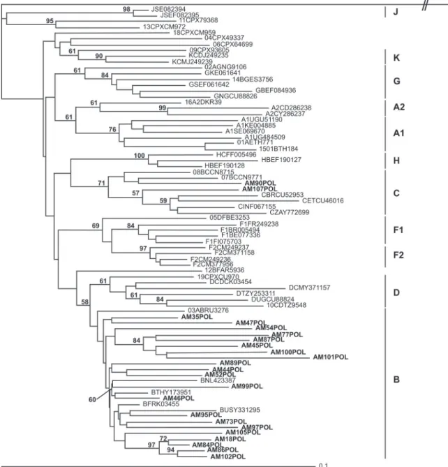
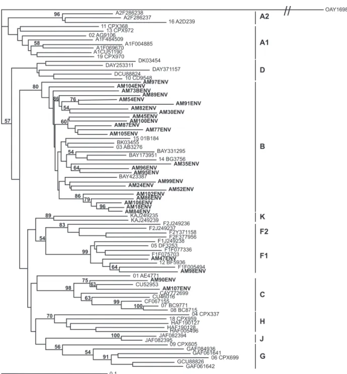
-ated. We analyzed 462, 706, and 498 bp of the gag, pol, and env regions, respectively (Table 1). The NJ trees constructed from the three regions show that subtype B is the most prevalent among Brazilian patients (Figures 1, 2, and 3).
The phylogenetic trees revealed two subtype C samples. Only the env and pol regions were characterized in one sample (AM090) because the gag region did not amplify in PCR (Table 3). In the other sample (AM107), all regions were characterized. These samples were submitted to boot scanning analysis, and the subtype C profiles were confirmed (data not shown).
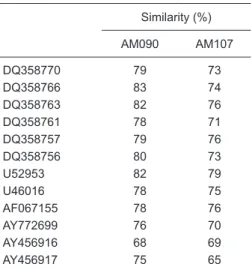
Subtype C has not been previously described in the Amazonas State, but has been isolated in other regions
of Brazil. Analyses of similarity were performed to com
-pare the env sequences found in the Southern region of Brazil (DQ358770, DQ358766, DQ358763, DQ358761, DQ358757, DQ358756, and U52953) and env sequences from different geographic regions, including Venezuela (AY456916 and AY456917), South Africa (AY772699), India (AF067155), and Ethiopia (U46016) (Table 4). This genomic region was selected for similarity analysis because it is the most variable region of HIV-1. These analyses showed that Amazonian C subtype isolates show high similarity to sequences from the Southern region, suggesting that HIV-1 subtype C may have disseminated to the North following a South to North gradient pattern.
The partial sequences of gag, pol, and env genes were obtained for 18 samples, while for 10 samples, only two regions (gag and env, gag and pol, or pol and env) were obtained. For three samples, only one region (gag, pol, or env) was obtained (Table 3). In agreement with the topology of the phylogenetic trees built for each gene, 17 samples were classified as subtype B and one as subtype C for the three genes. However, one sample was classified as subtype B for the gag and pol genes and subtype F for the env gene, suggesting the presence of one circulating
recombinant form.
Table 2. Distribution of HIV-1-seropositive blood
donors according to age and gender.
Age, median (range)
Male 31.7 (19-61)
Female 33.1 (23-49)
Gender, N (%)
Male 24 (77.4%)
HIV subtypes in the State of Amazonas, Brazil 107
The 828-bp concatenated sequence of probable CRF (sample AM047) was submitted to bootscanning using SimPlot with the following parameters: window size (300 bp), step (20 bp), algorithm (NJ), distance model (Kimura 2-parameter), and replicate number (100). The reference group/subtypes used for bootscanning were the M (A-D,
F-H, J, K) and O groups. The map generated by the Sim
-Plot software showed that the AM047 sample presented a
breakpoint (subtypes B and F) at position 525. At 230, the sequence is characterized as subtype B (95% reliability), while at position 720 it is characterized as subtype F (99% reliability; data not shown).
Discussion
Several HIV-1 subtypes, sub-subtypes, circulating re
combinant forms, and unique recombinant forms (URFs) have been described extensively worldwide (23). The HIV-1-positive prevalence among Brazilians is approximately 0.5%, ranging from 0.3 to 1.6% since 2000 (24). Most of the molecular and epidemiological characterization of HIV-1 in Brazil has concentrated in the Southeast, where HIV-1 subtype B is prevalent. Other Southern regions in the country have reported a predominance of the C subtype; however, the non-B and non-F subtypes may be neglected
in other regions of the country (25-27). A survey of blood donors between 1996 and 2004 (over 400,000 donations) showed that the HIV-1/2 seroprevalence is about 0.2% in the Northern region of Brazil (HEMOAM Foundation, Blood Bank of Amazon State, unpublished data). In agreement with other regions, the present study showed a higher prevalence of subtypes B and F among recent blood donors and the presence of one recombinant B/F form (AM047:
Bpol, Bgag, Fenv). Moreover, the present study also identified
HIV subtypes in the State of Amazonas, Brazil 109
two C subtype isolates (AM090: Cpol, Cenv; AM107: Cpol,
Cgag, Cenv). Subtype C is the most prevalent subtype of
HIV-1 worldwide and accounts for more than half of
infec-tions. Recently, one study performed in the States of Pará
and Amapá in Northern Brazil showed CRFs containing
subtype C (Cenv, Bpro) (21). Our data indicate, for the first
time, the possible presence of a “homogenous” subtype C
virus in the Amazon Basin. Completegenome sequencing
of AM090 and AM107 isolates is requiredto confirm this
hypothesis.
Two hypotheses may explain the route of HIV-1 sub
-type C transmission. First, although sub-type B is the most prevalent subtype in Brazil, subtype C is present mainly
in the Southern region (28).This subtype was previously
reported to be found in Argentina, Uruguay, Paraguay (29), and other countries in South America. Recently, Jones et
al. (30) reportedthat subtype C was introduced into South
America from Argentina, which acquired African strains.
However, other studies support the view that Brazil is the
dispersion center of subtype C in South America. The exact origin of subtype C cannot be determined due to the lack of epidemiological and historical data, sample problems and doubts regarding phylogeny (30). Subtype C is concentrated
in the Southern region of this continent and other states
of Brazil, including Espírito Santo (Southeast region) and Goiás (Center-West region) (17-19). Our analysis shows that the Northern subtype C isolates are more similar to the
Southern subtype C samples than in other regions, sup
-porting the ideathat subtype C has been disseminated in
Brazil following a South to North gradient pattern.
In 2005, subtypes C and B/C were confirmedin Venezu
-elan patients who took trips to Africa and Europe. One patient (subtype C) was infected in South Africa, and another patient (CRF B/C) had high sexual risk behavior and a history of trips to Portugal and South Africa (31). These subtypes are also described in other countries near the Amazon Basin, including Peru and Ecuador (32). The second hypothesis is that subtype C was recently introduced into the Northern
region of Brazil through countries that border this region.
In the present study, it was not possible to identify the transmission pattern of HIV-1 in the State of Amazonas. Additional studies analyzing the lifestyles of these patients, including their origin, history of trips and sexual transmission and sexual relationships with foreigners, are necessary to determine the route of HIV-1 subtype C transmission.
In Brazil, the subtypes B, F1 and C coexist in high risk groups and the prevalence differs only by geographical location (10,33). The coexistence of several subtypes in a region leads to the emergence of URFs and CRFs. While CRFs B/F1 are common in areas where subtypes B and F1 coexist, in the South of the country CRFs B/C are emerging (19) due to the presence of these two subtypes. The CRF B/F identified in the present study in Manaus corroborates the theory that these forms are recombining in different areas,
increasing the genetic variability of HIV-1 in Brazil. The present study reports one of the first global views of the molecular epidemiology of HIV-1 among Northern Brazilian isolates. We demonstrated by sequence analysis that HIV-1-infected individuals harbor the subtypes B, F and C, and different and/or complete genomic regions of HIV-1 should be analyzed in order to genetically characterize the Brazilian strains in this region. These studies will contribute to an understanding of the divergence of Brazilian strains of HIV-1 and the transmission patterns of this infection in our country. HIV-1 molecular epidemiology studies should further our understanding of the biological properties of HIV-1 and the mutations related to antiretroviral resistance and vaccine design.
Acknowledgments
We thank Rochele Azevedo, Andréia Monteiro Tarragô and Adriana Aparecida Marques for technical assistance. Research supported by CNPq, Fundação de Amparo à Pesquisa do Estado do Amazonas, Center for Cell-Therapy (CTC)/Fundação Hemocentro de Ribeirão Preto, and FAPESP.
Table 3. Regions of the HIV-1 genes sequenced in samples.
Sequenced regions Sample names
Three regions: gag, pol and env AM018, AM035, AM045, AM047, AM052, AM054, AM073, AM077, AM084, AM086, AM087, AM095, AM097, AM099, AM100, AM102, AM105, AM107 Two regions: gag and env or gag and pol or pol and env AM030, AM044, AM046, AM082, AM089, AM090, AM091, AM101,
AM104, AM106
One region: gag or pol or env AM024, AM096, AM098
Table 4. Similarity analysis using the env region
of HIV-1 isolates.
Similarity (%)
AM090 AM107
DQ358770 79 73
DQ358766 83 74
DQ358763 82 76
DQ358761 78 71
DQ358757 79 76
DQ358756 80 73
U52953 82 79
U46016 78 75
AF067155 78 76
AY772699 76 70
AY456916 68 69
HIV subtypes in the State of Amazonas, Brazil 111
References
1. Louwagie J, McCutchan F, Mascola J, Eddy G, Fransen K, Peerters M, et al. Genetic subtypes of HIV-1. AIDS Res Hum
Retroviruses 1993; 9: S147-S150.
2. Louwagie J, McCutchan FE, Peeters M, Brennan TP, Sanders-Buell E, Eddy GA, et al. Phylogenetic analysis of gag genes from 70 international HIV-1 isolates provides evidence for multiple genotypes. AIDS 1993; 7: 769-780.
3. McCutchan F, Louwagie J, Eddy G, Van Der Groen G, Piot P, Myers G, et al. Identification of multiple genetic subtypes of HIV-1 and evidence for geographic dispersal. J Acquir
Immune Defic Syndr 1993; 6: 685.
4. Simon F, Mauclere P, Roques P, Loussert-Ajaka I, Muller-Trutwin MC, Saragosti S, et al. Identification of a new human immunodeficiency virus type 1 distinct from group M and group O. Nat Med 1998; 4: 1032-1037.
5. Takebe Y, Kusagawa S, Motomura K. Molecular epidemiol
-ogy of HIV: tracking AIDS pandemic. Pediatr Int 2004; 46:
236-244.
6. Thomson MM, Najera R. Molecular epidemiology of HIV-1 variants in the global AIDS pandemic: an update. AIDS Rev
2005; 7: 210-224.
7. Los Alamos Laboratory. The circulating recombinant forms (CRFs). http://www.hiv.lanl.gov/content/sequence/HIV/ CRFs/CRFs.html.
8. Koch WH, Sullivan PS, Roberts C, Francis K, Downing R, Mastro TD, et al. Evaluation of United States-licensed hu
-man immunodeficiency virus immunoassays for detection of group M viral variants. J Clin Microbiol 2001; 39:
1017-1020.
9. Joint United Nations Programme On HIV/AIDS (UNAIDS) and World Health Organization (WHO). Latin America: AIDS epidemic update: Regional summary. http://data.unaids.org/ pub/Report/2008/jc1530_epibriefs_latinamerica_en.pdf. Ac
-cessed July 25, 2008.
10. Bello G, Guimaraes ML, Morgado MG. Evolutionary history of HIV-1 subtype B and F infections in Brazil. AIDS 2006;
20: 763-768.
11. Sabino EC, Shpaer EG, Morgado MG, Korber BT, Diaz RS, Bongertz V, et al. Identification of human immunodeficiency virus type 1 envelope genes recombinant between subtypes B and F in two epidemiologically linked individuals from
Brazil. J Virol 1994; 68: 6340-6346.
12. Guimaraes ML, dos Santos MA, Loureiro R, Galvao-Castro B, Morgado MG. High frequency of recombinant genomes in HIV type 1 samples from Brazilian southeastern and
southern regions. AIDS Res Hum Retroviruses 2002; 18:
1261-1269.
13. Couto-Fernandez JC, Eyer-Silva WA, Guimaraes ML, Chequer-Fernandez SL, Grinsztejn B, Delaporte E, et al. Phylogenetic analysis of Brazilian HIV type 1 subtype D strains: tracing the origin of this subtype in Brazil. AIDS Res
Hum Retroviruses 2006; 22: 207-211.
14. Sanabani S, Kleine NW, Kalmar EM, Diaz RS, Janini LM, Sabino EC. Analysis of the near full length genomes of HIV-1 subtypes B, F and BF recombinant from a cohort of 14 patients in São Paulo, Brazil. Infect Genet Evol 2006; 6:
368-377.
15. Sa Filho DJ, Sanabani S, Diaz RS, Munerato P, Brunstein A, Fusuma E, et al. Analysis of full-length human immunodefi
-ciency virus type 1 genome reveals a variable spectrum of subtypes B and F recombinants in São Paulo, Brazil. AIDS
Res Hum Retroviruses 2005; 21: 145-151.
16. Soares EA, Santos RP, Pellegrini JA, Sprinz E, Tanuri A, Soares MA. Epidemiologic and molecular characterization of human immunodeficiency virus type 1 in southern Brazil. J Acquir Immune Defic Syndr 2003; 34: 520-526.
17. Cabral VP, Cunha CB, Magalhaes EF, Pinto-Neto LF, Couto-Fernandez JC, Dietze R, et al. Human immunodeficiency virus type-1 subtypes of infected patients in Espírito Santo,
Brazil. Mem Inst Oswaldo Cruz 2006; 101: 881-885.
18. Stefani MM, Pereira GA, Lins JA, Alcantara KC, Silveira AA, Viegas AA, et al. Molecular screening shows extensive HIV-1 genetic diversity in Central West Brazil. J Clin Virol 2007;
39: 205-209.
19. Brennan CA, Brites C, Bodelle P, Golden A, Hackett J Jr, Holzmayer V, et al. HIV-1 strains identified in Brazilian blood donors: significant prevalence of B/F1 recombinants. AIDS
Res Hum Retroviruses 2007; 23: 1434-1441.
20. Couto-Fernandez JC, Silva-de-Jesus C, Veloso VG, Rachid M, Gracie RS, Chequer-Fernandez SL, et al. Human im
-munodeficiency virus type 1 (HIV-1) genotyping in Rio de Janeiro, Brazil: assessing subtype and drug-resistance as
-sociated mutations in HIV-1 infected individuals failing highly active antiretroviral therapy. Mem Inst Oswaldo Cruz 2005;
100: 73-78.
21. Machado LF, Ishak MO, Vallinoto AC, Lemos JA, Azevedo VN, Moreira MR, et al. Molecular epidemiology of HIV type 1 in northern Brazil: identification of subtypes C and D and the introduction of CRF02_AG in the Amazon region of Brazil.
AIDS Res Hum Retroviruses 2009; 25: 961-966.
22. Vicente AC, Otsuki K, Silva NB, Castilho MC, Barros FS, Pieniazek D, et al. The HIV epidemic in the Amazon Basin is driven by prototypic and recombinant HIV-1 subtypes B and F. J Acquir Immune Defic Syndr 2000; 23: 327-331. 23. Robertson DL, Anderson JP, Bradac JA, Carr JK, Foley B,
Funkhouser RK, et al. HIV-1 nomenclature proposal. Sci-ence 2000; 288: 55-56.
24. Joint United Nations Programme On HIV/AIDS (UNAIDS). Report on the global AIDS epidemic. http://www.unaids. org/en/KnowledgeCentre/HIVData/GlobalReport/2006/. Ac
-cessed July 25, 2008.
25. Brigido LF, Franco HM, Custodio RM, Oliveira CA, Ferreira JL, Eira M, et al. Molecular characteristics of HIV type 1 circulating in São Paulo, Brazil. AIDS Res Hum Retroviruses
2005; 21: 673-682.
26. Brigido LF, Nunes CC, Oliveira CM, Knoll RK, Ferreira JL, Freitas CA, et al. HIV type 1 subtype C and CB Pol recom
-binants prevail at the cities with the highest AIDS prevalence
rate in Brazil. AIDS Res Hum Retroviruses 2007; 23:
1579-1586.
27. Ferreira JL, Thomaz M, Rodrigues R, Harrad D, Oliveira CM, Oliveira CA, et al. Molecular characterisation of newly identified HIV-1 infections in Curitiba, Brazil: preponderance of clade C among males with recent infections. Mem Inst
Oswaldo Cruz 2008; 103: 800-808.
28. Soares EA, Martinez AM, Souza TM, Santos AF, Da Hora V, Silveira J, et al. HIV-1 subtype C dissemination in southern
29. Carrion G, Eyzaguirre L, Montano SM, Laguna-Torres V, Serra M, Aguayo N, et al. Documentation of subtype C HIV type 1 strains in Argentina, Paraguay, and Uruguay. AIDS
Res Hum Retroviruses 2004; 20: 1022-1025.
30. Jones LR, Dilernia DA, Manrique JM, Moretti F, Salomon H, Gomez-Carrillo M. In-depth analysis of the origins of HIV type 1 subtype C in South America. AIDS Res Hum
Retro-viruses 2009; 25: 951-959.
31. Castro E, Moreno M, Deibis L, de Perez G, Salmen S, Berru
-eta L. Trends of HIV-1 molecular epidemiology in Venezuela:
introduction of subtype C and identification of a novel B/C
mosaic genome. J Clin Virol 2005; 32: 257-258.
32. Montano SM, Sanchez JL, Laguna-Torres A, Cuchi P, Avila MM, Weissenbacher M, et al. Prevalences, genotypes, and risk factors for HIV transmission in South America. J Acquir
Immune Defic Syndr 2005; 40: 57-64.