Generation of BAC Transgenic Tadpoles
Enabling Live Imaging of Motoneurons by
Using the Urotensin II-Related Peptide
(
ust2b
) Gene as a Driver
Marion Bougerol1, Frédéric Auradé2,3‡, François M. Lambert4‡, Didier Le Ray4, Denis Combes4, Muriel Thoby-Brisson4, Frédéric Relaix2,3, Nicolas Pollet5, Hervé Tostivint1
*
1Evolution des Régulations Endocriniennes, UMR 7221 CNRS, Muséum National d’Histoire Naturelle, Paris, France,2UPMC/INSERM UMRS 974, CNRS FRE 3617, AIM, Paris, France,3INSERM, Avenir Team, Pitié-Salpêtrière, Paris, France,4UMR 5287 CNRS, Université de Bordeaux, INCIA,
Bordeaux, France,5Institute of Systems and Synthetic Biology, CNRS, Université d’Evry Val d’Essonne, Evry, France
‡These authors contributed equally to this work. *htostivi@mnhn.fr
Abstract
Xenopusis an excellent tetrapod model for studying normal and pathological motoneuron ontogeny due to its developmental morpho-physiological advantages. In mammals, the uro-tensin II-related peptide (UTS2B) gene is primarily expressed in motoneurons of the brain-stem and the spinal cord. Here, we show that this expression pattern was conserved in
Xenopusand established during the early embryonic development, starting at the early tail-bud stage. In late tadpole stage,uts2bmRNA was detected both in the hindbrain and in the spinal cord. Spinaluts2b+cells were identified as axial motoneurons. In adult, however, the
uts2bexpression was only detected in the hindbrain. We assessed the ability of theuts2b
promoter to drive the expression of a fluorescent reporter in motoneurons by recombineer-ing a green fluorescent protein (GFP) into a bacterial artificial chromosome (BAC) clone containing the entireX. tropicalis uts2blocus. After injection of this construction in one-cell stage embryos, a transient GFP expression was observed in the spinal cord of about a quar-ter of the resulting animals from the early tailbud stage and up to juveniles. The GFP expres-sion pattern was globally consistent with that of the endogenousuts2bin the spinal cord but no fluorescence was observed in the brainstem. A combination of histological and
electrophysiological approaches was employed to further characterize the GFP+cells in the
larvae. More than 98% of the GFP+cells expressed choline acetyltransferase, while their
projections were co-localized withα-bungarotoxin labeling. When tail myotomes were
in-jected with rhodamine dextran amine crystals, numerous double-stained GFP+cells were observed. In addition, intracellular electrophysiological recordings of GFP+neurons re-vealed locomotion-related rhythmic discharge patterns during fictive swimming. Taken
OPEN ACCESS
Citation:Bougerol M, Auradé F, Lambert FM, Le Ray D, Combes D, Thoby-Brisson M, et al. (2015) Generation of BAC Transgenic Tadpoles Enabling Live Imaging of Motoneurons by Using the Urotensin II-Related Peptide (ust2b) Gene as a Driver. PLoS ONE 10(2): e0117370. doi:10.1371/journal. pone.0117370
Academic Editor:Cedric Raoul, Inserm, FRANCE
Received:October 16, 2014
Accepted:December 22, 2014
Published:February 6, 2015
Copyright:© 2015 Bougerol et al. This is an open access article distributed under the terms of the Creative Commons Attribution License, which permits unrestricted use, distribution, and reproduction in any medium, provided the original author and source are credited.
Data Availability Statement:All relevant data are within the paper.
together our results provide evidence thatuts2bis an appropriate driver to express reporter genes in larval motoneurons of theXenopusspinal cord.
Introduction
Generation of transgenic lines with promoter-specific fluorescent reporter proteins has signifi-cantly advanced neurobiological research by enabling the visualization of neuronal subsetsin vivo. For genetic engineering of such animals, mice have long been the most frequently used ex-perimental vertebrate model [1]. However, organisms such as zebrafish andXenopushave re-cently emerged as alternative systems and are now being widely exploited to study human neurological diseases as well as to screen for potential therapeutics [2–5]. Several features of these species make them particularly amenable for neurodevelopmental and neurophysiologi-cal investigations including their rapid development rate as free-living larvae, their transparen-cy allowing easy visualization of internal structures and cells in live animals, and the general organization of their central nervous system (CNS) which is quite similar to other vertebrate species, including human. Moreover, high-quality and well annotated sequenced genomes exist for both species [6,7] and many readily available genome editing technologies have been suc-cessfully adapted to them [8–11].Xenopus, as a tetrapod, offers the added advantage of being much more closely related to mammals than the zebrafish. This proximity is especially impor-tant when studying the neural control of processes that have no genuine equivalent in fish, such as legged terrestrial locomotion [12].
For engineering useful transgenic reporter genes, a crucial step is to choose the correct se-quences which will drive the fluorescent reporter in the neuronal cell type of interest. In the case of motoneurons, the regulatory sequences of several genes encoding transcription factors involved in motoneuron specification, such asHB9(also namedMnx1),ISLET-1(ISL1) and OLIG2, were shown to be particularly well suited, especially in zebrafish [13–15]. InXenopus, generation of transgenic motoneuronal reporter lines usinghb9as a driver is currently in prog-ress [16, NP, unpublished results]. Despite their great interest, all these genes have the disad-vantage of also being expressed in other neurons than motoneurons, not only in the brain but also in the spinal cord. Typically,olig2is expressed within the progenitor domain that gives rise both to motoneurons and oligodendrocytes [17]. Likewise,isl1andhb9are expressed in motoneurons as well as in some sensory neurons and/or interneurons [18,19].
Urotensin II (UTS2) and urotensin II-related peptide, also known as urotensin 2B (UTS2B), are two structurally and phylogenetically related neuropeptides [20,21] that, at least in tetra-pods, are mainly expressed in motoneurons of the brainstem and the spinal cord [22–29]. UTS2 and UTS2B have been shown to be of biological importance, playing an important role in the regulation of behavior, neuroendocrine activities, and central and peripheral control of blood pressure and heart rate [30–33]. In mammals, all these effects are mediated by only one receptor known as UT, which is the reason why the proper effects of each peptide are often dif-ficult to discriminate [21]. Even though the functional significance of their motoneuronal ex-pression is currently poorly understood, UTS2 and UTS2B can be considered as two
interesting putative spinal motoneuronal markers.
Here, our aim was to test the ability of theuts2bpromoter to drive the expression of the green fluorescent protein (GFP) specifically in motoneurons. Up to now, functional studies on theuts2bpromoter have never been carried out inXenopus. Moreover, sequence analyses did not reveal any conserved motif which would be involved in the motoneuronal expression of
(FRM ARF20140128968;http://www.frm.org) to FML, from the PEPS IdEx de Bordeaux-CNRS (101SG/ 1024R) to DLR, and from Genopole and the AFM (Amphigentools) to NP. MB was the recipient of Ph.D grants from the French Ministry of Research and the Legs Prévost (MNHN). The funders had no role in study design, data collection and analysis, decision to publish, or preparation of the manuscript.
theuts2bgene. One significant limitation in generating transgenic reporter genes is that the regulatory elements that control the gene transcription can be scattered over large regions, therefore making their identification quite difficult and time consuming, especially when they are not conserved among species. This limitation can be overcome by placing the reporter gene into large-insert clones such as bacterial artificial chromosomes (BAC) that are thought to con-tain all the regulatory sequences driving its appropriate expression [34]. For this purpose, we recombined aGFPreporter gene into a BAC clone containing the entireX. tropicalis uts2b locus and employed a recently reported method of BAC injection intoX. laevisembryo [35]. In this method, the transgene construct is delivered as a large circular DNA which does not pro-mote its genomic integration. Yet, such large DNA constructs can be replicated and then main-tained as episomes over cell divisions. It is therefore believed that the reporter gene can be only transiently transcribed. Combining various histological and electrophysiological approaches, we observed that up to a quarter of injected embryos expressedGFPin a manner consistent with endogenousuts2bexpression in spinal cord,i.e.almost exclusively (up to 98%) in moto-neurons. Thus, our results showed thatuts2bis an appropriate driver to express reporter genes in theXenopusspinal motoneurons.
Materials and Methods
Animals
Studies were performed on the South African clawed toadX. laevisobtained from the Centre de Ressources Biologiques Xénope in France (CNRS and University of Rennes 1;http:// xenopus.univ-rennes1.fr/) and maintained at 20–22°C in aquaria exposed to a 12:12 h light/ dark cycle. Embryos were obtained by breeding adult frogs after injection of human chorionic gonadotropin (hCG) (Chorulon) (400 units/female and 200 units/male) or afterin vitro fertili-zation as described in [36]. Tadpoles were raised in Marc’s Modified Ringers (MMR) solution (100 mM NaCl, 2 mM KCl, 1 mM MgCl2, 2 mM CaCl2, 5 mM HEPES, pH 7.5). Animals were
sorted according to the developmental stages outlined by Nieuwkoop and Faber [37]. All pro-cedures were carried out in accordance with and approved by the local ethics committees: the Comité d’Ethique Cuvier du Museum National d’Histoire Naturelle (protocols # 68–019 to HT) and the Comité d’Ethique de Bordeaux en Expérimentation Animale (protocols # 3301100012-A to DLR).
Construction of the uts2b-EGFP BAC transgene
The BAC clone ALN0AAA14YB24 was obtained from a custom BAC library fromX. tropicalis Adiopodoume strain genomic DNA prepared in the pECBAC1 vector (NP, unpublished). All BAC clones from this library were end-sequenced and mapped to theX. tropicalisgenome se-quence version 7.1. We crossed the mapping information of theuts2bgene obtained from Xen-base (scaffold_5:5523406–5531650) and identified three BAC clones overlapping this genomic region. The BAC ALN0AAA14YB24 was the most interesting because both of its end se-quences that could be unambiguously aligned on the genome. Thus ALN0AAA14YB24 con-tained theoretically*164 kilo-base pairs (kbp) ofX. tropicalisgenomic DNA including the wholeuts2btranscription unit (scaffold_5:5387394–5551119, Genbank: JS994624.1 and JS988846.1). We then estimated ALN0AAA14YB24 BAC DNA insert size by pulsed-field gel electrophoresis after a NotI restriction digest. A single NotI restriction fragment of around 180 +/−10 kbp was obtained, in agreement with the theoretical expectation. Thus this BAC
contains most, if not all, regulatory elements necessary to recapitulate the endogenous expres-sion ofuts2b in vivo.
The open reading frame of theuts2bgene was replaced by an enhanced GFP (EGFP).FRT-kanamycin-FRT.2polyA cassette by recombineering-based cloning strategy [38]. Two homolo-gy arms of roughly 550 bp were chosen and amplified by PCR (seeS1 Tablefor the primer se-quences) and assembled in pSKT with NcoI in the middle so that the ATG contained in the NcoI site corresponds touts2bATG with minor alterations of the 5’UTR (2 bases substitution upstream of the ATG). The expression cassette was inserted between the recombination arms in the NcoI pivotal site by in-fusion PCR using the In-Fusion HD Cloning kit (Clontech; seeS1 Tablefor the primer sequences) resulting in the positioning of the EGFP ATG in place of the uts2bATG. BAC ALN0AAA14YB24 DNA was electroporated into recombineering strain SW105 [39]. BAC DNA from resulting clones was analyzed for BAC integrity by restriction profile using EcoRI+NotI and EcoRV+NotI. A good clone was then electroporated with the re-combination cassette. Kanamycin resistant recombinant clones were analyzed by restriction
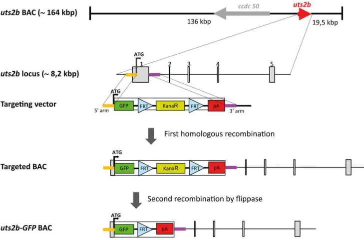
Fig 1. Strategy for constructing the recombinantuts2bBAC containinguts2b-EGFP expression cassette.TheX. tropicalis uts2bBAC clone, ALNOAAA14YB24, contains approximately 164 kbp genomic DNA. Theuts2bgene is approximately 8.2 kbp long as represented by a red arrow, and it comprises 5 exons. The targeting vector was designed to contain twouts2bgenomic DNAs on its either side (denoted 5’- and 3’-arms), that allows a specific homologous recombination between the targeting vector and theuts2bBAC DNA at the genomic region surrounding the first exon. Homology arms for the first exon ofuts2bwere ligated to both ends of an EGFP.FRT-Promotor Bacterial Minimal.kanamycin-FRT.2pA expression cassette. Through the first homologous recombination, the expression cassette was inserted in place of the firstuts2bexon in theuts2bBAC clone. Then, the kanamycin resistance cassette was selectively eliminated by expressing Flipase, leading to theuts2b-EGFP BAC clone containing only the EGFP cDNA expression cassette under control of theuts2bpromoter. Finally, note that theccdc50gene, located close to theuts2blocus, was deleted from theuts2bBAC to avoid possible artifacts due to its overexpression (not shown).
profile for proper cassette integration. A clone satisfying the criteria was then grown on arabi-nose to induce the Flp recombinase which led to excision of the kanamycin cassette.
In addition touts2b, the ALN0AAA14YB24 clone contains the complete locus ofccdc50 which encodes for an effector of EGF-mediated cell signaling shown to be required for survival of some cell types [40–42]. To avoid possible artifacts due to its overexpression,ccdc50was in-activated in the reporter BAC through the insertion in the coding sequence of exon1 of stop co-dons on the 3 reading frames and multimerized polyA signals, further resulting in the
destruction of the exon1 splice donor site (seeS1 Tablefor the primer sequences used).
Generation of transgenic animals
BAC DNA was purified with Nucleobond BAC100 (Macherey-Nagel, Düren, Germany) and dialyzed against microinjection buffer (10 mM Tris-HCl, pH 7.5, 0.1 mM EDTA, 30 µM sper-mine, 70 µM spermidine, 100 mM NaCl) and stored at 4°C [35,43]. On the day of injections, DNA was diluted to 100 ng/µl in sterile water, and further diluted in sperm dilution buffer (250 mM sucrose, 75 mM KCl, 0.5 mM spermidine trihydrochloride, 0.2 mM spermine tetra-hydrochloride, pH 7.3–7.5) to the appropriate concentration (5 pg/nl) for injections. Embryos were injected using a Microinjector 5242 (Eppendorf, Germany) at the one-cell stage with 4 nL of the solution containing the BAC DNA.
Synthesis of the riboprobes for
in situ
hybridization
To generate theuts2bprobe, a PCR fragment of 638 bp was amplified from adultX. laevis brain and spinal cord RACE-ready cDNA (seeS1 Tablefor the primer sequences) then sub-cloned into pGEM-T easy (Promega, Charbonnières, France). Sense and antisense digoxigenin (Dig)-labeled probes were synthesized from the linearized plasmid with the RNA polymerases T7 and Sp6, respectively, using the RNA Labeling Kit (Roche Diagnostics, Mannheim, Germany).
In situ
hybridization
In situhybridization was performed as previously described [44,45] on whole tadpoles (up to stage 40), dissected CNS (from stages 48 to 60) or CNS sections (18 µm). To facilitate observa-tions, whole CNS preparations were longitudinally incised with fine-tip scissors and opened up like a book. To revealuts2bexpression, probes were detected with anti-Dig antibodies conju-gated to alkaline phosphatase followed by a chromogenic reaction using a solution of BM Pur-ple (Roche Diagnostics) or Fast Red (Roche Diagnostics) as substrates. Alternatively, in particular to ascertain transcript co-localization, probes were detected with antibodies conju-gated to horseradish peroxidase and were revealed by Tyramide Signal Amplification using Tyramide-FITC or -TAMRA as substrates. The specificity of theuts2bprobe was verified using the senseuts2bprobe as a negative control.
Immunofluorescence
for 1h30 at room temperature with the fluorescently labeled secondary antibody, and washed again five times 10 min in PBS. The primary antibodies were goat
anti-choline-acetyltransferase (ChAT; 1:100, Millipore) [46] and rabbit anti-GFP (1:300 dilution, Life tech-nologies). The secondary antibodies were donkey anti-rabbit and anti-goat IgGs coupled to Alexa Fluor 488 and Alexa Fluor 546, respectively (1:500, Life Technologies). The specificity of the GFP antibody was verified on wild type animals, while the specificity of secondary antibod-ies was established by omitting the primary specific antibodantibod-ies (data not shown).
α-bungarotoxin staining was performed on whole-mount stage 57 tadpoles fixed in 4% PFA
for 1 h at room temperature according to Ymlahi-Ouazzani et al. [47]. Animals were incubated in Alexa 594-conjugatedα-bungarotoxin (10μg/ml; Life Technologies) overnight at 4°C, then
rinsed six times 20 min in PBS, Triton X-100 0.1% at room temperature.
Combined fluorescent
in situ
hybridization and immunofluorescence
In situhybridization was performed before immunohistochemistry as described above and re-vealed using FITC- or TAMRA-conjugated tyramide. After several washes in PBS, tissues were submitted to immunochemistry as described above.
Neuronal retrograde tracing
Animals were anesthetized in a 0.05% MS-222 water solution and transferred to a Sylgard-lined Petri dish to perform intramuscular dye injections. Spinal motoneurons were retrograde-ly labeled from various muscles at different critical developmental stages. Hereafter, motoneu-rons labeled from tail at stage 50 or 55 will be referred to as axial motoneumotoneu-rons, motoneumotoneu-rons labeled from dorsal trunk at stage 60 as thoracic dorsal motoneurons and motoneurons labeled from hindlimb buds at stage 50 and leg muscles at stage 55 or 60 as appendicular motoneurons [48–50]. First, the skin was dried before making a tiny incision to expose the muscles of inter-est. Crystals of fluorescent dextran amine dyes (Invitrogen) were applied intramuscularly with an insect pin. Either 3 kD rhodamine or 10 kD Alexa fluor 647 were used to be visually compat-ible with the GFP fluorescence wavelength in transgenic animals. Surplus dye was washed out with an excess of cold Ringer solution (75 mM NaCl, 25 mM NaHCO3, 2 mM CaCl2, 2 mM
KCl, 0.5 mM MgCl2, and 11 mM glucose, pH 7.4). Tadpoles recovered from anesthesia in a
water tank and were kept alive for a week to allow tracer migration into the motoneurons. Thereafter, spinal cords were dissected and fixed in 4% PFA for 12 h at 4°C. Neuronal retro-grade tracing experiments combined with whole-mount fluorescent immunochemistry treat-ment (for GFP detection) were carried out as previously described in the immunofluorescence section. Preparations were incubated in a 20% sucrose solution (in PB 0.1%) for 24h at 4°C, then embedded in a tissue-tek solution (VWR-Chemicals) and frozen at−45°C in isopentane.
30 µm cross-sections were made using a Leica Cryostat.
Electrophysiology
integrated (time constant 100 ms; Neurolog System). Spinal GFP+motoneurons were visual-ized within the whole-mount spinal cord (dorsal-side opened) using a differential interference contrast with an infrared video camera and a standard epifluorescent illumination system (FITC filter). Simultaneously to the spinal ventral root recordings, whole-cell patch-clamp re-cordings of GFP+motoneurons were performed with a borosilicate glass patch-clamp electrode (pipette resistance = 5–6 MO; Clark GC 150TF; Harvard Apparatus) and filled with a solution containing 100 mM K-gluconate, 10 mM EGTA, 2 mM MgCl2, 3 mM Na2ATP, 0.5 mM
NaGTP, 10 mM HEPES, pH 7.3. The intracellular signal was acquired using an Axoclamp 2A amplifier (Molecular Devices). All electrophysiological signals were computer-stored using a digitizer interface (Digidata 1440; Pclamp10 software; Molecular Devices) and analyzed off-line using the Clampfit software (Molecular Devices).
Image acquisition
Samples stained by fluorescent probes and/or antibodies were acquired using laser scanning confocal microscopy (Zeiss LSM 510) at wavelengths of 405, 488, 543 and 633 nm. Stacks of 10 to 30 confocal images with 1–10μm z-step intervals were generated with 10x/0.5 air and 20x/
0.75 oil objectives. Final images presented in figures were obtained by orthogonal projection of entire stacks with artificial fluorescent colors using ZEN (Zeiss), Fiji [51] and Adobe Illustrator (Adobe Systems) softwares.
Quantification of immunostained cells in wild type and transgenic
tadpoles
Image stacks with 2–6μm z-step intervals were performed from 72–120μm-thick
whole-mount dissected and opened spinal cords. Quantification of cells was carried out in a domain between the 12thand 15thsegments that contained only axial motoneurons. To assess the uts2b/ChAT co-localization in wild type tadpoles, cells were counted in a region of 634 × 1000μm width (n = 1). Note that the results obtained from this single specimen were globally
consistent with those provided in three other ones. For GFP/uts2band GFP/ChAT co-localizations in transgenic tadpoles, cells were counted in regions of 317 × 1500μm width
(n = 3, for each combination). Immuno-stained cells were manually counted in every plan of the z-stack by using the collaborative bioimage informatics platform, Icy [52].
Results
Expression pattern of the
uts2b
gene during development of wild
type
X. laevis
adulthood (data not shown). No hybridization signal was observed with the senseuts2b ribop-robe (Fig. 2H).
To better characterize theuts2b+cells in the spinal cord, we performed single fluorescentin situhybridization using theuts2bprobe followed by immunohistochemistry labeling against ChAT, a marker of cholinergic neurons. As depicted inFig. 3A,uts2b+cells were restricted to the ventro-medial part of the cholinergic area in the spinal cord of stage-50 tadpoles and they were all found to express ChAT (Fig. 3B), in agreement with their putative motoneuronal na-ture.uts2bmRNA was visualized in a little more than half of all the ChAT+cells analyzed.
Selective retrograde motoneuron labeling was performed with intramuscular application of fluorescent dextran amine dyes, in order to unravel which motoneuron pools expresseduts2b. Axial motoneurons were labeled from the first 10–12thtail myotomes (Fig. 4A) whereas appen-dicular motoneurons were labeled from posterior leggluteus magnusandsemimenbranosus muscles (Fig. 4B), both in stage 55 tadpoles. A significant proportion of the axial retrogradely labeled motoneurons appeared to containutsbmRNA (yellow arrowheads;Fig. 4A3). In con-trast, none of the retrogradely labeled appendicular motoneurons expressedutsb(Fig. 4B). Note that some axial motoneurons retrogradely labeled did not containuts2bmRNA (red ar-rowheads;Fig. 4A3) suggesting thatuts2bis expressed in only a subset of these neurons.
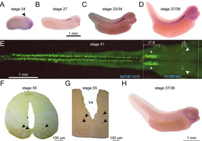
Fig 2.uts2bmRNA is restricted to the ventral spinal cord and hindbraininX. laevistadpoles.Distribution of theuts2bmRNA revealed byin situ hybridization withuts2bantisense probe on whole tadpoles (A–DandH), dissected and opened CNS (E), or CNS sections (F,G) at stages 24 (A), 27 (B), 33/ 34 (C), 37/38 (D) 51 (E), and 55 (F,G).H. Stage 37/38 tadpole hybridized withuts2bsense probe used as negative control.A–DandH. Lateral views of tadpoles with dorsal side up and rostral to the right.E. Dorsal view of whole-mount preparation of hindbrain-spinal cord (CNS). Note that in this preparation, the more ventral structures are closer to the midline.FandG. Coronal sections at the level of the spinal cord (F) and the posterior part of brainstem (G) with dorsal up. Arrowheads point touts2bmRNA detection in stage 24 tadpole and in F and G coronal sections. White arrows designate the region of nuclei of the trigeminal (V) nerve; asterisks denote the region of nuclei of the vagus/hypoglossal/accessory (IX–XI) nerves. r, rhombomere; V4, fourth ventricule.
Generation of
uts2b
-GFP transgenic
X. laevis
tadpoles
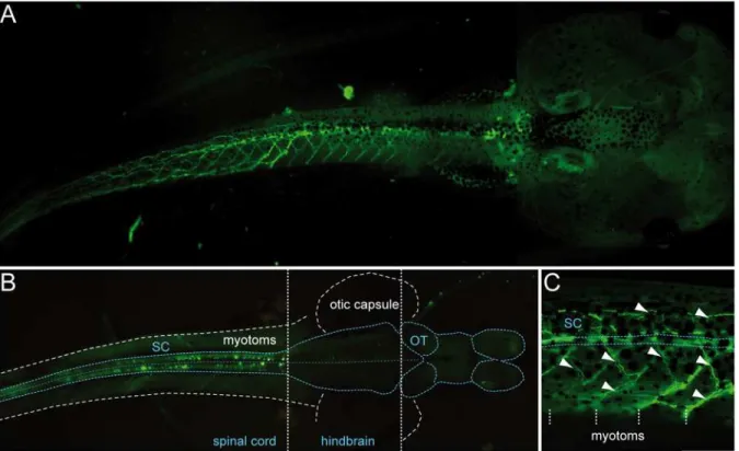
Since we found thatuts2bwas constitutively active in cells that correspond to spinal motoneu-rons,uts2bpromoter was a good candidate to use to drive the expression of GFP in transgenic Xenopusmotoneurons. To generateuts2b-GFP transgenic tadpoles, we used a BAC clone that carried the wholeuts2bgenomic region including the upstream and downstream regions. GFP cDNA was substituted in the first exon of the coding sequence of the uts2b gene, in the BAC clone (process steps summarized inFig. 1). To test the recombineereduts2b-GFP BAC for ex-pression in transgenic animals, circular recombineered BAC DNA was injected into one-cell stageX. laevisembryos, as previously described by Fish et al. (2012). 24.9% of theuts2b-GFP transgenic tadpoles (out of the 2061 injected embryos that were still alive at stage 40 in total) exhibited the expected GFP expression pattern with a robust, extensive rostro-caudal distribu-tion of spinal GFP+cells (Fig. 5A) with labeling of soma (Fig. 5B) and neuronal ramifications (Fig. 5A,C). 18.4% of transgenic tadpoles showed a reduced GFP expression profile, restricted to the soma or to a smaller part of the spinal cord. In contrast, 3.3% showed only ectopic ex-pression of GFP while 53.3% were totally devoid of fluorescence. It is worth noting that in a number of the transgenic tadpoles, the fluorescence was expressed in just one lateral half of the body (data not shown).The GFP expression was seen in spinal neurons without ambiguity, namely both in their somata and axonal projections, from stage 40, until at least stage 62–63. However, the first fluorescence signal could be detected as early as 32, but only as small dots in the rostral spinal cord. It is noteworthy that in some animals, spinal GFP+cells could be
Fig 3. Spinaluts2b+cells are cholinergic neurons inX. laevistadpoles.Confocal images of combined fluorescent in situ hybridization of uts2b mRNA (A1,B1) and ChAT immunolabeling (A2,B2) in a whole-mount dissected and opened spinal cord preparation of stage 50 wild type tadpole.A3,B3. Merged image obtained when uts2b and ChAT stainings were superimposed. Dorsal view with rostral up. Note that in this preparation, the more ventral structures are close to the midline (dashed line). The boxed region inA3is shown at higher magnification inB1–B3.
Fig 4. Spinaluts2b+cells ofX. laevistadpoles project to tail myotomes.Confocal image of combined fluorescentin situhybridization ofuts2bmRNA (A1,B1) and retrograde labeling of spinal axial (A2) and appendicular (B2) motoneurons in stage 55 tadpole whole-mount spinal cord preparations.A3,B3. Merged images ofuts2bstaining and retrograde labeling. All images display dorsal view of hemi-cords, with the rostral side up (dashed-line on the left indicates the midline). Axial motoneurons (Ax MN) were labeled with rhodamine dextran dye (RDA) injected into tail myotomes while appendicular motoneurons (Ap MN) were labeled from posterior leg muscles with Alexa Dextran 647 dye (A.D. 647; see upper scheme on the left panel). The drawing on the left panel illustrates the localization of Ax MN and developing Ap MN in larval spinal cord at stage 55. Yellow arrowheads indicate double stained cells. Red arrowheads indicateuts2b—retrograde labeled cells. sp.sgt 7–10, spinal segments 7 to 10.
detected until stages 65–66. Due to their ventro-medial distribution in the spinal cord (Fig. 5) and their typical morphology (i.e.multipolar soma with ventral and dorsal dendrites and an axon projecting ventrally and caudally toward the axial musculature) it is likely that the GFP+ cells of theuts2b-GFP transgenic tadpoles were spinal motoneurons, as compared to the previ-ously describedX. laevistadpole motoneurons [49,54]. Surprisingly, no GFP+cells were de-tected in the hindbrain (Fig. 5B), in contrast to the endogenousuts2b(Fig. 2E, G).
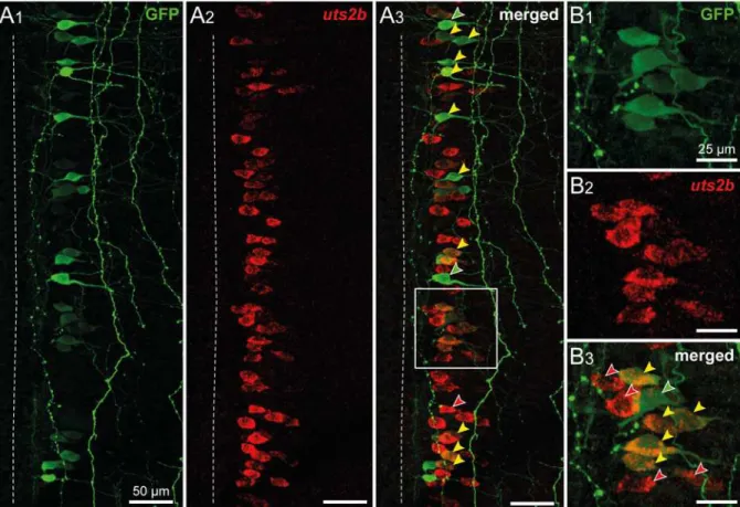
To further compare the GFP and endogenousuts2bexpression patterns, we performed an in situhybridization using theuts2bantisense probe combined with an anti-GFP immunos-taining in the stage 50uts2b-GFP transgenic tadpoles. 82.0 ± 7% of the spinal GFP+cells (n = 3 animals, with ~ 85 GFP+cells counted per animal) were alsouts2b+(yellow arrowheads;
Fig. 6A3, B3). However, 46 ± 9% of theuts2b+cells (n = 3 animals, with ~ 160uts2b+cells counted per animal) did not contain GFP (red arrowheads;Fig. 6A3, B3).
Neurochemical and neuroanatomical characterization of the GFP
+cells
in transgenic
X. laevis
tadpoles
In order to examine the putative motoneuronal nature of GFP+spinal neurons, co-localization of ChAT immuno-fluorescence was investigated inuts2b-GFP transgenic tadpoles exhibiting a strong fluorescence both in the spinal somata and axon projections (Fig. 7). Double immuno-labeling against GFP and ChAT demonstrated that almost all spinal GFP+neurons expressed ChAT at all developmental stages tested. Indeed, at stage 50, 98.2 ± 0.5% (n = 3 animals, ~140
Fig 5. Most of the fluorescence visible in transgenicuts2b-GFPX. laevistadpoles occurs in cells located in the spinal cord and in motor axon
projections. A. GFP fluorescence imaging of a representative transgenic tadpole at stage 58. The tail is seen in lateral view while the head is seen in dorsal view.B. GFP expression at the level of a dissected and opened whole-mount CNS of a stage 50 transgenic tadpole. The CNS was optically exposed by removing the dorsal part of the tail and the top of the head. Note that GFP+cells are restricted to the spinal cord. Dorsal view, rostral to the right.
C. Detail ofA showing GFP+motor axon projections (arrowheads) extending towards axial musculature. White dashed vertical lines indicate myotome boundaries. SC, spinal cord. OT, optic tectum.
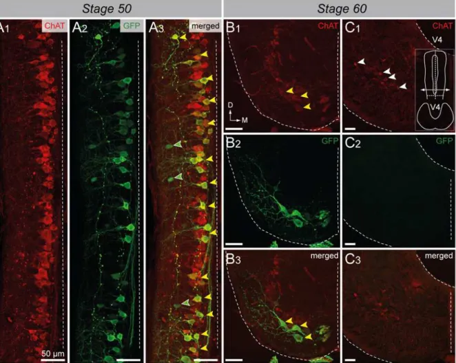
GFP+cells counted per animal) of the GFP+cells were ChAT+. Conversely, 45.0 ± 5.4% (n = 3 animals, ~300 ChAT+cells counted per animal) of the ChAT+neurons were also GFP+(yellow arrowheads;Fig. 7A3), indicating that theuts2b-driven GFP expression was restrained to a spe-cific subpopulation of cholinergic neurons, as reported earlier for endogenousuts2b expres-sion. At stage 60, all spinal ventro-medial GFP+neurons were also found to express ChAT (yellow arrowheads;Fig. 7B3). Here too, we did not observe GFP+cells in the hindbrain (Fig. 7C).
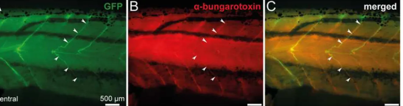
Axonal projections of spinal GFP+cells were further characterized in stage 57 transgenic tadpoles, usingα-bungarotoxin staining to locate the neuromuscular junctions in the axial
musculature. As shown inFig. 8,α-bungarotoxin fluorescence revealed clusters of nicotinic
acetylcholine receptors (Fig. 8B) that were co-localized with GFP+axon terminals (Fig. 8C). Such a specific neuromuscular arrangement was, again, supportive of a motoneuronal nature for GFP+neurons.
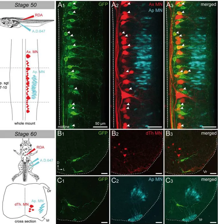
Selective retrograde motoneuron labeling was performed as previously described in wild-type specimens in order to unravel which motoneuron pools expressed GFP from pre- to post-metamorphicuts2b-GFP transgenicXenopus. Axial motoneurons were labeled from the first 10–12thtail myotomes at stage 50 (Fig. 9A); thoracic dorsal motoneurons were labeled from dorsalis truncimuscles at stage 60 (Fig. 9B) whereas appendicular motoneurons were labeled
Fig 6. Theuts2bexpression pattern is partially reproduced by GFP in transgenicuts2b-GFPX. laevistadpoles.Confocal images of combined immuno-labeling of GFP+neurons (
A1,B1) and fluorescentin situhybridization ofuts2bmRNA (A2,B2) in a whole-mount spinal cord preparation of stage 50uts2b-GFP tadpole.A3,B3. Merged image obtained when GFP anduts2bstainings were superimposed. Dorsal view with rostral up. Only the hemi-cord is shown. The dashed-line on the left side represents the midline. Note that in this preparation, the more ventral structures are closer to the midline. The boxed region inA3is shown at higher magnification inB1–B3. Yellow arrowheads denote cells that express both GFP anduts2b. Red arrowheads designate uts2b+/GFP−cells and green arrowheads designateuts2b−/GFP+cells.
from hindlimb buds at stage 50 (Fig. 9A) and from thegluteus magnusandsemimenbranosus leg muscles at stage 60 (Fig. 9C). A significant proportion of the axial motoneurons retrograde-ly labeled appeared to be GFP+(white arrowheads;Fig. 9A). This demonstrated that theuts2b -GFP transgene was expressed in tail myotome-innervating motoneurons in pre-metamorphic stages 50–58, corroborating the above ChAT immuno-labeling patterns (Fig. 7). In contrast, none of the retrogradely labeled thoracic dorsal (Fig. 9B) or appendicular (Fig. 9A, C) moto-neurons expressed GFP.
Electrophysiological characterization of the GFP
+cells in transgenic
X. laevis
tadpoles
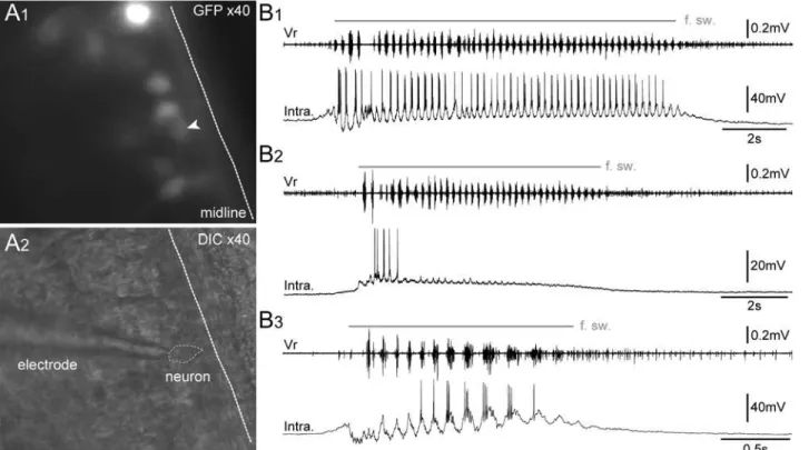
GFP+spinal neurons were specifically targeted for patch-clamp recordings (Fig. 10A) in order to functionally characterize their electrophysiological activity during the spontaneous fictive
Fig 7. Spinal GFP+cells of transgenicuts2b-GFPX. laevistadpoles mainly express ChAT.Confocal images of combined immuno-labeling of ChAT+
(A1) and GFP+(A2) neurons in a whole-mount spinal cord preparation of stage 50 transgenic tadpole.A3. Merged image obtained when ChAT and GFP
stainings were superimposed. Dorsal view with rostral up. Only the hemi-cord is shown. The dashed-line on the right side represents the midline. Note that in this preparation, the more ventral structures are closer to the midline. Confocal images of combined immuno-labeling of ChAT+(B1–C1) and GFP+(B2–C2)
neurons in lumbar spinal cord (B) and brainstem (C) cross-sections of stage 60uts2b-GFP tadpoles.B3,C3. Merged images obtained when ChAT and GFP stainings were superimposed. The cross-section inC1–3originates from the caudal hindbrain (rhombomeres 7–8) where IX-XI motor nuclei are located. Yellow arrowheads designate GFP+/ChAT+cells; green arrowheads designate GFP+/ChAT−cells; White arrowheads designate GFP−/ChAT+cells. D,
dorsal; M, medial; V4, 4thventricule.
swimming (Fig. 10B) inin vitroisolated spinal cord preparations. All recorded GFP+neurons (n = 17) were rhythmically active in coordination with the fictive swimming bursts of activity recorded from axial ventral motor roots (Vr). Three main intracellular discharge patterns were observed:i)some neurons (n = 5) were depolarized at the onset of the fictive swimming and fired rhythmically coupled with Vr bursts throughout the episode duration (Fig. 10B1);ii) some neurons (n = 4) stopped firing after the 3–4 first Vr bursts despite the persistence of the swimming activity, however, these neurons continued to exhibit rhythmic depolarizations in strict coordination with the following Vr bursts (Fig. 10B2);iii)the other neurons (n = 8) were hyperpolarized at the beginning of the fictive swimming episode and started to fire in coordina-tion with the Vr rhythmic bursts with a delay after the swimming onset (Fig. 10B3). For all re-corded GFP+neurons, the membrane potential was between‑53 and‑68 mV. These
electrophysiological results are consistent with locomotor-related firing patterns previously de-scribed in motoneurons of larvalXenopus[55] and zebrafish [56–58].
Taken together, neurochemical, anatomical and electrophysiological results demonstrate that GFP+spinal neurons represent a heterogeneous subpopulation of axial motoneurons in-nervating tail muscles.
Discussion
The aim of this study was to test the ability of the regulatory sequences of theuts2bgene to drive expression of reporter genes inXenopusmotoneurons. Expression ofuts2bin motoneu-rons of the brainstem and spinal cord has been formally demonstrated in several species in-cluding the mice [28], chicken and zebrafish (HT, unpublished results). In mice, it has been shown that a majority of spinal motoneurons simultaneously expressesuts2band its paralog uts2[27]. InX. laevis, Konno et al. [29] reported the occurrence of UTS2/UTS2B-immunoreac-tive motoneurons in the brain and spinal cord but due to the technique employed they were not able to discriminate the cells expressing specifically each peptide. Here, we provide evi-dence that in pre-metamorphic tadpole, spinaluts2b+cells are motoneurons, as revealed by the fact that they express ChAT and can be retrolabeled from axial muscles. The onset of theuts2b gene expression (stage 24), which appears shortly after differentiation of the first motoneurons (up to stage 22) [59], is consistent with this view, even if at earliest stages, the motoneuronal nature of theuts2b+cells remains to be confirmed. At later stages,uts2bmRNA was detected in retrolabled axial motoneurons but not with appendicular motoneurons suggesting thatuts2bis specifically expressed in axial motoneurons innervating tail muscles.
Fig 8. Spinal GFP+axon motor projections of transgenicuts2b-GFPX. laevistadpoles co-localize withα-bungarotoxin, a marker of postsynaptic
neuromuscular junctions.Combined fluorescence of GFP (A) and immuno-labeling ofα-bungarotoxin (B) at the level of spinal axon motor projections in a whole-mount stage 57 transgenic tadpole.C. Merged image obtained when GFP fluorescence andα-bungarotoxin staining were superimposed.
Postsynaptic nicotinic acetylcholine receptors labeled byα-bungarotoxin and GFP+motor neuron axons overlap (arrowheads). Lateral views, rostral to the left.
Fig 9. Spinal GFP+cells of transgenicuts2b-GFPX. laevistadpoles project to tail myotomes.Confocal image of combined immuno-labeling of spinal GFP+neurons (
A1) and retrograde labeling of spinal motoneurons (A2) in a stage 50uts2b-GFP tadpole whole-mount spinal cord preparation.A3. Merged image obtained when GFP staining and retrograde labeling were superimposed. Dorsal view with rostral up. Only the hemi-cord is shown. The dashed-line on the right side represents the midline. Axial motoneurons (Ax MN) innervating myotomes were labeled from tail muscles with rhodamine (RDA) dextran dye whereas appendicular motoneurons (Ap MN) innervating limbs were labeled from hindlimb buds with alexa dextran 647 dye (A.D. 647; see scheme on the left panel). The drawing at the top left panel illustrates the medio-lateral localization of Ax MN and developing Ap MN in larval spinal cord at stage 50. Yellow arrowheads indicate double stained cells. Cross-section confocal image of combined immuno-labeling of spinal GFP+neurons (B1,C1) and retrograde labeling of dorsal thoracic motoneurons (dTh MN,B2) and Ap MN, (C2) in stage 60 metamorphosinguts2b-GFP tadpole.B3,C3. Merged images obtained GFP staining and retrograde labeling were superimposed. dTh MN were labeled fromdorsalis truncimuscles with RDA whereas Ap MN were labeled from groups of extensor and flexor of the hindlimb with A.D. 647 (see scheme on the left side). The cross-section drawing at the bottom left panel illustrates the dorso-ventral localization of dTh MN and Ap MN in the future adult spinal cord at stage 60. sp.sgt 7–10, spinal segments 7 to 10, Vr, ventral root. D, dorsal; L, lateral.
The absence ofuts2b+cells in the spinal cord of adult specimens was quite surprising since uts2bmRNA in spinal cord was detected by RT-PCR [29]. Our results indicate thatuts2b ex-pression pattern ofX. laevisin adult is very similar to that found in rat. Indeed, the rat spinal cord has been shown to contain onlyuts2but notuts2bmRNA [25].
TheX. laevis uts2bexpression pattern suggests the presence, in its promoter, of regulatory ele-ments that are able to drive motoneuron-specific expression. Since BAC technology makes it possible to generate transgenes even when the promoter region of a gene is unknown, which was the case here, we decided to generate transgenic tadpoles expressing the EGFP reporter from a BAC containing the entireX. tropicalis uts2bgene including more than 130 kbp of upstream and almost 20 kbp of downstream regulatory elements. BAC transgenesis has been successfully used for little over a decade in mouse [38], and recently has been more readily applied to other species such as zebrafish [60]. InXenopus, trials of BAC transgenesis have been reported in only two studies so far [35,61]. We opted for the method developed by Fish et al. [35] due to its simplicity and high efficiency. Up to 25% of theuts2b-GFP transgenic tadpoles globally exhibited the ex-pected pattern of fluorescence in our case, admittedly somewhat lower than that reported in the original publication (i.e.60%) but nonetheless a very acceptable rate. The delay between the first uts2bsignal (stage 24) and the first GFP fluorescence signal (stage 32) may correspond to the time needed to complete both translation and then maturation of GFP [62].
Some other discrepancies were nevertheless observed between the GFP and endogenous uts2bexpression patterns. No fluorescence was expressed in the hindbrain, suggesting that
Fig 10. GFP+cells exhibit a typical pattern of motoneuronal electrical activity during spontaneous fictive swimming episodes.Visualization of spinal GFP+neurons at x40 in fluorescence condition (A1) and infrared condition (A2) in a whole-mount isolatedin vitropreparation of brainstem-spinal cord with dorsal-side opened. Patch-clamp intracellular recordings (bottom traces) of spinal GFP+neurons (Intra.) either with a persisting (B1), a non-persisting firing
pattern (B2), or a slow-delayed firing pattern (B3) during spontaneous fictive swimming episodes (f. sw.). The upper trace inB1–B3represents the
extracellular fictive swimming activity recorded simultaneously in a ventral motor root (Vr). DIC, differential interference contrast. The white arrowhead points to the GFP+neuron targeted for patch clamp recording.
some of the regulatory elements of theX. tropicalis uts2bgene, crucial for its expression in the brainstem, were not present in the BAC, or not functional inX. laeviscells. At the spinal cord level, manyuts2b+cells expressed GFP. However, we also observed a number ofuts2b+cells that did not express GFP, as well as some GFP+cells that did not expressuts2b. Theuts2b+ /GFP- cells could be cells lacking theuts2b-GFP BAC DNA, or containing only a very small number of copies. Alternatively, theseuts2b+/GFP−cells could represent newly differentiated neurons in which GFP had not the time to accumulate, since motoneurogenesis continues to be active during a large part of the larval period (up to stage 52) [63]. The occurrence of GFP+/ uts2b-cells might potentially reflect temporal variations inuts2bexpression in motoneurons, i.e.motoneurons that had temporarily ceased to expressuts2bbut still contained GFP. Howev-er, the fact that all spinal GFP+cells of ouruts2b-GFP transgenic tadpoles were motoneurons is supported by several lines of evidence:i)they were exclusively located in the ventral horns of the spinal cord;ii)they were almost all ChAT+;iii)they could be stained by retrograde labeling from axial muscles;iv)they exhibited locomotion-related rhythmic discharges during sponta-neous fictive swimming episodes andv)the axon terminals co-localized withα-bungarotoxin.
However, as mentioned previously foruts2bin wild-type, only a fraction of the motoneurons appeared to express GFP. Retrograde labeling reveals that these neurons correspond to axial motoneurons, which innervate tail muscles, but not to appendicular or thoracic motoneurons. In this respect, the GFP expression pattern is perfectly consistent with endogenousuts2b ex-pression in spinal cord of wild-type tadpoles. The fact that very few spinal GFP+cells could be identified in early post-metamorphic juveniles also supports this view, since axial motoneurons elimination starts during metamorphosis as the tail regresses [64–66]. The higher number of spinal GFP+cells in transgenic juveniles compared to that ofuts2b+cells in wild-type ones is probably due to the higher stability of EGFP [67].
We used a transgenesis method in which the BAC DNA is injected into one-cell stage em-bryos as a circular DNA molecule with no mechanism to mediate genomic integration [35]. It is therefore believed that the injected DNA can be only transiently transcribed unless it is repli-cated in a similar fashion as the nuclear DNA. Since BAC DNA can contain a large amount of genomic DNA, it is more efficiently replicated [35]. In their original study using apax6-GFP BAC, Fish et al. [35] actually reported loss of transgene expression around stage 50 in most of the injected embryos. In the present study, we observed that in most transgenic tadpoles, GFP was still significantly expressed up until stage 62–63 and, as mentioned above, GFP+cells could be even observed in stage 66 juveniles. Thus, the decrease in GFP expression observed during metamorphosis may not be due to any BAC loss but, rather, reflects the endogenous expression pattern ofuts2b. Maintenance of the transgene expression for such a long time may be ex-plained by BAC DNA episomal replication and a particular stability of the intracellular envi-ronment, perhaps related to the post-mitotic state of motoneurons [68].
Conclusion
Supporting Information
S1 Table. Sequences of the primers used for PCR amplifications. (DOCX)
Acknowledgments
We thank Gérard Benisti forXenopuscare, Drs Ghislaine Morvan-Dubois, Laurent Coen, Natacha Kenigfest (MNHN) for helpful discussions and Pr Barbara Demeneix (MNHN) for her support. We are grateful to Feng Quan, Sébastien Le Mével (MNHN) and Laura Cardoit (University of Bordeaux) for excellent technical assistance, to Margaret B. Fish (University of Virginia, USA) for precious advice regarding BAC injections and to Bilal Mughal (MNHN) for English proofreading of the manuscript. We also thank the teams of the“Plateforme d’Imagerie Cellulaire Pitié Salpêtrière”and the“Bordeaux Imaging Center”for their support.
Author Contributions
Conceived and designed the experiments: MB HT. Performed the experiments: MB FA FML DLR MTB. Analyzed the data: MB FA FML DLR MTB HT. Contributed reagents/materials/ analysis tools: DC FR NP. Wrote the paper: MB FML HT.
References
1. Abe T, Fujimori T (2013) Reporter mouse lines for fluorescence imaging. Dev Growth Differ 55:390–405. doi:10.1111/dgd.12062PMID:23621623
2. Becker TS, Rinkwitz S (2012) Zebrafish as a genomics model for human neurological and polygenic disorders. Dev Neurobiol 72:415–428. doi:10.1002/dneu.20888PMID:21465670
3. Babin PJ, Goizet C, Raldúa D (2014) Zebrafish models of human motor neuron diseases: advantages and limitations. Prog Neurobiol 118:36–58. doi:10.1016/j.pneurobio.2014.03.001PMID:24705136
4. Pratt KG, Khakhalin AS (2013) Modeling human neurodevelopmental disorders in theXenopus tad-pole: from mechanisms to therapeutic targets. Dis Model Mech 6:1057–1065. doi:10.1242/dmm. 012138PMID:23929939
5. Schmitt SM, Gull M, Brändli AW (2014) EngineeringXenopusembryos for phenotypic drug discovery screening. Adv Drug Deliv Rev 69–70:225–46. doi:10.1016/j.addr.2014.02.004PMID:24576445
6. Howe K, Clark MD, Torroja CF, Torrance J, Berthelot C, et al. (2013) The zebrafish reference genome sequence and its relationship to the human genome. Nature 496:498–503. doi:10.1038/nature12111
PMID:23594743
7. Hellsten U, Harland RM, Gilchrist MJ, Hendrix D, Jurka J, et al. (2010) The genome of the Western clawed frogXenopus tropicalis. Science 328:633–636. doi:10.1126/science.1183670PMID:
20431018
8. Detrich III HW, Westerfield M, Zon LI (2011) The zebrafish: genetics, genomics and informatics. Aca-demic Press, San Diego. 300 p.
9. Hwang WY, Fu Y, Reyon D, Maeder ML, Tsai SQ, et al. (2013) Efficient genome editing in zebrafish using a CRISPR-Cas system. Nat Biotechnol 31:227–229. doi:10.1038/nbt.2501PMID:23360964
10. Vouillot L, Thélie A, Scalvenzi T, Pollet N (2014) Genomics and Genome Engineering inXenopus. In: Kloc, M, Kubiak, JZ, editors.XenopusDevelopment. John Wiley & Sons, Inc. pp 383–402.
11. Nakayama T, Fish MB, Fisher M, Oomen-Hajagos J, Thomsen GH, et al. (2013) Simple and efficient CRISPR/Cas9-mediated targeted mutagenesis inXenopus tropicalis. Genesis 51:835–843. doi:10. 1002/dvg.22720PMID:24123613
12. Grillner S, Jessell TM (2009) Measured motion: searching for simplicity in spinal locomotor networks. Curr Opin Neurobiol 19:572–586. doi:10.1016/j.conb.2009.10.011PMID:19896834
14. Higashijima S, Hotta Y, Okamoto H (2000) Visualization of cranial motor neurons in live transgenic zeb-rafish expressing green fluorescent protein under the control of the islet-1 promoter/enhancer. J Neu-rosci 20:206–218. PMID:10627598
15. Shin J, Park HC, Topczewska JM, Mawdsley DJ, Appel B (2003) Neural cell fate analysis in zebrafish using olig2 BAC transgenics. Methods Cell Sci 25:7–14. doi:10.1023/B:MICS.0000006847.09037.3a
PMID:14739582
16. Pelzer D, McDermott L, Dorey K (2014) Characterisation of a transgenicXenopustropicalis line (Hb9-GFP) for the analysis of motor neuron regeneration. 15thInternationalXenopusConference. Pacific
Grove, USA.
17. Park HC, Shin J, Appel B (2004) Spatial and temporal regulation of ventral spinal cord precursor specifi-cation by Hedgehog signaling. Development 131:5959–5969. doi:10.1242/dev.01456PMID:15539490
18. Korzh V, Edlund T, Thor S (1993) Zebrafish primary neurons initiate expression of the LIM homeodo-main protein Isl-1 at the end of gastrulation. Development 118:417–425. PMID:8223269
19. Seredick SD, Van Ryswyk L, Hutchinson SA, Eisen JS (2012) Zebrafish Mnx proteins specify one mo-toneuron subtype and suppress acquisition of interneuron characteristics. Neural Dev 7:35. doi:10. 1186/1749-8104-7-35PMID:23122226
20. Tostivint H, Quan FB, Bougerol M, Kenigfest NB, Lihrmann I (2013) Impact of gene/genome duplications on the evolution of the urotensin II and somatostatin families. Gen Comp Endocrinol 188: 110–117. doi:
10.1016/j.ygcen.2012.12.015PMID:23313073
21. Tostivint H, Ocampo Daza D, Bergqvist CA, Quan FB, Bougerol M, et al. (2014) Molecular evolution of somatostatin and urotensin II systems. J Mol Endocrinol 52:T61–86. doi:10.1530/JME-13-0274PMID:
24740737
22. Coulouarn Y, Lihrmann I, Jégou S, Anouar Y, Tostivint H, et al. (1998) Cloning of the cDNA encoding the urotensin II precursor in frog and human reveals intense expression of the urotensin II gene in moto-neurons of the spinal cord. Proc Natl Acad Sci USA 95: 15803–15808. doi:10.1073/pnas.95.26.15803
PMID:9861051
23. Coulouarn Y, Jegou S, Tostivint H, Vaudry H, Lihrmann I (1999) Cloning, sequence analysis and tissue distribution of the mouse and rat urotensin II precursors. FEBS Lett 457: 28–32. doi: 10.1016/S0014-5793(99)01003-0PMID:10486557
24. Dun SL, Brailoiu GC, Yang J,Chang JK,Dun NJ (2001) Urotensin II-immunoreactivity in the brainstem and spinal cord of the rat. Neurosci Lett 305:9–12. doi:10.1016/S0304-3940(01)01804-3PMID:
11356295
25. Sugo T, Murakami Y, Shimomura Y, Harada M, Abe M, I et al. (2003) Identification of urotensin II-related peptide as the urotensin II-immunoreactive molecule in the rat brain. Biochem Biophys Res Commun 310:860–868. doi:10.1016/j.bbrc.2003.09.102PMID:14550283
26. Pelletier G, Lihrmann I, Vaudry H (2002) Role of androgens in the regulation of urotensin II precursor mRNA expression in the rat brainstem and spinal cord. Neuroscience 115:525–532. doi:10.1016/ S0306-4522(02)00413-XPMID:12421619
27. Pelletier G, Lihrmann I, Dubessy C, Luu-The V, Vaudry H, et al. (2005) Androgenic down-regulation of urotensin II precursor, urotensin II-related peptide precursor and androgen receptor mRNA in the mouse spinal cord. Neuroscience 132:689–696. doi:10.1016/j.neuroscience.2004.12.045PMID:
15837130
28. Dubessy C, Cartier D, Lectez B, Bucharles C, Chartrel N, et al. (2008) Characterization of urotensin II, distribution of urotensin II, urotensin II-related peptide and UT receptor mRNAs in mouse: evidence of urotensin II at the neuromuscular junction. J Neurochem 107: 361–374. doi:10.1111/j.1471-4159. 2008.05624.xPMID:18710417
29. Konno N, Fujii Y, Imae H, Kaiya H, Mukuda T, et al. (2013) Urotensin II receptor (UTR) exists in hyaline chondrocytes: a study of peripheral distribution of UTR in the African clawed frog,Xenopus laevis. Gen Comp Endocrinol 185: 44–56. doi:10.1016/j.ygcen.2013.01.015PMID:23399967
30. Douglas SA, Dhanak D, Johns DG (2004) From‘gills to pills’: urotensin-II as a regulator of mammalian cardiorenal function. Trends Pharmacol Sci 25:76–85. doi:10.1016/j.tips.2003.12.005PMID:15102493
31. Vaudry H, Do Rego JC, Le Mevel JC, Chatenet D, Tostivint H, et al. (2010) Urotensin II, from fish to human. Ann N Y Acad Sci 1200: 53–66. doi:10.1111/j.1749-6632.2010.05514.xPMID:20633133
32. Vaudry H, Leprince J, Chatenet D, Fournier A, Lambert DG, et al. (2014) International union of basic and clinical pharmacology: urotensin II, urotensin II-related peptide and their receptor: from structure to function. Pharmacol Rev (in press).
34. Long X, Miano JM (2007) Remote control of gene expression. J. Biol. Chem., 282:15941–15945. doi:
10.1074/jbc.R700010200PMID:17403687
35. Fish MB, Nakayama T, Grainger RM (2012) Simple, fast, tissue-specific bacterial artificial chromosome transgenesis inXenopus. Genesis 50:307–315. doi:10.1002/dvg.20819PMID:22084035
36. Sive HL, Grainger RM, Harland RM (2007) Inducing ovulation in Xenopus laevis. CSH Protoc pdb: prot4734.
37. Nieuwkoop PD, Faber J (1994) Normal table of Xenopuslaevis(Daudin). New York: Garland Publish-ing, Inc. 282 p.
38. Copeland NG, Jenkins NA, Court DL (2001) Recombineering: a powerful new tool for mouse functional genomics. Nat Rev Genet 2:769–779. doi:10.1038/35093556PMID:11584293
39. Lee EC, Yu D, Martinez de Velasco J, Tessarollo L, Swing DA, et al. (2001) A highly efficient Escheri-chia coli-based chromosome engineering system adapted for recombinogenic targeting and subcloning of BAC DNA. Genomics 73:56–65. doi:10.1006/geno.2000.6451PMID:11352566
40. Blagoev B, Ong SE, Kratchmarova I, Mann M (2004) Temporal analysis of phosphotyrosine-dependent signalling networks by quantitative proteomics. Nat Biotechnol 22:1139–1145. doi:10.1038/nbt1005
PMID:15314609
41. Kratchmarova I, Blagoev B, Haack-Sorensen M, Kassem M, Mann M (2005) Mechanism of divergent growth factor effects in mesenchymal stem cell differentiation. Science 308:1472–1477. doi:10.1126/ science.1107627PMID:15933201
42. Farfsing A, Engel F, Seiffert M, Hartmann E, Ott G, et al. (2009) Gene knockdown studies revealed CCDC50 as a candidate gene in mantle cell lymphoma and chronic lymphocytic leukemia. Leukemia 23:2018–2026. doi:10.1038/leu.2009.144PMID:19641524
43. Schedl A, Larin Z, Montoliu L, Thies E, Kelsey G, et al. (1993) A method for the generation of YAC trans-genic mice by pronuclear microinjection. Nucleic Acids Res 21:4783–4787. doi:10.1093/nar/21.20. 4783PMID:8233827
44. Parmentier C, Hameury E, Lihrmann I, Taxi J, Hardin-Pouzet H, et al. (2008) Comparative distribution of the mRNAs encoding urotensin I and urotensin II in zebrafish. Peptides 29: 820–829. doi:10.1016/j. peptides.2008.01.023PMID:18403048
45. Djenoune L, Khabou H, Joubert F, Quan FB, Nunes Figueiredo S, et al. (2014) Investigation of spinal cerebrospinal fluid-contacting neurons expressing PKD2L1: evidence for a conserved system from fish to primates. Front Neuroanat 8: 26. doi:10.3389/fnana.2014.00026PMID:24834029
46. Lopez JM, Smeets WJ, Gonzalez A (2002) Choline acetyltransferase immunoreactivity in the develop-ing brain ofXenopus laevis. J Comp Neurol 453:418–434. doi:10.1002/cne.10419PMID:12389211
47. Ymlahi-Ouazzani QJ, Bronchain O,Paillard E,Ballagny C,Chesneau A, et al. (2010) Reduced levels of survival motor neuron protein leads to aberrant motoneuron growth in aXenopusmodel of muscular at-rophy. Neurogenetics 11:27–40. doi:10.1007/s10048-009-0200-6PMID:19517146
48. Forehand CJ, Farel PB (1982) Spinal cord development in anuran larvae: I. Primary and secondary neurons. J Comp Neurol 209:386–394. doi:10.1002/cne.902090409PMID:6982287
49. van Mier P, van Rheden R, ten Donkelaar HJ (1985) The development of the dendritic organization of primary and secondary motoneurons in the spinal cord ofXenopus laevis. An HRP study.Anat Embryol 172:311–324. doi:10.1007/BF00318979PMID:4061871
50. Roberts A, Walford A, Soffe SR, Yoshida M (1999) Motoneurons of the axial swimming muscles in hatchlingXenopustadpoles: features, distribution, and central synapses. J Comp Neurol 411:472–486. doi:10.1002/(SICI)1096-9861(19990830)411:3%3C472::AID-CNE9%3E3.0.CO;2-BPMID:10413780
51. Schindelin J, Arganda-Carreras I, Frise E, Kaynig V, Longair M, et al. (2012) Fiji: an open-source plat-form for biological-image analysis. Nat Methods 9:676–682. doi:10.1038/nmeth.2019PMID:22743772
52. de Chaumont F, Dallongeville S, Chenouard N, Hervé N, Pop S, et al. (2012) Icy: an open bioimage in-formatics platform for extended reproducible research. Nat Methods 9:690–696. doi:10.1038/nmeth. 2075PMID:22743774
53. Straka H, Gilland E, Baker R (1998) Rhombomeric organization of brainstem motor neurons in larval frogs. Biol Bull 195:220–222. doi:10.2307/1542849PMID:9818376
54. van Mier P, Joosten HW, van Rheden R, ten Donkelaar HJ (1986) The development of serotonergic raphespinal projections in Xenopus laevis. Int J Dev Neurosci 9:465–475. doi:10.1016/0736-5748(86) 90028-6
56. Ampatzis K, Song J, Ausborn J, El Manira A (2013) Pattern of innervation and recruitment of different classes of motoneurons in adult zebrafish. J Neurosci 33:10875–10886. doi:10.1523/JNEUROSCI. 0896-13.2013PMID:23804107
57. Gabriel JP, Ausborn J, Ampatzis K, Mahmood R, Eklöf-Ljunggren E, et al. (2011) Principles governing recruitment of motoneurons during swimming in zebrafish. Nat Neurosci 14:93–99. doi:10.1038/nn. 2704PMID:21113162
58. McLean DL, Fan J, Higashijima S, Hale ME, Fetcho JR (2007) A topographic map of recruitment in spi-nal cord. Nature 446:71–75. doi:10.1038/nature05588PMID:17330042
59. Hartenstein V (1993) Early pattern of neuronal differentiation in the Xenopus embryonic brainstem and spinal cord. J Comp Neurol 328:213–231. doi:10.1002/cne.903280205PMID:8423241
60. Suster ML, Abe G, Schouw A, Kawakami K (2011) Transposon-mediated BAC transgenesis in zebra-fish. Nat Protoc 6:1998–2021. doi:10.1038/nprot.2011.416PMID:22134125
61. Kelly LE, Davy BE,Berbari NF,Robinson ML,El-Hodiri HM (2005) RecombineeredXenopus tropicalis BAC expresses a GFP reporter under the control of Arx transcriptional regulatory elements in transgen-icXenopus laevisembryos. Genesis 41:185–191. doi:10.1002/gene.20113PMID:15789419
62. Tsien RY (1998) The green fluorescent protein. Annu Rev Biochem 67:509–44. doi:10.1146/annurev. biochem.67.1.509PMID:9759496
63. Schlosser G, Koyano-Nakagawa N, Kintner C (2002) Thyroid hormone promotes neurogenesis in the Xenopusspinal cord. Dev Dyn 225:485–498. doi:10.1002/dvdy.10179PMID:12454925
64. Hughes A (1961) Cell degeneration in the larval ventral horn ofXenopus laevis(Daudin) J Embryol Exp Morphol 9:269–284. PMID:13716620
65. Prestige MC (1967) The control of cell number in the lumbar ventral horns during the development of Xenopus laevistadpoles. J Embryol Exp Morphol 18:359–387. PMID:5587106
66. Marsh-Armstrong N, Cai L, Brown DD (2004) Thyroid hormone controls the development of connec-tions between the spinal cord and limbs duringXenopus laevismetamorphosis. Proc Natl Acad Sci USA 101:165–170. doi:10.1073/pnas.2136755100PMID:14691251
67. Zhang G, Gurtu V, Kain SR (1996) An enhanced green fluorescent protein allows sensitive detection of gene transfer in mammalian cells. Biochem Biophys Res Commun 227:707–711. doi:10.1006/bbrc. 1996.1573PMID:8885998
68. Lee SK, Lee B, Ruiz EC, Pfaff SL (2005) Olig2 and Ngn2 function in opposition to modulate gene ex-pression in motor neuron progenitor cells. Genes Dev 19:282–294. doi:10.1101/gad.1257105PMID: